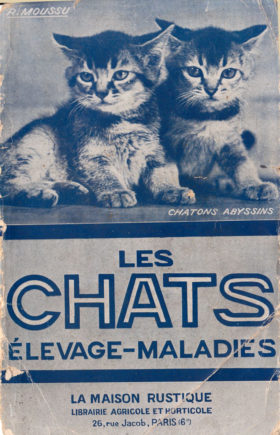
CATS - BREEDING AND DISEASES
By R. MOUSSU
Head of Clinic at the National Veterinary School of Alfort.
1938
TABLE OF CONTENTS
Reproduction.
Choice of breeding stock.
Oestrus
Mating
Maternal impregnation
Pregnancy
Pregnancy hygiene
Kittening
Difficulties in kittening
Breeding.
Care of the young
Weaning
Mother care
Artificial breastfeeding
Hygiene and post-weaning care
Feeding
Castration
Male castration
Castration of the female
Skin hygiene
Diseases of the musculoskeletal system.
Fractures
Joint diseases.
Sprains
Dislocations
Traumatic arthritis
Diseases of the tendons and muscles.
Sectioned tendons
Wounds
Abscess
Diseases of the foot.
Tearing and fracture of the claws.
Foreign bodies.
Diseases of the digestive system.
Diseases of the mouth
Stomatitis
Dental tartar
Foreign bodies
Labial ulcer
Pharyngitis
Obstruction of the oesophagus
Diseases of the stomach, intestines, liver and peritoneum.
Stomach indigestion
Foreign bodies in the stomach
Gastroenteritis
Constipation
Bowel obstruction
Jaundice
Ascites
Poisoning
Poisoning by spoiled food
Phosphorus poisoning
Arsenic poisoning
Intestinal worms
Ascarids
Tapeworms
Disorders of the anus and rectum
Imperforate anus
Rectal prolapse
Diseases of the respiratory system.
Nasal cavity disorders
Coryza
Epistaxis
Disorders of the bronchi and lungs.
Acute bronchitis
Bronchopneumonia
Chronic bronchitis
Pulmonary emphysema
Diseases of the pleura
Pleurisy
Diseases of the nervous system.
Eclampsia
Epilepsy
False pregnancy
Diseases of the genitals
Funiculitis
Swollen breasts
Mastitis
Metritis
Mammary tumours
Diseases of the urinary system.
Congestion of the kidneys
Acute nephritis
Chronic nephritis
Bladder diseases
Cystitis
Urinary lithiasis. Calculus
Hernias
Umbilical hernia
Ventral hernia
Skin diseases.
Eczema
Pityriasis
Syphiloid lesions
Scabies
Ringworm
Skin parasites, external:
Lice
Fleas
Harvest mites
Ear disease.
Auricular catarrh
Ear haematoma
Ear mites
Eye diseases.
Conjunctivitis
Keratitis
Dislocation of the eye
Protruding third eyelid
Infectious diseases.
Infectious angina
Contagious coryza
Infectious gastroenteritis
Typhus
Vaccination
Distemper
Rabies
Tuberculosis
PART ONE.
CHAPTER 1. REPRODUCTION
1. Choosing Breeding Stock.
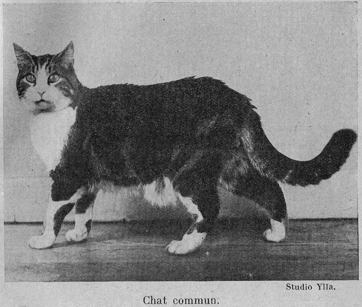
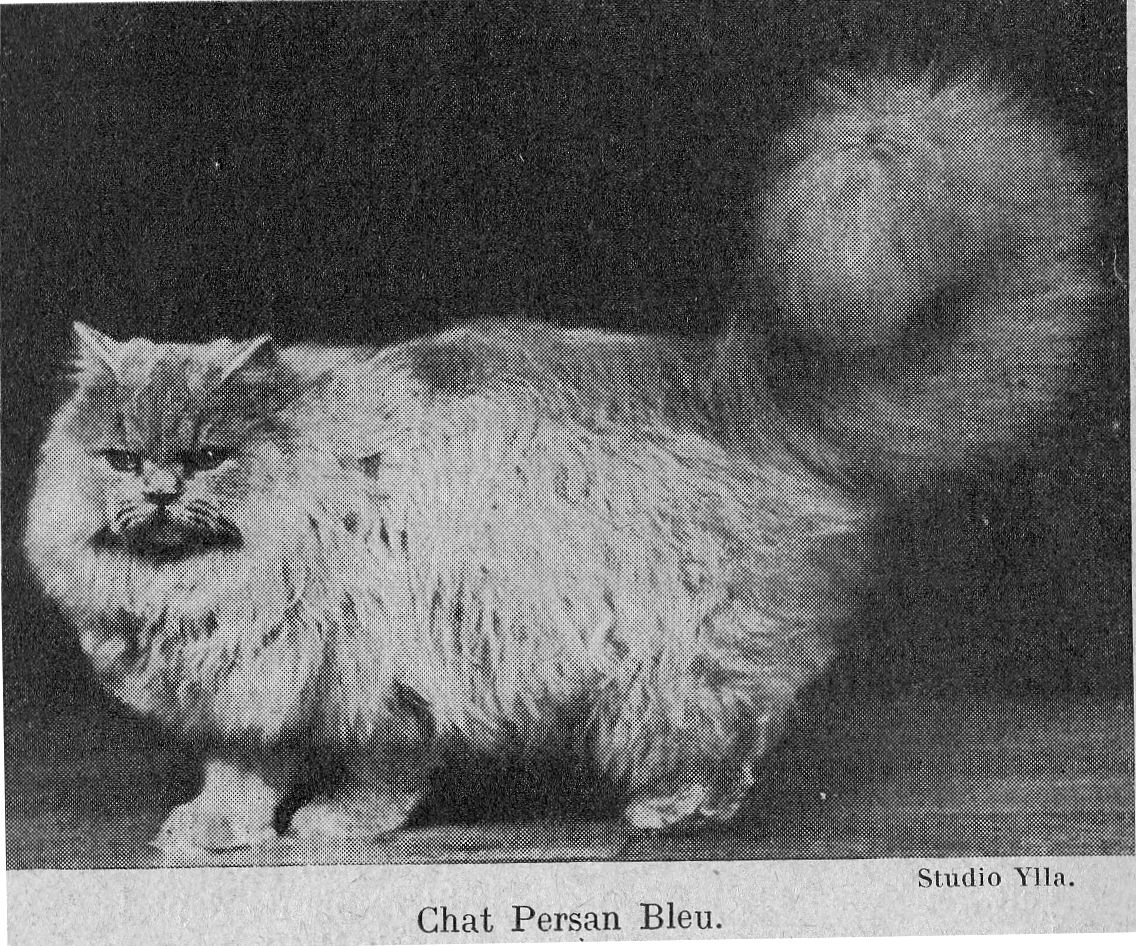
We are not concerned here with the racial aspect that quite rightly preoccupies owners who carefully research, in the subjects chosen for breeding, the characteristics of the breed which interests them, but we consider here any incompatibilities linked to conformation or state of health of the breeding animals.
Cat breeds have a uniformity of size and development not found in any other species; dogs, horses, even cattle have dwarf breeds and giant breeds, but these are unknown in cats. Any normally constituted adult female cat can safely be mated to an adult male cat of any breed.
Pelvic deformities resulting from fractures or rickets are rare in cats, fortunately moreover because these create serious, sometimes insurmountable, obstacles to parturition and they are always a formal contraindication to reproduction in the affected animal.
The health status of breeding animals is much more important as many parasitic and, in particular, infectious diseases are transmitted through a single short-term contact. I will speak further about how contagious some infectious diseases are and I know of catteries that have been decimated after sending a cat to an establishment where a serious disease was rife or had raged earlier. This is because here, in fact, it is necessary to take into account not only the state of health of the chosen breeding animal but also the health situation of the entire cattery because certain deadly diseases can be transmitted by infected carriers that appear pictures of good health. The general rule is that it is necessary to abstain for a few months from any contact with a cattery where there has been significant mortality.
Some animals, male or female, are poor breeders; they are either sterile or very poorly fertile (produce only one or two young). These are found especially in very selective catteries where the animals lack exercise and live in conditions far removed from a natural life.
2. Reproduction.
Heat. —- The first heat occurs in the female from the 6th to the 10th month and then reappears regularly three times a year.
This results in swelling of the vulva which is congested and exudes a little serous bloody fluid. This discharge is always much less abundant than it is in dogs, cats that "stain" are the exception. At the same time their character changes, the female becomes more affectionate, more cuddly and, above all, she almost continuously utters mournful, deeply unpleasant meows. If she is not closely watched, she will runs away in search of a male, whom she calls with her special cries, and it is often only after a few days of running loose that she returns to her master's house, her desires Satisfied but skinny, mud-stained, and frequently bearing bites and wounds that bear witness to her recent passionate struggles.
The heat duration is 15 to 20 days if she is not mated. As a general rule, only well-developed females that have finished growing should be delivered to the male.
Coupling. This most often happens in the wild and usually ends in a boxing matched due to the pain that the female experiences as a result of the special anatomical arrangement of the cat's penis.
In controlled catteries it is sufficient to take the female into the presence of the chosen male; mating takes place two or three times in a 24-hour period. From this moment the heat decreases in intensity, and after a few days of regularly decreasing meowing everything returns to normal.
Maternal impregnation. - It has been claimed that the first fertilization leaves indelible traces in the female and that a purebred female whose first pregnancy is fathered by a nondescript cat will subsequently produce offspring that resemble the father of that first litter, even if the subsequent litters are sired by a male of her own breed. This is telegony or maternal impregnation, and it is still considered an indisputable phenomenon by many breeders of cats and dogs. The laws of inheritance have dealt with this mistaken opinion, which is most emphatically overturned by science.
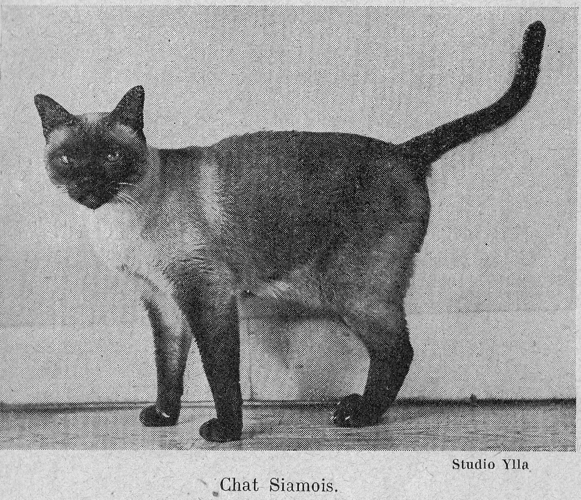
Pregnancy. - The average duration of pregnancy is 58 to 59 days, however there are important breed-related variations which are important to know: in Siamese in particular, prolonged pregnancy is the rule and birth often does not occur until the 65th or even the 68th day.
The signs of pregnancy are slight and often undetectable during the first month. It is usually only from the end of the fifth week that her belly becomes larger and droops a little, and that she becomes calmer and less exuberant. These symptoms are clearly marked at this period when there are 4 or 5 foetuses making the diagnosis of pregnancy easy.
However, we must be careful not to rely completely on changes in belly size. To confirm the pregnancy, it is essential to feel the foetuses by palpation of the abdomen. To do this, the cat is placed on a raised surface, a table for example, and immobilized with the left hand which grasps the nape and base of the neck. The lower part of the belly, immediately in front of the pelvis, is compressed with the right hand; the thumb on one side, the fingers on the other. The exploration must be slow, gradual and gentle. Towards the end of the 5th week it is possible to feel slightly elongated globular masses arranged in two lines; these are the foetuses.
When pregnancy approaches full term, the signs become more and more marked: the mammary glands develop, the cat is heavy, moves slowly, and often remains lying down. Soon the mammary glands begin to secrete, and by pressing lightly the end of the teats a drop of thick syrupy greyish liquid is expressed: colostrum which indicates that birth is near. At this time the vulva is thickened, congested, soiled with a little albuminous mucus. Now is the time to settle her in a clean, calm, slightly dark place, on dry cloths where she can give birth in peace.
Pregnancy hygiene. - During pregnancy, the cat needs hardly any special care, on the it is contrary important not to modify her habits and to let her play and frolic because the exercise is particularly beneficial not only to good foetal development, also to the gradual and complex changes that take place within the pelvis in order to facilitate the rapid expulsion of the foetuses.
Accidents during pregnancy do not seem to bother the cat, so to speak, and abortion is rare; I have often seen cats give birth to full-term, live kittens after falling from the 1st, 2nd, or even 3rd floor shortly before the birth.
The diet must meet all the needs of the pregnant female, who must obtain from her ration the elements necessary for the foetuses to develop. It is necessary to give her fresh meat, boiled fish and cow's milk rich in lime salts. Two meals a day are sufficient during the 1st month, but later on when the belly is obviously enlarged it is better to give 3 meals per day to prevent compression of the abdominal and thoracic organs.
Birth. - As soon as her labour pains start, the cat lies down in a circle, her head close to her vulva. From time to time violent contractions of the uterus and abdominal muscles push a kitten towards the entrance of the pelvis. After a short lull, the pains become more intense and more sustained and the kitten soon appears at the opening of the vulva and is expelled. The mother cuts the cord by biting it and takes a moment of respite following the birth to pull her young towards her and lick it. A few less violent contractions expel the placenta to be expelled; the cat makes disposes of these by eating them. Soon the previous steps are repeated as many times as there are kittens to be born. Each birth occurs fifteen to twenty minutes apart and the number of young varies from 1 to 7 or 8 with an average of 4, and the whole birthing process takes two to five hours.
When everything is going normally there is no need to intervene. It is good, however, to watch discreetly as there are females, especially first-time mothers, who devour their young as soon as they are born. In this case, it is necessary to seize the kittens as soon as they emerge, dry them carefully, wrap them in a woollen cloth and place them out of the reach of the mother. When the delivery is over, the kittens can be made to suckle and can then be left to the mother, all danger being over.
Birthing difficulties. – Birthing difficulties and dystocia are unusual in the cat; my previous statement about their uniform conformation and size across the different breeds explains its rarity. Incorrect presentations and positions are of no practical importance and it intervention is usually only necessary in the case of a large kitten due to there being a small number of foetuses, or if the mother becomes exhausted and weak. During these difficult births she becomes worn out by her fruitless efforts, the kittens’ waters are quickly lost and the uterus presses down on them making it difficult, if not impossible, to move them. The female quickly becomes exhausted and stops pushing.
It is therefore important to intervene quickly because infection quickly sets in and 36 to 48 hours after the start of labour the chances of success are already very limited. This intervention must be methodical; you must: 1. Inject a fatty or mucilaginous substance into the uterus: ordinary oil, a decoction of flax seeds [linseed] which lifts the uterus off the foetuses, makes them mobile and lubricates the birth canal. 2. Rouse uterine contractions. Among the methods that can be used for this purpose, the most effective are subcutaneous injections of pituitary extracts that are found commercially under various names: pituilobin, pituitrin, etc. in titrated and dosed ampoules. Their use is only required as a precaution, but it is absolutely mandatory to ensure that the cervix is dilated and that there is nothing blocking the exit of the kitten. 3. Help the action of her efforts to expel the kitten by performing traction on it. These pulls are barely possible unless it is already partially expelled. Grasp it with your whole hand, through a dry cloth, and make progressive, intermittent pulls in time with the mother's efforts. As well as applying straight traction it is also necessary apply slightly oblique pulls to the left and right alternately.
These instructions cover almost all cases of obstructed labour in cats, however it is possible to encounter more serious situations which require either embryotomy - the extraction of a stuck kitten after fragmenting it - or hysterotomy or caesarean section. These are serious interventions, but are capable of saving the mother and sometimes even the kitten, if they are carried out quickly while the female is not yet exhausted or seriously infected, and it is important not to waste any time and to not wait until it is too late.
The events following normal childbirth are straightforward and simple; a few hours after giving birth the mother regains her cheerfulness and does not require any special care. After giving birth, a slight odourless bloody vulvar discharge is observed for 10 to 12 days, which corresponds to lochia in humans; it does not require any special care and is not a sign of infection unless the discharge become smelly.
In some circumstances, the pregnancy is accidental, such as when a purebred female has been impregnated by a nondescript male, the consequences must quickly be disposed of. The young are immediately destroyed either by drowning in water or, better, by suffocation with household gas under a bell jar. An effort must be made to stop the production of milk. To do this, administer a purgative (a teaspoon of castor oil in the morning on an empty stomach) and apply a cotton ball soaked in a 4% solution of crystallized alum to the nipples. This bandage is attached at the back and changed twice in 24 hours. For 3 to 4 days give only vegetable broth to eat.
CHAPTER II
REARING
The number of kittens is variable; litters of 3 and 4 are most common but it can range from 1 to 8 kittens. First-time mothers and older cats frequently have small litters. If you want to have beautiful kittens without tiring the mother too much, you should only leave her with a limited number of babies.
At birth, kittens have their eyes closed. Their limbs are short, and they move with difficulty by crawling around the mother who leaves them only during her meals during the first week.
Care of kittens. - During the first days of their existence, young kittens suckle, sleep, develop rapidly and require little special care. As in young puppies, umbilical infections are uncommon, however it is always advisable to disinfect the umbilical cord and the umbilical wound daily, after the cord has fallen away, by applying iodinated glycerine for 10 to 12 days following birth.
Around the 10th day, the eyes open and the youngsters begin to frolic and play. When they are strong and vigorous towards the end of the 4th week, the mother no longer has enough milk so you should start providing some milk 2-3 times a day.
Weaning. - This is a delicate procedure on which the kitten’s future often depends. It must be done gradually, smoothly and not suddenly. They must abandon maternal nutrition only gradually and you must take each step only if the youngster is ready for it.
Begin weaning around the 5th week in the following way. Separate the young from the mother and place them in a bright, well-ventilated room, large enough for them to play and frolic, at a constant medium temperature. At the beginning return them to the mother 3 or 4 times a day for feeding and after feeding offer them whole milk in a perfectly clean dish. Each week reduce the number of maternal feedings by one and give the milk supplement, with the addition of pulped raw meat (passed through the grater to rid it of tendons, nerves, etc.), one teaspoon per day per kitten. Weaning is complete around the age of 2-and-a-half to 3 months, the young are strong, vigorous and resilient.
When we follow this process, the mother’s milk production is almost dried up by the time of weaning; it is usually sufficient to reduce her ration slightly to completely halt lactation. If it persists abnormally to the point of becoming troublesome it is sufficient to administer a purgative (a teaspoon of castor oil) and put her on vegetable broth for 48 hours.
Care of the mother. – Suckling kittens is a severe test for the mother who must, throughout its duration, receive a rich diet to promote lactation. It is necessary to give 3 meals per day consisting of fresh raw meat, boiled fish and as much milk as she wants.
Another important precaution to observe is that of ridding the mother of any intestinal parasites she may have, especially ascarids. If this is neglected, their eliminated eggs contaminate the nipples and the young are infected during suckling. Around the middle of gestation and then each month give the mother:
25 centigr freshly ground areca nut in a raw meatball.
Artificial feeding. - In some cases, - large litter, sick or dead mothers, — breastfeeding is not possible. The ideal in such circumstances is to find a wet nurse of the same species or even of a different species, but it is not common to have access to one so artificial feeding is often needed. This is a method that can give satisfactory results, but only if you use milk that is as similar as possible to the mother’s milk.
Many authors recommend sweetened, watered down cow's milk. This is a mistake, because cat's milk, like bitch's milk, is richer in fat and albuminoid matter and lower in sugar than cow's milk. It is, compared to cow's milk, more like unsweetened condensed milk. Commercial unsweetened condensed milk used as it comes out of the box is fine. It is given from a bottle fitted with a thin nipple with a fairly large hole.
During the first two weeks give a lukewarm feed every three hours (a teaspoonful of unsweetened condensed milk). After the third week, give four feeds while increasing the volume administered to fully meet the needs of the kitten. At the end of the sixth week, begin weaning by proceeding as described above. Naturally, the bottle must be kept strictly clean and scalded before each feeding.
Hygiene and care after weaning. - The suckling period is over. I have already said how important it is; it is on this and properly conducted weaning that the youngster’s future often depends. Well-suckled and correctly weaned babies are strong, vigorous, resistant, and perfectly equipped to resist the diseases which threaten them, and which wreak especial havoc among the weak and the sickly.
Feeding. - The cat is a carnivore - we often tend to forget this, and while some seem to be content with a diet of bread and vegetables, this is because they can hunt and correct their diet by eating their prey. For those who live strictly in apartments, such a diet would be disastrous.
Right from weaning it is necessary to start giving cooked and raw meat, boiled fish and sardines in oil which, along with raw meat, constitute the cat’s normal meals and do not need to be otherwise varied. Give four meals a day, from the third to the fifth month, three meals up to six months and finally a meal morning and evening; give pure water or milk for drinking. This diet ensures regular growth and contains all the elements necessary for building a normal skeleton and well-developed muscles.
Growth disturbances are much rarer in cats than in dogs and the regular use of cod liver oil, which is a common and very useful practice in dogs, is far from essential for cats. However, in slightly stunted youngsters, a daily dose of half a teaspoonful given on raw meat stimulates growth and activates development. Light and exercise are also factors that play an important role in skeletal development and growth, the youngsters should be kept in a well ventilated room, with open windows, well lit, at medium and constant temperature, where they can frolic.
Castration. - The breeding season is often particularly unpleasant for owners. Cats, male and female, leave the house, often returning injured, sometimes infected (scabies, etc ...). Those who are well guarded and cannot escape, utter intolerable mournful meows for days and nights. Then there is the prospect of having to destroy a litter of kittens two or three times a year, something unpleasant for sensitive people and for others. Castration eliminates all these drawbacks. It is a common operation in males and becoming increasingly so in females.
Male castration. - The male is castrated around the age of four to five months; the testicles are small, the operation is not very painful and the consequences are particularly harmless.
The subject to be operated must be fasted for twelve hours. Restraint is particularly important as it is essential for the operator and his assistants to protect themselves from the patient's claws and teeth. To immobilize the subject, we can use makeshift means, two of which - the blanket, and the stove-pipe - are common. In the first case, the cat is rolled up in a thick blanket wrapped around his body two or three times, securely holding the head and all four limbs. The tail region is kept free so that the testicles can be grasped. When using the stove-pipe we take a pipe of 1 m. to 1.5 m. long, the diameter being such that the cat engaged in it cannot turn around. With the pipe placed vertically, the cat is engaged with its head forward and an assistant grasps the tail and hind legs which alone remain outside. These are makeshift means which can be of service when one does not have a special restraint device at his disposal, something which is common in all veterinary clinics these days and which allows the animal to be completely immobilized in a position that is not distressing for him.
With the subject immobilized, local anaesthesia is given by three 1/2 cc injections of a 1% stovaine solution, one in the middle part of the sac and one in each of the testicular cords. With the left hand, the operator pushes the testicles into the bottom of the bursa and holds them there; with his right hand, he incises the superficial covering along the midline, then with two strokes of the scalpel, one on the right, the other on the left, he frees the testicles. He places two tightly clamped pliers on the two cords, half a centimetre apart, the upper clamp is held fixed with the left hand, while the lower, operated with the right hand, twists the cords until they break. The upper clamp is removed, the severed cords enter the inguinal tract, the superficial wound is dabbed with tincture of iodine, and the operation is completed without a drop of blood.
Treatment after the operation is as simple as possible, the same evening the patient can take a little milk, the following days he is returned to his normal diet. Morning and evening, the wound is dabbed lightly with tincture of iodine; in three or four days it has healed without suppuration, and everything is finished.
Castration of the female. - The castration of the female, ovariectomy, is much more delicate. Here again, it is important to work on young females, 6 or 7 months old; any later, and especially in females who have previously had kittens, the aftermath of the operation is much more delicate. Personally, I recommend taking action a few days after the first heat has passed.
The cat to be operated on, which must be in perfect health, is prepared by 24 to 48 hours of exclusively milk diet with a light purgative each day (5 grams of manna ash syrup in hot milk). Two surgical techniques can be used:
1. Surgery along the white line [midline]. - Requires general anaesthetic. Ether anaesthesia or mixed anaesthesia: basic anaesthesia with Soneryl by intraperitoneal injection supplemented by ethyl chloride by inhalation, are the least harmful.
The patient is fixed on her back, the abdominal wall is shaved or plucked. We perform a sub-umbilical laparotomy in which we search for the ovaries. These are removed using two forceps: a fixed upper, and a lower one with which we twist until the ovarian pedicles rupture. The abdominal cavity is then closed by a double suture: one deep to the muscle using tangut and one on the skin using silk. We finish by applying an aseptic dressing.
2. Surgery along the flank. - This is the one I prefer for two reasons: 1. It only requires local anaesthesia, as cats are very sensitive to general anaesthesia which can cause them frequent and serious complications; 2. The abdominal organs do not come outside of the body, which may not be the case with the first method no matter how carefully it is done, and the dangers of infection are greatly reduced.
The cat lies on her left side, the flank area is shaved or plucked. Everything is prepared for a perfectly aseptic operation, as in the previous case, by the way. Local anaesthesia is performed by a staged injection of a 2% stovaine solution into the angle formed by the last rib and the line of the lumbar transverse processes. A 3 cm vertical incision is made on the skin and the muscle wall; it is important that the muscular incision be straight and clean, without loosening the tissues.
The peritoneum is punctured with the spatula of a grooved probe. The right index finger inserted into the abdomen searches for the ovaries, the superficial one located close to the opening is found without difficulty, it is brought out and fixed with a clamp. Then we search for the deep ovary by following the uterine horns and slowly pull this out. The two ovaries fixed in the same forceps are removed by twisting. The abdomen is closed with a double muscle and skin suture as in the previous method and an aseptic dressing is applied.
The patient is given some milk the next day and is fed gradually over the following days. Healing should take place without significant rise in temperature; if fever appears, the dressing should be lifted. Very often the external wound then appears perfect and may even be scarred. Care should be taken not to be misled by this favourable appearance and it is important to widely open the skin wound under which we usually discover a purulent, foetid, diphtheroid-like pus, which should be thoroughly cleaned, disinfected and ventilated.
The results of surgery vary with the conditions of the operation. The factors for success are 1. a clean, straight musculo-cutaneous incision, causing minimal tissue damage; 2. rapid intervention. The success rate is around 80 to 85%.
Skin hygiene. - Cats are naturally clean, they groom themselves carefully every day and this is a sign of good health. There is no question of bathing them, their well-known hatred of water does not allow it, but it is necessary to brush short-haired cats daily and to carefully comb longhaired cats. If this precaution is not taken, the cat swallows its hair while grooming and this can cause serious complications.
PART TWO
CHAPTER 1. DISEASES OF THE LOCOMOTOR SYSTEM
Fractures. - The cat's agility and skill are proverbial. Despite these abilities, which are not found to the same degree in any other domestic animal, accidental injuries to the musculoskeletal system, during falls, for example, are not unknown.
Fractures are among the most common. Most are limb fractures; pelvic fractures are unusual. As for fractures in other regions, these are rare, and their severity is such that they are only rarely treated.
The symptoms vary depending on the location of the fracture, those of the limbs result in very obvious lameness, with lack of function of the limb, which is not used for support and swings loosely when the cat moves. On exploration, the hand feels a dry crepitation due to friction - bone ends - at the fracture site. Movement causes a very sharp pain. Open fractures are accompanied by tearing of the muscles and skin with the fracture site being exposed to the outside. In free-range cats, we find trap fractures which cause severe crushing damage to the bones and the soft tissues that cover them.
Limb fractures heal quickly, sometimes even without intervention, especially in youngsters. But it may not heal straight and the application of a bandage is essential in any case.
Treatment. - We must intervene quickly. With the two ends of the fractured bone brought back into contact, they must be kept there using a bandage that immobilizes not only the broken bone, but also the upper and lower joints. Surround the limb with a strong layer of cotton wool, this precaution is essential to prevent excessive compressions and injuries to the protruding parts of the limb and cover the cotton wool with a substance that becomes hard and resilient when dry; this produces a kind of external skeleton that replaces the ruptured internal skeleton. This reinforcement is made up of bandages impregnated with a substance that hardens quickly. In cats, one can use either plaster bandages, or preferably, primed, light tarlatan bandage [starched muslin]. These are soaked in water and two or three layers are arranged over the area to be immobilized to provide sufficient strength. After 10 to 15 minutes, the dressing is hard, rigid and keeps the ends of the fractured bone perfectly in contact. If we are dealing with an open fracture, complicated by a wound open to the outside at the fracture site, it is important not to cover the opening of the wound with the dressing; the wound must be treated daily as instructed for an infected wound. It is essential that the dressing is not too tight, which would result in a hot and painful swelling of the extremity the next day, requiring the immediate removal of the dressing in order to avoid gangrene of the foot. To facilitate the rapid formation of the bone scar, it is best to administer 4 to 5 drops of an oily solution of irradiated ergosterol (vitamin D) each day. Fractures caused by traps almost always require amputation. The fracture dressing must be left in place for fifteen to eighteen days for youngsters, twenty to twenty-five days for adults. To remove it, simply cut the bandage with strong scissors.
DISEASES OF THE JOINTS
Sprains. - The incomplete and momentary displacement of the bone surfaces constituting a joint. This is accompanied by more or less severe lesions of peri-articular tissues, ligaments, muscles, etc. A sprain is almost always the consequence of a serious fall; it is mostly seen in the knee (radiocarpal joint) and shoulder (scapulohumeral joint).
The area is swollen, hot, sensitive, forced movement of the joint is painful and provokes a defensive reaction, lameness is always present.
Treatment. - It is necessary, from the outset, to calm the inflammation and pain by applying hot wet dressings renewed every two or three hours, or of l’eau blanche compresses. As soon as it is less sensitive, massage the area with camphor oil morning and evening, lightly to start with and massages at the beginning, then with increasing pressure and force the patient to walk. [L’eau blanche - diluted lead acetate, an astringent.]
Dislocations. - Rarer than sprains, these are rare accidents in cats, and are caused in the same way as sprains, but more brutally.
In a sprain. the articulating surfaces spontaneously come back into place, but in a dislocation they remain in an abnormal position and no longer come together exactly; joint movements are extremely difficult or impossible. There is intense lameness. The joint is painful and distorted; this is easily judged by comparison with the corresponding part on the other side. In addition, and this is a crucial sign, forced movements imposed on the joint are always extremely small in their amplitude.
Treatment. - Rapid intervention is also a necessity; the longer one waits, the more difficult it is to treat, and the danger of complications increases.
Treatment involves reducing the dislocation and immobilizing the joint. To reduce dislocation, it is necessary to slightly spread the displaced articular surfaces by pulling in opposite directions on both ends of the limb and, at the same time, acting on the joint with pressures that returns the bones to their normal position. This procedure is difficult, painful, it provokes defensive actions from the patient and, in many cases, it can only be carried out with the help of general anaesthesia which suppresses the pain and relaxes the muscles.
Once the dislocation is reduced, the joint is immobilized by applying a rigid bandage, as in the case of a fracture (plaster bandage or primed tarlatan). After a week, the bandage is removed, and camphor oil massages are given for a few days.
Traumatic arthritis. - Fairly common in cats, where it appears in almost all cases as a result of bites from other cats, especially during fights in the mating period. The cat’s long, sharp canines make penetrating wounds, real punctures which, when made at a joint, can be the starting point of arthritis. The hock and knee joints are the most frequently affected. The primary wound is complicated by infection which progresses from outside to inside and invades the whole joint. It is important to point out right away that the infection is not fatal, and that it can often be prevented by careful and vigorous disinfection of the initial wound. If this precaution is not taken, the arthritis is more likely to progress, resulting in very severe lameness due to swelling of the whole joint, and in a discharge of bloody pus from the wound. The patient is feverish (39.5 – 40 degrees C), thin, and eats little. The progression is quite slow, soon new fistulas appear around the joint; like the first, they are stained with pus and blood; sometimes one of them dries up but soon one or more appear, spontaneous healing is rare.
Treatment. - Never neglect bite wounds in cats; disinfect them carefully by applying strong antiseptics - tincture of iodine, ether – this prevents many serious complications, in particular traumatic arthritis. In such cases, cut the hair around the wound as soon as possible, remove any foreign bodies that may contaminate it, brush carefully with iodine tincture on the outside and right inside the wound and apply a bandage. If the cat remains cheerful, eats well, and is not feverish, the bandage can be removed after four to five days and all danger is over.
It is necessary to tackle overt arthritis with antiseptic baths morning and evening, by immersing the limb in a hot solution of chlorinated water (one tablespoon of bleach extract for one litre of boiled water). After the bath, dry the area, instil a few drops of iodine tincture into the depth of the wound and apply a padded dressing.
If there are several fistulas around the joint, it is necessary to perform deep disinfection by lukewarm antiseptic injections, in particular with iodized water. A small glass syringe is used for this purpose and the liquid is injected strongly to flush it out through the various fistulas. In all cases, we end with the application of a bandage.
DISEASES OF THE TENDONS AND MUSCLES
Division of tendons. - Relatively rare, these accidents are mainly the result of cuts from sharp objects (broken bottle, flint). The flexor tendons on the posterior surface of the cannons and fingers are the most frequently affected. At the level of the wound, we can see the ends of the severed tendons; the extremity of the limb is no longer supported by the ruptured tendons and deviates from its normal position.
Treatment. - Tendon sutures are difficult to perform and most often give way. Fortunately, they can be dispensed with and recovery is easily obtained by applying a bandage after careful disinfection. Trim the hair, clean the wound very carefully with chlorinated water and meticulously disinfect with tincture of iodine. Apply a bandage that fixes the limb in the position of the tendon’s action to bring the severed ends together and into contact with each other; in the particular case of flexor tendons, the dressing must be applied so as to immobilize the limb, with the joints flexed. If suppuration appears, which is very often, give a lukewarm antiseptic bath every day, taking care to keep the dressing in place between each bath. Healing takes eight to ten days.
Wounds. - Wounds are not uncommon in cats; I have already mentioned the frequency of bite wounds, which are exceptionally severe in this species. They heal poorly, slowly and are the starting point for frequent and serious complications. They can be of concern on all parts of the body, but they are mostly found in two well-defined regions: the limbs and the base of the tail. Wounds in these areas are almost always bite wounds, are rarely simple and benign, and are almost always complicated by recurrent fistulas, which often last for weeks or even months. These fistulous sores discharge fluid pus mixed with blood, they often extend deeply, and it is not uncommon for suppuration to spread to the tendons or even the bones.
Treatment. – This all leads to the need for early and vigorous treatment of wounds in cats. Treatment includes washing the area (cutting hair, removing foreign bodies) and careful disinfection, by applying a strong antiseptic, preferably tincture of iodine, all followed by the application of a wadded bandage. Suturing of accidental wounds is not recommended in cats, as this species is prone to an infectious complication characterized by the formation of dirty grey, friable false diphtheroid membranes which are bathe in very smelly fluid pus. This is a very serious complication, often fatal, which mostly affects sutured and air-tight wounds. In conclusion: treat wounds in the open by daily disinfection followed by the application of a bandage. Fistulous wounds on the limbs and at the base of the tail must be debrided along their entire length and disinfected as before. When they are old and suppuration has spread to the bone, they often do not heal without curettage.
Abscess. - These are complications of wounds and may occur anywhere on the body where the skin was previously damaged. In cats, the postauricular region is frequently invaded by abscesses due to otocariasis (see page 90).
The area where an abscess develops is swollen, tender, slightly warm, oedematous. After a few days, pus collects in the central part which is easily depressed by finger pressure, and there is a feeling of fluctuation.
Treatment. - If the abscess is left alone, the suppuration spreads and sometimes causes severe damage. At the beginning, when the swelling is hard and tender, and the pus has not yet formed, the abscess can be brought to a head by rubbing with tincture of iodine in the morning and evening. Soon the fluctuation appears and pus is formed, it is then necessary to puncture the abscess, evacuate the pus and brush the cavity daily with a strong antiseptic solution.
DISEASES OF THE FOOT
Torn or fractured claws. - This is a fairly rare occurrence, however, it may occur if the cat gets caught by its claws and tries to free itself. It is accompanied by lameness and on exploring the toes one finds a torn or broken claw.
Treatment. - Just tidy the nail wound by cutting with scissors and disinfect it thoroughly to prevent infection.
Note, incidentally, that you should never cut a cat's nails, but should provide him with a thick cloth fixed on a board; he does his nails there and wears them regularly.
Foreign bodies. - The penetration of foreign bodies (steel splinters, flint, thorns ...), deep into the tissues of the sole of the foot is not uncommon. The result is sharp inflammation, sometimes followed by an abscess, and the onset of lameness that is always severe.
The first task is to extract the foreign body with small forceps. It is sometimes difficult, and it is often necessary to perform local anaesthesia with a few drops of 2% stovaine solution. After extracting of the foreign body use hot antiseptic baths and apply antiseptic wet dressings.
CHAPTER II - DISEASES OF THE DIGESTIVE SYSTEM
Diseases of the mouth
Stomatitis. - Inflammation of the oral mucosa is quite rare in cats as a primary disease; it is most often localized either on the tongue or inside the cheeks.
The causes are variable, but are mainly caused by injuries to the mucosa by foreign bodies (bones, pins, needles, bone splinters). Distortion of the teeth, especially dental calculus, are sometimes complicated by stomatitis.
Stomatitis begins with pain and difficulty in holding and chewing foods. Salivation soon appears in the form of threads, often tinged with blood, which flow from the corners of the lips. On exploration, with the mouth wide open, we see a more or less limited area that is red, congested and painful. Often at its centre there is the trace of a foreign body that can be seen more or less deeply embedded in the submucosal tissues. In a few cases, it develops in the form of an abscess which can spread to most of the tongue which becomes huge, congested, and painful, and protrudes from the mouth between the teeth. Swallowing is impossible, the patient is depressed, prostrate, feverish, and completely unable to eat. A methodical and gentle exploration of the region often identifies a deeply implanted foreign body.
Treatment – It is first essential to remove any foreign bodies. It is necessary to operate with the patient very solidly immobilized. With the mouth held open, grasp the foreign body with forceps and carefully extract it. In some cases, it is necessary to perform general anaesthesia. In the following days, wash the mouth with a 4% solution of chromic acid morning and evening.
Tooth tartar. - Especially in older cats, the molar teeth are frequently covered in a yellowish, irregular lime deposit which, in extreme cases, ends up coating the entire dental arch. The teeth loosen, become wobbly, the gingival mucosa becomes red and irritated, and the patients eats with difficulty. There is a foul smell from the mouth.
Treatment. - At the beginning, when the deposit is scarce, it can be removed by washing it daily with a 1% hydrochloric acid solution. If the deposit is voluminous, this method is not enough, and it is necessary to remove the tartar using a blunt-ended curette and finish with daily acid washes.
Foreign bodies. - Foreign bodies in the mouth are common in cats. These are sharp bodies - pins and especially needles, or sometimes hooks - that implant themselves in soft tissue or blunt bodies - such as bones - that get stuck in molar teeth.
As soon as the foreign body is in place, the patient stops eating, keeps his mouth open, and salivates profusely. Every now and then he paws at his mouth, as if trying to extract the offending item. On direct examination, the body of the object is usually seen implanted in the tongue or attached to the molars. Sometimes the needle, for example, has penetrated deeply and it can only be detected by methodical digital exploration, finally, exceptionally, it is essential to resort to radioscopy.
Treatment. - There is only one treatment: extract the foreign body. It is, depending on the case and the place where it is fixed and the depth of penetration, a benign, serious or very serious operation, which very often can only be carried out with the help of general anaesthesia.
Labial ulcer. – This is a special affliction of cats, quite common, which is seen in all breeds. It begins on the upper lip, sometimes at its middle part, sometimes to one side side, by a wound which mainly involves the inner edge of the lip. This greyish, dull wound does not appear painful, the patient eats, is cheerful, and grooms himself. But the lesion, in most cases, tends not to heal; on the contrary it spreads and seems to be regularly maintained by repeated irritation of the cat’s rough tongue, and soon presents as an arched wound with thickened edges, which clearly alters the patient’s physiognomy. Sometimes it spreads to the nostrils. Even then, its general condition is not changed, but the patient is in pain. This ulcer has been said to be contagious between cats, but this has not been demonstrated and is insignificant in practice. It has also been claimed that the patient can inoculate himself, by licking himself in different regions, especially on the underside of the belly. This is again a mistake due to confusion with a different ailment.
Treatment. - It is necessary to intervene as soon as possible, because the ulcer is tenacious and only yields to attentive and prolonged care. At the start, healing is frequently obtained by dabbing twice daily, morning and evening, with the following topical treatment:
Methylene blue - 0.50 gr
Glycerine - 5 gr
Distilled water - 5 gr
If this method fails, or if the intervention is delayed and the lesion is widely developed, cauterization with the tip of a thermocautery should be used. It is sometimes necessary to repeat this cauterization. A scab forms at the cauterized surface, which clears up after a few days, leaving a vivid red sore which is treated until healed by dabbing it with the methylene blue solution above.
Pharyngitis. - Common angina is rare in cats; it is seen in youngsters, especially following colds, in spring and fall.
Symptoms are depression and a medium intensity fever around 39.5 C. The appetite is suppressed or greatly diminished, swallowing is difficult, the throat area is sensitive to pressure. In severe cases, the eyes cry, the conjunctiva is thickened, congested, and soiled with purulent discharge. Coughing is not uncommon, especially during meals. When you open the mouth widely, you can see the red, turgid pharyngeal mucosa, dotted here and there with white spots in severe cases.
Treatment. - It is necessary to treat the throat directly through the mouth by twice-daily applications of an antiseptic mouthwash. The following formula gives good results:
Methylene blue - 1 gr.
Salicylic acid - 0.50 gr
Glycerine - 10 gr
Distilled water - 10 gr
On the outside, treat superficial inflammation under the throat by vigorous rubbing with tincture of iodine or by a poultice, followed in both cases by a padded wrap. A few drops of gomenolated oil [Melaleuca viridiflora oil] two or three times a day in the nostrils creates an antiseptic and soothing environment favourable to healing.
Obstruction of the oesophagus. - Foreign bodies that enter the oesophagus are not uncommon; depending on their nature and size, they cause either complete obstruction or simple oesophageal lesions with signs of oesophageal spasms; the first is mainly caused by bones, the second is most often caused by needles or chicken bones. The symptoms vary somewhat; complete occlusion appears suddenly as soon as the foreign body is trapped in the oesophagus. The restless patient makes violent efforts to swallow, brings his front limbs towards his mouth or jugular groove, as if to drive away the body which bothers him. At the same time, the saliva which is no longer swallowed flows in long threads from the corner of the lips. If the foreign body has stopped in the cervical region, before the entry to the chest, it is easily detected on palpation of the neck, the left side of which is also slightly deformed, confirming the diagnosis. However, it is more difficult when the object stops in the thoracic portion of the oesophagus and it is often necessary to use radioscopy to determine the site of the obstruction. Probing the oesophagus would give the same information, but it is difficult to do on a cat. Sharp foreign bodies - needles, chicken bones - cause slightly different problems. Here again the trouble appears suddenly. The obstruction is always partial, and swallowing is still possible, but it is painful and each passage of food requires effort and visible contraction of the cervical muscles. Palpation of the area is painful; it allows us, in certain cases, to detect the presence of a thin, rigid body, a few centimetres long: a needle. Problems swallowing persist for a few days, then a hot and painful swelling appears at a localized point in the cervical region, which corresponds to a developing abscess. If left unchecked, the abscess opens to the outside, most often after a few days, and the foreign body can find a way out that allows rapid healing. This is what usually happens when the needle is unthreaded, whereas those with a length of thread travel less easily and cause the formation of a fistula which only heals after extracting the offending item.
Treatment. – The foreign body must be extracted by oesophagotomy surgery. It is a grave, serious procedure that requires general anaesthesia. However, the results are generally good when it involves the cervical part of the oesophagus, which is easily accessible. The situation is quite different when the foreign body is stuck in the thoracic portion of the oesophagus, for it can only be retrieved by means of laparotomy and an incision into the stomach that allows backwards passage into the oesophagus; here the prognosis is particularly gloomy.
Diseases of the stomach, intestines, liver and peritoneum
Stomach indigestion. - Rather rare, stomach indigestion occurs in gluttonous, hungry subjects, who load their stomachs beyond its physiological limits. It is also seen following ingestion of spoiled, fermented food.
It appears shortly after eating; the patient is depressed, seems uneasy, he has mild dyspnoea and he makes frequent efforts to vomit. Palpation of the lower abdominal region shows the presence in its anterior part of a bulky, globular, tense mass with pasty contents: this is the distended stomach.
Treatment. – Urgently induce emptying of the stomach by provoking vomiting. It is often sufficient to press down the base of the tongue with the handle of a spoon. If this method is ineffective, the subcutaneous injection of:
Apomorphine - 5 milligrammes.
Distilled water - 5 cc.
is quickly effective.
Put the patient on a water diet for 24 to 48 hours, during which time give a teaspoonful of the following every three hours:
Soda citrate - 1 gramme.
Lemon syrup - 50 grammes.
Distilled water - 50 grammes.
Foreign bodies in the stomach. - Foreign bodies usually stop in the oesophagus, but if they are not too large they can reach the stomach, where they can stay for a longer or shorter time if their dimensions do not allow them to enter the intestine.
They are sometimes well tolerated, but they more often cause irritation, symptoms of gastritis, and vomiting which attracts attention. If a case of gastritis does not yield to the logical course of treatment, it is necessary to consider the possible presence of foreign bodies in the stomach and to submit the patient to radioscopic examination which generally clarifies the situation.
Treatment. - When these foreign bodies cause serious disturbances – vomiting or weight loss - it is necessary to intervene and extract them by gastrotomy. This is a serious operation, but if done well it is often followed by success.
Gastroenteritis. - Common inflammation of the stomach and intestines is relatively common. Although gastritis and enteritis can develop separately, in most cases they are related.
Gastroenteritis is often the consequence of a poorly comprised diet, an irregular diet, or may even be caused by ingesting fermented or spoiled food.
Symptoms vary depending on whether gastritis or enteritis predominates. When the stomach is first affected, mucous, foamy, or bilious vomiting is the dominant sign. This is accompanied by strong thirst, often with intolerance of liquids, fever around 39 C, dejection and constipation. The latter lasts only a short time and very soon more or less marked diarrhoea appears: liquid, foetid, five, six or even more times per day.
The patient quickly loses weight, becomes exhausted, no longer cleans himself and within a few days presents a state of marked decline.
Treatment. - Treat as soon as possible. The intervention should address two symptoms: vomiting and diarrhoea.
Put patients on a water diet: Vals-Saint-Jean water at will and administer three or four times a day a teaspoonful of the following potion:
Soda citrate - 2 grammes.
Lemon syrup - 50 grammes.
Distilled water - 50 grammes.
Against diarrhoea, take three or four times a day, between the previous administrations, a spoonful of:
Bismuth sub-nitrate - 4 grammes.
Lactic acid - 4 grammes.
Quince syrup - 50 grammes.
Distilled water - 100 grammes.
To combat dehydration and decline of patients, give subcutaneous injections of lukewarm physiological serum (9 parts per thousand salt water) 20 to 30 cc., repeated daily. After recovery, gradually and slowly return to the normal diet.
Constipation. - Frequently observed in sedentary cats living exclusively in apartments, especially neutered cats. The constipated patient makes infrequent small quantities of hard faeces. When the constipation is long-standing, the patients are chronically intoxicated, have a dull, brittle coat, and are indolent and flabby.
Treatment. - Diet matters as much, if not more, than drug treatment. Do not overuse purgatives as these tire the intestine. If constipation is persistent, administer enemas using a small, oiled rubber bulb: one tablespoon of petroleum jelly in two tablespoons of lukewarm water. They can be repeated every two hours. If the result is insufficient, it can be supplemented by taking a purgative: a teaspoon of castor oil. Finally, to obtain regular stools, regularly administer a teaspoon of paraffin oil in the morning on an empty stomach two or three times a week.
The diet should be particularly watched, regular portions of milk, boiled unsalted fish, sardines in oil and a little raw meat should form the basis of the diet.
Bowel obstruction. - This is the stopping of intestinal function when the lumen is obstructed by either a foreign body, a tumour, or impacted stool. Bowel obstruction appears either slowly (obstinate constipation, tumour) or suddenly (foreign body); it results in the stopping of intestinal functions (complete suppression of defecation) and general disorders: dejection, depression, which are the consequence of intoxication. Finally, vomiting with the smell of faeces is a very important sign. Whenever such vomiting coincides with the suppression of bowel movements, one should consider intestinal obstruction. On palpation of the abdomen, one sometimes, but not always, feels a mobile mass of variable shape and volume. In doubtful cases, radioscopy after administration of an opaque meal can determine the existence and location of the blockage.
Treatment. - Bowel obstruction is always a very serious condition, which always requires rapid intervention. This intervention varies with the nature of the blockage; coprostasis (impacted faeces) should be treated like constipation, with the remark, however, that if ordinary means fail, surgery must be resorted to without delay. Surgery is the only treatment for neoplastic [tumour] blockages or foreign bodies. Surgery consists of a sub-umbilical laparotomy on the anesthetized patient through which one performs either an enterectomy (resection the part of the intestine invaded by the tumour) or an enterotomy (opening of the intestine for extraction of the foreign body). In both cases, it is a serious, even grave, operation, which was until recently considered fatal in most cases. Intravenous injections of hypertonic serum (20 parts per thousand sodium chloride solution), 10 to 15 cc. shortly before surgery, markedly improves the results.
Jaundice or icterus. - Relatively rare in cats, jaundice in this species is almost always the consequence of an obstacle to the evacuation of bile; it is a serious ailment which is accompanied by intense intoxication and rapid decline of the patients.
It presents as very characteristic signs: inappetence, depression, vomiting, constipation with discoloured stools, putty-coloured. At the same time, and this is the main symptom, the skin and especially the mucous membranes of the mouth and eye are a more or less intense yellow colour, sometimes sulphur yellow or saffron yellow. There is usually no fever, and sometimes even hypothermia.
Treatment. - A cat with jaundice must be kept well covered in a room at a mild and regular temperature. Each day, give an injection of physiological serum or lukewarm glucose serum under the skin, 15 to 20 cc. Morning and evening, administer a teaspoonful of the potion:
Sodium sulphate - 5 grammes.
Sodium salicylate - 2 grammes.
Sodium bicarbonate - 5 grammes.
Sodium benzoate - 3 grammes
Rhubarb syrup - 50 grams
Distilled water - 100 grammes
In severe cases, in severely dejected patients, administer a daily subcutaneous injection of 1 to 2 cc. of camphor oil.
Ascites. - A chronic pathological condition characterized by the accumulation of fluid in the peritoneum without inflammation of the latter. It is not a disease, but a symptom common to different ailments. It is mostly seen in older cats, caused by damage to various organs which, in order of frequency, are the liver, heart and lung. It can also be the consequence of tuberculous lesions or tumours.
Its course is irregular, sometimes it accumulates slowly over two to three weeks, sometimes it appears within a few days. When the effusion is abundant, the belly is deformed, enlarged, especially in its lower parts. Examination finds no sensitivity. Gently tapping the lower part of the abdominal wall with fingertips causes the liquid to move, which slightly lifts the stomach wall on the opposite side: this is the flow movement that can be seen, and above all felt very well with the hand lying flat.
This percussion of the lower regions of the belly produces a dull sound up to a horizontal line which indicates the upper limit of the liquid. When this fluid is abundant, it pushes against the diaphragm and limits its expansion movements; breathing is difficult, short, rapid. To reduce this discomfort, the patient avoids lying down and remains standing for longer than normal.
Treatment. - Ascites always suggests tuberculosis so tuberculinization [TB test] is required. In all cases, an abdominal puncture should be performed to drain the collected fluid. This puncture is done using a thin sterilized trocar in the middle of a line joining the umbilicus to the fold of the flank. Wounding the intestine is not a worry because it floats in the upper part of the liquid. The characteristics of the liquid provide interesting information; it is clear, limpid, serous in ascites of cardiac or hepatic origin; it is haemorrhagic in cancerous ascites; it is sometimes white, resembling milk, in certain tuberculous ascites. This last characteristic is found almost exclusively in cats and is quite significant.
Drain the liquid slowly, taking care to interrupt the flow from time to time by closing the cannula of the trocar.
When the ascites is not of tuberculous origin, one can try to cause absorption of the liquid by administering each day two to three teaspoons of:
Calcium chloride - 5 grammes.
Currant syrup - 50 grammes.
Distilled water - 100 grammes.
But these methods are most often insufficient, the fluid re-accumulates and more or less frequent successive punctures are necessary, the patients become exhausted and most end up wasting away over a period of time.
Poisoning
Poisoning by spoiled food. - Especially seen in free range cats rummaging in garbage where they may encounter rotting meat scraps. The larger the amount ingested, the more serious the resulting poisoning.
The problems appear quickly; symptoms are depression, which leads to prostration, vomiting, and profuse foetid diarrhoea which may be bloody. The stomach is sensitive, painful on palpation, the pulse is weak and racing, the pupils are greatly dilated. Fever, 39.5-40 C, is the general rule at first, but in severe cases this gives way to hypothermia which precedes the final throes and which usually coincides with paralysis, especially of the hindquarters. The progression is always rapid, and death can occur within hours.
Treatment. - We can do little more than support the patient and help him fight the poison. In particular, the heart must be supported by subcutaneous injections of camphor oil (1 to 2 cc depending on his size). Simple physiological serum, caffeinated or glucose, injected under the skin at a dose of 20 to 40 cc., increases blood pressure, activates the functioning of the kidney and facilitates the elimination of toxic products. These sera must be injected hot at 38 or 39 C if the temperature of the patient is below normal. General tonics, particularly strong, slightly alcoholic black coffee (a teaspoon of rum or brandy in an ordinary cup), help stimulate the organic defence.
Phosphorus poisoning. - This is rare until after the use of phosphate paste or rat poison for killing rodents. Rodents will die all over the place and their corpses may be eaten by cats, which get poisoned this way.
Patients completely lose their appetite, are weak, staggering, and sometimes seem paralyzed. The oral mucosa is swollen. The exhaled breath smells like garlic. Vomited material can be seen in the dark by its phosphorescent reflections.
Treatment. - Act as soon as possible. Induce vomiting by administering 0.50 gr. ipecac in lukewarm water. Do not give milk as this is a solvent for phosphorus and facilitates absorption. Turpentine is the counter-agent of choice; alcohol is added to it as a general stimulant. You can use the following potion:
Oil of turpentine - 4 grammes.
Brandy - 6 grammes.
Water - 50 grammes.
by teaspoonfuls, two or three a day.
Arsenic poisoning. - This occurs under the above conditions when arsenical wheat is used for the killing small rodents and mice whose corpses can be eaten by cats.
It manifests as depression and very marked weakness, severe colic, salivation and paralysis which shortly precedes death.
Albuminous water (an egg white beaten in a little water) at a dose of one teaspoonful every 10 to 15 minutes is the most effective antidote.
Intestinal worms.
Intestinal worms are less frequent in cats than in dogs, but are not uncommon, especially in young cats where they often cause more or less serious disorders.
Ascarids. - Roundworms (ascaris mystax) are round, whitish, tapering worms, one end of which is slightly curved and swollen; their length varies from 4 to 10 centimetres with a diameter up to 1 millimetre to 1.5 millimetres in the middle region. They are found in the small intestine and are especially common among young cats.
Infestation occurs through the digestive tract, especially from drinking water. Adult worms lay eggs which develop on the ground, if the eggs are ingested by a cat, they produce parasites which develop into adults in the intestine. Nursing kittens can become infested during feedings if the mother is a carrier of adult parasites whose eggs, expelled with her faeces, contaminate the nipples. It even seems that infection of the young can occur through the blood during intrauterine life if the mothers have worms, hence the need to rid breeding females of their parasites.
Ascariasis causes disorders that vary with the intensity of the infestation. These are sometimes only vague, imprecise symptoms: slight weight loss with irregular appetite, poor general condition, prickly hair. In other cases, the intestinal irritation from the worms causes epileptiform seizures which exhaust the patients. Sometimes the parasites travel up into the stomach, causing vomiting, during which some of the parasites are ejected. Exceptionally, roundworms gathered in a ball cause intestinal obstruction. Finally, they irritate the digestive muscle, impair nutrition, and cause enteritis with persistent diarrhoea.
Treatment. - Santonin remains the drug par excellence for ascariasis in cats, at a dose of 2 to 5 centigrammes, administered two days in a row, in the morning on an empty stomach, in a small meatball; it gives excellent results. Complete treatment with a purgative (one teaspoon of castor oil) four hours after the last dose.
Tapeworms. - These are ribbon-shaped flatworms made up of several segments. They are sometimes seen in young cats but are mainly parasites of adult cats.
They usually cause only mild illness that would go unnoticed if not for the presence of parasitic debris in the faeces. Their importance, however, is not negligible, it is mainly due to their mode of evolution during which we observe the adult form that exists in the cat, and a larva, which develops in the viscera or muscles of a second host. One of these larval forms, that of the echinococcal tapeworm, lives in different species, including humans in whom it causes serious illnesses that sometimes necessitate serious surgery.
The cat becomes infected by consuming animal debris, sometimes fish, infected with the larval form which developed from the eggs shed by the cat carrying the adult parasites.
The main tapeworms of the cat are:
Taenia crassicollis, 35 to 40 centimetres long, made up of segments up to 1 centimetre long. The larval form develops in the livers of rats and mice, where it takes on the appearance of small translucent loops commonly known as "water balls".
Taenia echinococcus is very short, only half a centimetre long, and formed of only a few segments. Its larval form, which can be enormous, presents with the appearance of a large vesicle that invades the liver and lungs of various animals and humans.
Botriocephalus felix is 15 to 20 centimetres long, its head is broad and flattened. Its larval form develops in some freshwater fish and cats become infected by eating raw fish.
Treatment. - All these signs show the need to rid cats of their tapeworms.
Freshly powdered areca nut administered after a 24-hour milk diet, two days in a row, in the morning on an empty stomach, in a meatball at a dosage of 0.25 gramme to 0.50 gramme, regularly gives excellent results. Follow with a purgative (one teaspoon of castor oil) three to four hours after the last dose.
Disorders of the anus and rectum.
Imperforate anus. - A congenital condition that is inevitably fatal if not remedied quickly.
The first signs do not appear until a few days after birth. The newborn looks normal, sucks well, but soon his belly gets bigger and he stops feeding. On palpation, the stomach is stretched slightly hard. At the same time, we can observe unsuccessful defecation efforts. This can be explained by examining the anal region, which shows the absence of the anus.
If the kitten has no particular value, it is not advisable to save it. Rarely, especially valuable kittens, it is possible to operate. But surgery should only be attempted in the simplest cases, when the intestine distended by excrement protrudes under the tail which is evidence of attempts to expel the faeces. It is then sufficient, after local anaesthesia, to use a scalpel to puncture the unyielding projection formed by the distended intestine. The puncture opens the intestine to the outside and creates the anus. In more complex cases, where the terminal bulb of the rectum is farther from the anal region, the operation is too delicate in cats to hope for success.
Prolapse of the rectum. This is quite common in cats during inflammatory conditions of the intestine which cause violent and repeated attempts to defecate; it is mostly seen in youngsters with enteritis.
The rectum moves gradually, first the congested, reddish mucosa, appears on the outside where it forms a protruding bulge. Then, as expulsive efforts continue, the rectum, in all its thickness, is pushed out to a greater or lesser length. At this degree, the herniated rectum appears as a bulky, irregular, pleated, blackish-red sausage, presenting at its lower end an infundibulum representing the lumen of the rectum. Soon the congestion gets worse, the organ is soiled, excoriated, and after two or three days gangrenous lesions may appear. The patient quickly becomes exhausted, urination is impaired and serious complications can occur if left unchecked.
Treatment. - Washing the herniated organ is the first thing to do. All foreign bodies are removed - straw, hair, scabs, excrement, etc. - and a large wash with lukewarm physiological serum is carried out. Then you must attempt reduction. The patient is carefully immobilized, the hindquarters greatly raised; the rectum is oiled or vaselined and, by methodical pressure exerted with the hands in the immediate vicinity of the anus, the ectopic rectum is gradually pushed back into position. This reduction must be done slowly, methodically, and patiently, carefully avoiding injury to the organ whose strength is greatly reduced. When the reduction is complete, a lukewarm enema is administered which straightens the rectum and puts everything back in place. Now it is a matter of preventing recurrence and it is essential to suture the anus. The best suture is the purse suture performed around the finger; by pulling on the ends of the thread, we limit at will the anal opening, which allows defecation while preventing a repeat of the prolapse.
Sometimes, however, under the pressure of efforts to defecate, the suture gives way and the situation occurs again. Everything has to be started over. We then have the choice between two methods which must be used in the order in which they are given here.
1st method. - After reduction of the prolapse, disinfect the margins of the anus with tincture of iodine, then, using a hypodermic needle inserted into the sides of the rectum to 2 or 4 centimeters deep, while the index finger is introduced into the rectum to prevent an accidental prick, inject 1 cc of absolute alcohol on each side.
2nd method. - To be used only when all other means have failed, for treating repeated prolapse. This consists, after general anaesthesia, of performing a laparotomy in the flank, reducing the prolapse by pulling forward the prolapsed section of intestine and performing a colopexy by fixing the terminal part of the intestine to the muscular wall of the abdominal wound by two or three stitches which involve only the serous and muscular layers of the intestine.
Finally, in particularly serious cases, when there is a tear or gangrene of the herniated rectum, amputation of the stump remains the ultimate recourse. It is a serious operation which can however be followed by success.
CHAPTER III
DISEASES OF THE RESPIRATORY SYSTEM
Nasal cavity disorders.
Coryza. - Inflammation of the mucous membrane of the nasal cavities is quite common in cats, it develops especially in the acute form due to colds or irritation caused by dust and certain irritating gases. Cats of any age can be affected.
It is important to distinguish the trivial coryza, detailed here, which manifests as isolated cases, from contagious coryza, which spreads rapidly, and which often takes many victims in large catteries.
The patient with coryza is slightly depressed, he presents with frequent sneezing which results in mucopurulent discharge which soils the nostrils where they dry out forming small cracked scabs; the cat still has an appetite and there is no fever. Coryza should not be neglected, otherwise the inflammation sets in at the base of the nasal cavities and becomes chronic.
The treatment is not very convenient because, in general, the cat does not lend itself well to the interventions indicated in such a case. Inhalations which thins the phlegm, facilitates its expulsion and calms inflammation is poorly tolerated by cats. However, whenever possible, it should be used. This are easily achieved by throwing a few eucalyptus leaves into a bowl of boiling water and making the patient breath the vapours. It can be supplemented with instillations [i.e. nasal drops] of gomenolated oil. But, of all the treatments tried, the one that gave me the best results was instillation, morning and evening, of a few drops of a solution of silver nitrate at 1 p 450, taking care to keep the nostril openings raised to allow the liquid to penetrate deeply. This treatment is particularly indicated when the coryza is prolonged and shows signs of becoming chronic.
Epistaxis. - Nose bleeds are unusual, even after severe trauma with a fall on the nose. I have only seen them as a result of polyps in the nasal cavities, usually unilateral polyps, which cause a slight flow of blood along with marked respiratory discomfort. These polyps are located either in the immediate vicinity of the nasal opening, where they appear as a rounded, reddish, often ulcerated and bleeding buds, or deeper and safe from the most careful exploration. In the latter case, the diagnosis is based on low key nasal haemorrhage, difficulty breathing and the unilateral nature of these symptoms.
Treatment is exclusively surgical, it is necessary to remove the polyps. The surgery is easy to perform when the lesion is close to the nostril, but is more delicate when the polyp is deeper, it then requires trepanation of the nasal cavities, which can only be done under general anaesthesia.
Bronchial and lung disorders.
Acute bronchitis. - Inflammation of the bronchial mucosa is mainly due to colds. It is relatively rare and is seen mainly in young cats.
It is announced by depression, loss of gaiety, decreased appetite and medium intensity fever: 39 C, 39.5 C. The situation soon becomes clearer with the appearance of two characteristic signs: coughing and shortness of breath. The cough is quite strong, hacking, hoarse at first, then it becomes wet and is accompanied by the expulsion of mucus which is most often swallowed it passes through the pharynx. Coughing fits are often followed by retching. Shortness of breath, which results in markedly fast breathing, becomes especially marked during and after coughing fits. It becomes clearer that there is deeper inflammation; in severe forms, when the fine bronchi are affected, the dyspnoea is intense, the cat breathes through its mouth and this is accompanied by labial breath where the lips are raised with each exhalation. There is also greyish, muco-purulent nasal discharge which dries up around the entrance of the nostrils, forming scabs which aggravate the cat’s respiratory discomfort.
Percussion of the chest does not give very precise information, but auscultation reveals mucous rattles whose characters vary with the extent of the lesions.
Treatment. - Place the patient in a room at medium and constant temperature, support him with a selective diet: raw meat, fish, etc.
Combat the inflammation by using mustard plasters or strongly counter-irritant flaxseed meal poultices (equal parts of flaxseed flour and mustard flour). The poultice is placed in a handkerchief is applied to both sides of the chest, completely surrounding it, and is secured at the back with two safety pins. It is left in place for 20 to 25 minutes and renewed morning and evening. Between poultices, a cotton dressing to the chest.
Inhalations (see p. 54), when they can be used, aid expectoration, followed by instillations of gomenolated oil into the entry of the nostrils.
To dry up the bronchial secretions, administer morning and evening a teaspoonful of:
Terpine syrup - 50 grammes.
Tolu syrup - 50 grammes.
Linden syrup - 50 grammes.
If the temperature is elevated above 39.5 C and there is intense dyspnoea, support the heart with a daily subcutaneous injection of 1 cc of camphor oil.
If treatment is started quickly, the cat soon improves; the symptoms gradually reduce, and in eight to ten days it has run its course.
Broncho-pneumonia. – There is concomitant inflammation of the bronchi and lung tissue from the outset and, in most cases, it is a complication of distemper [in this case Moussu means cat flu], or is an extension of acute bronchitis.
In the latter case, the symptoms of bronchitis are particularly acute, the temperature exceeds 40 C, the patients, very depressed, completely stop eating, breathing is particularly rapid and difficult with an intense labial murmur, which reflects respiratory failure. The discharge is profuse, sometimes slightly bloody. All of these serious symptoms translate into a worrying situation which, in fact, very often ends in death after a few days.
Treatment. - Acting quickly and energetically are the prerequisites for success and they are far from being sufficient.
All the methods mentioned for acute bronchitis must also be implemented here: revulsion by application of mustard poultices morning and evening, cardiac tonics, and particularly camphor oil, represent the most effective means which should not be neglected. These actions are completed by disinfecting the respiratory tract (instillations of gomenolated oil), inhalations and anything that can help support the patient (force-feeding with spoonfuls of milk, meat juice, etc.).
A fixation abscess (injection of 1/2 cc of turpentine under the skin of the lower part of the base of the neckline) is also a powerful method to reserve for adults. [An obsolete treatment in which an abscess was artificially created with the intent of drawing the infection to a single, treatable site.]
Chronic bronchitis. - Most often seen as a residue of neglected or insufficiently treated acute bronchitis. The acute symptoms disappear, the fever subsides, the appetite returns to normal, the patient regains his gaiety, as well as normal breathing at least when at rest. But there is a persistent heavy, wet cough, without nasal discharge, because the phlegm released during the coughing efforts is swallowed when it enters the throat.
Chronic bronchitis is accompanied either by excellent general condition, or even by mild obesity, especially in castrated cats, or on the contrary by unexplained weight loss. In the latter case, we must consider the possibility of tuberculosis (p. 108).
Treatment. - The treatment of chronic bronchitis is rather poor, moreover the disease (apart from that of tuberculous origin) is unpleasant rather than dangerous. However, you can act on the bronchial secretions by administering the following potion:
Terpine - 1 gramme.
Tolu syrup - 50 grammes.
Distilled water - 100 grammes.
Alcohol – Q.S. [sufficient quantity for the preparation of the drug].
which, at a dose of 1 teaspoonful morning and evening, over a period of ten days, dries the bronchi and reduces the abundance of phlegm.
Pulmonary emphysema. - Chronic bronchitis leads almost inevitably, sooner or later, to pulmonary emphysema which it is very difficult to distinguish from it. The cough is again the dominant symptom; it is hacking, dry, small. Rare in animals at rest, it occurs mainly during activity when the cat runs and jumps, and also under the influence of a temperature change, especially when the fresh morning air enters a closed room during night. It is sometimes accompanied by retching and always by rapid breathing which disappears with rest.
The cat’s general condition does not change, there may often even be a slight tendency to obesity, appetite and gaiety are not altered in any way.
The treatment is much more palliative than curative, the cure is hardly to be hoped for because emphysema causes irreversible changes, and all is possible is to decrease the frequency of coughing fits.
For this purpose, administer a teaspoonful morning, noon and evening, of:
Bromoform - 2 grammes.
Tincture of aconite - XX drops.
Diacode syrup - 15 grammes.
Distilled water - 150 grammes.
Diseases of the pleura.
Pleurisy. - Inflammation of the pleura appears either as a primary disease, under the influence of cold - this is unusual - or as a secondary illness, during distemper [cat flu in this context] or tuberculosis. Any pleurisy which is not a complication of distemper must be suspected to be tuberculosis and the patient must be tested for tuberculin.
Pleurisy begins with general signs - fever in the region of 40 C, depression, loss of appetite - which are complicated after 48 hours by respiratory symptoms: small, nagging, painful cough, dyspnoea and rapid breathing. If we palpate, or better if we press the chest, this usually provokes pain and defensive movements in the patient. The methodical percussion of the thoracic cavity shows a dull sound in the lower region, while in the upper region there is a normal or even exaggerated resonance; the dividing line of these two zones is horizontal in a standing patient. The dullness is caused by fluid accumulated in the downward sloping parts of the pleura. There are two more important symptoms to note: discordant breathing where the sides widen when the chest is raised, whereas normally the movements of the chest and the side are in the same direction; and frequent vomiting which may be confused with digestive disease.
The diagnosis can be made by performing thoracentesis, which is the puncture of the pleura between two ribs, in the lower part of the dull-sounding area, using a sterilized needle. This puncture gives a discharge to either clear or slightly bloody fluid (tuberculous pleurisy), or to pus (pleurisy of distemper).
Pleurisy is a very serious condition which requires the destruction of patients if it is tuberculous in origin and which often kills patients in the purulent form.
Treatment. – Bypassing [drawing the fluid away] using poultices applied morning and evening, supporting the heart using camphor oil (a daily injection of 1 cc), and force-feeding the patient are the most useful means. If, despite these treatments, the dyspnoea remains intense, thoracentesis is required. It must be done slowly to avoid syncope, which is always a possibility. Discharging the liquid immediately relieves the patient who breathes more freely, but this liquid tends to re-accumulate quickly and to prevent this, the pleura must be disinfected by an intra-pleural injection of a lukewarm solution of 1% boric acid. Boric water is injected through the needle left in place after thoracentesis. The 10 to 20 cc injection should be done slowly, and the liquid evacuated after 5 to 10 minutes of contact. This double operation - thoracentesis and antiseptic lavage of the pleura, which may need to be repeated several times - makes it possible to obtain cures even in the most serious cases.
Eclampsia. - An acute disease of pregnancy or rather of the beginning of lactation, which usually appears from the second to sixth days after giving birth; exceptionally, it can appear in the days preceding childbirth. It seems that it is, as in other species, the consequence of pregnancy intoxication.
It progresses in the form of convulsive seizures, without loss of consciousness or awareness which, most often, are only separated by calm periods without a complete return to health.
The seizure begins with a sudden rapid breathing followed very quickly by falling to the ground and by clonic and tonic contractions of certain muscle groups. The limbs stiffen, contract, and shake irregularly with intense jerks. These symptoms can last up to 24 hours without the patient ever losing consciousness or awareness; the eyes remain sharp and the patient recognizes the people around her.
Treatment. - The crisis can end in death if we do not intervene, but an injection of 10% calcium gluconate under the skin is sufficient to quickly eliminate all problems. In the absence of this heroic method, the patient can be saved in almost all cases by administering chloral syrup from the codex: a teaspoonful every quarter of an hour.
Epilepsy. - A chronic disease characterized by fits with convulsions, loss of consciousness and awareness. It is said to be grand mal when it cannot be linked to any specific cause, this form is quite rare in cats in whom epilepsy is almost always a symptom of (in order of frequency): otocariasis (see p.90) and of intestinal parasites, particularly ascarids. We should investigate these primary conditions in cats with epileptiform seizures.
The seizure is onset or complete. In the first case, the patient stands still, blinks, his eyes twirl, the facial muscles are shaken by tremors, the head is carried high or sideways, the jaws make chewing motions with or without salivation. The patient moves a little forward or to the side but remains standing.
A complete seizure starts the same way, but the signs are more intense and soon the patient falls to the ground, grinds his teeth, salivates and his muscles twitch violently. There is a complete loss of recognition and awareness. The seizure ends with involuntary urination and defecation; the patient gets up looking haggard, dazed, and walks hesitantly or staggers. The duration of the attacks varies from a few seconds to a few minutes and occur very irregularly.
Treatment. – Where examination of the patient reveals a causal condition - otocariasis, ascariasis - this must be treated.
More rarely, when the origin is unknown, half a Gardenal [phenobarbital] tablet comprising 1 centigramme morning and evening makes it possible to space out or even eliminate seizures. During seizures, patients should be left calm, without any interference.
False pregnancy. – Rare in cats, false pregnancy appears after an oestrus without fertilization. Although there has been no fertilization, most of the signs of normal pregnancy are seen to appear up to and including the preliminary symptoms of labour which appear around the time when labour would normally occur if there had been fertilization. At this point, the vulva swells, becomes oedematous, the mammary glands are congested, and colostrum can be expressed from the nipples. The cat isolates herself, prepares a nest and frequently goes a day or two without eating. The normal term past, everything returns to normal without delivery, but milk secretion may persist.
False pregnancy has no other issues except to halt lactation.
To dry up the milk, it is necessary to give a purgative (a teaspoon of castor oil), to apply lead acetate lotions or a solution of 4% alum to the nipples 2 or 3 times daily and to give only vegetable broth for 2 to 3 days.
CHAPTER V
Diseases of the Genitals.
Funiculitis [inflammation of the spermatic cord]. - As a general rule, male castration performed correctly with simple cleanliness is a benign operation which hardly upsets the patient; a few days later, healing is complete without suppuration. In rare cases, however, infection of the testicular cord – funiculitis - occurs, which manifests itself in two ways:
In the first case, the most common, which appears especially on long-haired cats, whose surgical wound is difficult to clean and is easily infected, the scrotal region increases in size, the patient is slightly hampered when walking and eats poorly. These symptoms appear three to four days after castration. If we explore the swollen area, we find under the felted, clumped hairs white pus and an ulcerated surface about the size of a ten-centime coin.
More rarely, the castration wound seems to close up normally, but opens after a few days due to irritation from the scratchy tongue of the cat licking itself. The ends of the severed testicular cords appear red, turgid and congested, and bleed whenever the patient licks himself.
Treatment. – In the first case this is simple; when there is an abscess, it is sufficient to wash the area, cut the hair, remove the necrotic tissue and dab the wound morning and evening with iodized alcohol to get fast healing.
In the second case, which corresponds to true funiculitis, the inflamed ends of the cords have to be removed using two forceps which are operated exactly as for the castration itself. Once this small procedure is done, it will only take a few dabs with iodized alcohol for a few days to obtain a complete cure.
Breast congestion. - Breast congestion is a physiological change which represents the first important stage of the transformations which occur in the breasts prior to lactation. It is only a concern in cats whose kittens have been removed, and who are not suckling.
It manifests in the swelling of the mammary glands which become turgid and tender with hard, protruding nipples. It is almost always accompanied by a somewhat marked deterioration in the general condition: a little fever and slight discomfort walking.
Intervention is needed to prevent infection, also possible, as well as subsequent complications such as milk retention.
All the problems caused by breast congestion quickly yield to the administration of a purgative (a teaspoon of castor oil), twice daily lotions with a 4% alum solution, combined with a water diet (vegetable broth) excluding any other food for three to four days.
Mastitis. - In most cases this is a complication of breast congestion due to infection. Inflammation of the breast almost always develops into the suppurative form, either with localized suppuration and formation of an abscess or, on the contrary, of diffuse suppuration of an entire breast.
In the first case, the congested breast develops a very inflamed, painful red area that protrudes from the surrounding tissue. After 2 to 3 days, the skin in the central part of this inflamed area becomes purplish; palpation detects fluctuation which indicates an accumulation of pus. If left unchecked, the abscess bursts and discharges thick creamy pus.
In diffuse mastitis the symptoms are less intense, tissue is less turgid, less painfully sensitive and the suppuration that affects all the glandular tissue remains diffuse and does not form an abscess. By pressing on the breast, rather fluid dark grey pus is discharged from the nipple. The cat’s general condition is hardly changed.
Treatment. - In all cases, the inflammation needs to be calmed. Morning and evening, rub with:
Camphor sprayed - 5 grammes.
Belladonna extract - 7.5 grammes.
Vaseline - 25 grammes.
Lanolin - 25 grammes.
When the pus forms an abscess, quickly puncture this and start the healing process with twice daily antiseptic lotions of boiled water with Javel [chlorine] extract added (one tablespoon in an ordinary glass).
In the diffuse form, the most expeditious means is to remove the affected breast. Performed under local anaesthesia, it is a simple operation that quickly results in permanent recovery.
Metritis. - Inflammation of the uterus develops in two forms: 1. Acute metritis almost always following a long, difficult labour and especially after obstetric manoeuvres. It appears quickly, 12, 24 or 48 hours after kittening. Symptoms are chills, high fever of 40 C and above, and depression. Soon we see bloodstained, foul-smelling discharge from the vulva. The belly is painful.
Progression is always rapid, and many patients die within 2 to 3 days. Sometimes the general symptoms decrease in intensity while the discharge persists and develops into a chronic condition.
2. Chronic metritis is sometimes only the termination of the previous form, but its appearance is most often unrelated to a previous birth and can even occur shortly after oestrus in cats that have not mated. It manifests either as a continuous purulent discharge, or as an intermittent discharge in the form of profuse flows lasting for several days. Between these flows, the discharge stops and pus accumulates in the uterus, distending it and causing abnormal and progressive development of the belly similar to that seen during pregnancy.
Treatment. – There is little treatment for acute metritis. Above all, efforts should be made to fight the infection, prevent the onset of sepsis and support the patient.
Make a fixation abscess (inject 1 cc of turpentine under the skin of the lower part of the base of the neckline). Give a daily subcutaneous injection of 1 cc camphor oil. Local disinfection using antiseptic uterine irrigation facilitates the discharge of pus and reduces the signs of intoxication, this should be done morning and evening. Performed with a soft gutta probe of the type used for probing the urethra in dogs or humans, this does not present serious difficulties. Iodized water (one tablespoon of tincture of iodine in one litre of lukewarm saline) works best. Never neglect to support the patient by giving hot milk and a little strong, slightly alcoholic coffee (a teaspoon of rum or brandy in an ordinary cup).
Thanks to this care, we sometimes see the general symptoms subside and disappear; the discharge may also disappear, completing the cure, but more frequently it may also persist and develop into the chronic form.
It is difficult to fully cure chronic metritis. I am in favour of surgical treatment which alone seems capable of giving a complete and definitive cure. Antiseptic injections that are difficult to give for an extended period are ineffective. Recently, subcutaneous injections of pituitary extracts have been recommended, they work remarkably well by causing the uterus to contract and discharge the purulent contents. Following this treatment, the discharge disappears for a while, but my observations indicate that recurrences are frequent, if not the rule, sooner or later.
Hysterectomy, removal of the uterus, is a serious procedure, but remains the treatment of choice for chronic metritis.
Mammary tumours. – Mammary tumours are not rare in female cats, even in those who have never had kittens. Any of the mammary glands can be affected, but my observations indicate the anterior glands (pectoral breasts) are the most frequently affected, the posterior glands (inguinal breasts) are next, while tumours are rarer in the abdominal mammary glands.
Feline mammary tumours are most often malignant cancers; they should never be ignored, and it is important to remember that it is best to operate quickly.
These tumours are irregular, mamillated, rather poorly defined, often with an ulcerated surface when they are old. In most cases, the lymph nodes are also affected, specifically scapular or inguinal node inflammation. They may initially be compatible with a good general condition, but sooner or later they generally cause weight loss, anaemia and wasting.
Early intervention, which involves the removal of both the tumour and also the corresponding affected lymph node, is the only practical treatment. When it is not performed too late, it is almost always successful.
CHAPTER VI. DISEASES OF THE URINARY SYSTEM
Kidney congestion. - It is rarely a primary condition and is almost always the result of an infectious disease or poisoning. But in certain cases its symptoms are particularly clear and take on such a marked predominance over the other concomitant signs that a special description of renal congestion appears justified.
Three signs dominate the symptoms of kidney congestion. Despondency, the patient becomes depressed and does not move, he refuses to eat; urination is infrequent and only a few drops are expressed, these are often slightly blood-stained; and finally, the kidneys are sensitive which is shown by sharp reactions in response to pressing the lumbar region. One can also see congestion of the mucous membranes, the conjunctiva is particular easy to examine and is red and thickened; the pulse hard, tense, and violent.
These are the principal symptoms which cannot be confused except with acute nephritis, an inconsequential error as the treatment is almost the same in both cases.
Treatment. - Combat kidney pain by applying morning and evening to the lumbar region of a warm, slightly counter-irritant poultice (3 parts flaxseed flour, 1 part mustard flour); support the heart with a daily injection of 1 cc of camphor oil under the skin.
To activate kidney function, give an exclusively milk diet reinforced by the administration of one gramme of lactose morning and evening in a spoonful of milk. The reappearance of urination and especially its abundance is a sign of healing.
Acute nephritis. - Acute inflammation of the kidney may be difficult to differentiate from kidney congestion because these condition often go hand in hand, congestion being only the first stage of nephritis.
Like congestion, this is the consequence of irritation caused by microbial toxins (infectious diseases) or by external toxins most often absorbed through the digestive tract.
The symptoms of congestion preceding nephritis are followed by partial return of urination, but less than normal, then a general malaise sets in, characterized by marked weakness, rapid weight loss, prickly hair, hollow eyes sunk into their sockets, appetite almost completely suppressed, keen thirst. There is often vomiting and diarrhoea.
The urinalysis, if done, will show albumin, sometimes in considerable amounts.
Treatment. - Everything previously mentioned for kidney congestion also applies here. As a result of this treatment, two possibilities are possible: an apparent recovery or transition to a chronic state.
Chronic nephritis. - Previous causes (infections, intoxications) in massive, sudden doses, can cause acute nephritis, but when these intervene more insidiously by less conspicuous, repeated actions, they lead to chronic inflammation of the kidney, chronic nephritis, which especially afflicts the elderly whose kidneys become progressively inefficient.
Chronic nephritis sets in slowly and sneakily, and when it there are obvious clinical signs, the kidney damage that has progressed without appreciable repercussions is already serious and usually incurable. It is a sign of wear and tear in these extremely important organs.
Chronic nephritis is chronic poisoning by retention of waste products that the kidneys can no longer eliminate. It manifests as poor general condition: fatigue, abnormal weakness, progressive weight loss, anaemia, intense thirst, and frequent and abundant urination of thin, clear, colourless urine. In general, there is also a foetid, sometimes ammoniacal odour from the mouth, and a small, hard, rapid pulse. In severe cases, diarrhoea and vomiting appear. The decline is more or less rapid, but most patients succumb to wasting within a few months.
Treatment. - Very poor, because the doctor, unlike a mechanic, does not have spare parts to replace worn out parts such as kidneys with chronic nephritis.
All we can do is limit the development of toxic products and activate their elimination. To do this, institute a diet consisting of milk and a little white meat. Support the action of the heart by injections of 1 cc camphor oil every other day, and a dose containing:
Theobromine – 0.25 grammes
Lactose – 2 grammes.
Bladder diseases
Cystitis. - Inflammation of the bladder is either acute or chronic, it occurs due two causes: 1. the infection which is usually secondary, the infection becomes localised to the bladder following either vaginitis or metritis, which spread upwards, or to nephritis which progresses downwards; 2. Poisoning, and in this case cystitis almost always develops in conjunction with nephritis.
Acute cystitis manifests as urinary disorders: frequent urination, scanty urine with thick mucus, often bloodstained. The patient is depressed, colicky, and eats poorly. Palpation of the bladder through the abdomen is painful and causes protests and defensive movements.
In the chronic form, the general symptoms are reduced or suppressed but the urine is thick, more or less dark, sometimes purulent with a pronounced ammoniacal odour.
Treatment. - Fighting the primary disease (metritis, nephritis, eliminating poison) is the first requirement. A milk diet (milk cut with a quarter of Vittel water) is particularly beneficial. - To fight inflammation of the bladder, antiseptics are administered which are eliminated by the kidney in particular:
Uroformin - 1 gramme.
Sodium benzoate - 3 grammes.
Tolu syrup - 50 grammes.
Oil of turpentine - XX drops.
Distilled water- 100 grammes.
A teaspoonful morning, noon and evening.
Washing the bladder with a 1% boron solution is only indicated in severe cases. It is also not without its drawbacks and in any case requires perfect asepsis.
Urinary lithiasis. Calculi. - Bladder and urethral stones are quite rare in cats. They are especially serious in males where they can cause fatal obstructions. They are especially seen in adults, cystitis being a predisposing cause.
Obstruction of the urethra results in restlessness, colic, and continuous and unsuccessful attempts to urinate. When the obstruction is partial, there is slight urine flow, it trickles out drop by drop. Palpation of the bladder shows it to be distended, enormous, and protruding into the abdomen.
The diagnosis is made by probing the bladder. A small, soft gutta probe, previously vaselined is introduced into the urethra, it bumps into the calculus whose existence and situation are thus confirmed.
Bladder stones are result in less obvious signs, the main one being frequent, scanty, painless urination. Attention is drawn to the urinary tract and on palpation of the bladder, we sometimes perceive one or more foreign bodies that are hard, resistant and usually regular. But it is only the large stones that are easily felt, the smaller ones generally go unnoticed.
Treatment. - There is only one treatment: extraction of the previously identified stone by catheterization of the urethra. The procedure must at once, otherwise there is a risk of a ruptured bladder, which is usually fatal. After general anaesthesia, the tissue covering the urethra, and the canal itself, is debrided by the probe and we remove the stone. A two-plane suture closes the urethra and the skin, and healing is usually uncomplicated. The extraction of bladder stones is more complex, it is necessary to approach the bladder via the abdominal route using a sub-umbilical laparotomy. The bladder, which has previously been drained of all urine, is incised at its upper face, the stone or stones are removed and the bladder is closed with a double suture: the first crossing the lips of the bladder wound, the second, intramuscularly, burying the previous one. The abdomen is closed as in all cases of laparotomy and an aseptic dressing is applied.
HERNIA
Umbilical hernia. - This is the most common hernia in cats and presents with special characteristics in this species. It is a congenital hernia, that is, it exists at birth, and in most cases is a large hernia which often has the characteristics of a true rupture. Unlike in dogs or humans, it is not the consequence of incomplete closure of the umbilical ring but the result of a trophic disorder where the development of the abdominal wall is arrested. I have often observed them; in cats of a few months old they appear as hernial rings of 2, 3 and 4 centimetres in diameter. These openings are generally circular, and their edges are thinned and no very strong, which complicates surgery.
The hernia results in a deformation lower abdomen by a more or less bulky protruding hemispherical mass. Reduction is easy, and with gentle pressure, the protruding organs, which are almost always the bowel and omentum, are returned into the abdomen.
Treatment. - Surgery is the only possible treatment. After general anaesthesia, the skin is incised and the hernial sac isolated, taking care not to open it. The hernia is reduced and the ring, the edges of which are roughened, is closed with an extra-peritoneal suture. The suture is the difficult part of the operation because it is important to sufficiently encroach into the abdominal wall that the suture is strong and unlikely to give way. The skin is sutured and a clean dressing is applied. Healing normally takes 8 to 10 days.
Ventral hernia. – This is rare and is hardly noticed except following violent trauma which ruptures the abdominal wall. It results in a deformity of the belly, usually sited in the lateral part of the posterior region of the abdomen. There is a squishy, painless mass that is can usually be pushed back into the abdomen by methodical pressure, which then makes it possible to feel the rupture.
Treatment. - Operate at once, otherwise the protruding organs, in particular the omentum, adhere to the muscle tear and these adhesions complicate surgery. General anaesthesia is essential as is perfect asepsis. The skin incision reveals the protruding organs to be returned to the abdomen. The muscle tear is closed with a strong tangut suture, then the skin is sutured and a compression bandage applied.
The deformities formed by fatty masses which frequently develop in the posterior region of the lower abdominal wall in castrated males may, on simple inspection be mistaken for hernias; they must not be confused with abdominal hernias.
SKIN DISEASES
Eczema. - Eczema corresponds to an imprecisely defined group of pathological conditions with external symptoms. Any skin disease that is not parasitic or microbial is classified as eczema.
Eczema symptoms reflect auto-intoxication, that is, intoxication of the body with its own wastes. This intoxication can originate as an exaggerated production of toxic products, as insufficient neutralization or elimination of these products, or as abnormal sensitivity of the body.
Underlying it may be a faulty diet, digestive disturbance, a functional disorder of the liver, kidney, or a sensitivity specific to the patient. This shows how complex the issue is and how delicate the diagnosis of the primary cause is.
Eczema is especially common in adults, heavy eaters and sedentary cats. It is common in winter in lazy, overweight cats who stay permanently near the fire or radiator. The abuse of pastry, cakes, and confectionery is a frequent cause of eczema. Elderly cats with inefficient kidneys and chronic nephritis often suffer from eczema, and it should be noted that the onset of eczema often corresponds to an improvement in their general condition.
From a clinical point of view, eczema presents in two forms: acute eczema and chronic eczema.
Acute eczema is rare; it is seen especially in young cats in the form of irregular localized plaques the size of a 2-franc coin to that of a 5-franc coin. At this stage the skin is red, swollen and sensitive. Soon small, short-lived papules appear which may go unnoticed because, due to scratching, they rupture and release a clear mucous fluid which agglutinates the hairs and sticks them to the skin. From this moment the previously intense itching becomes much less marked, the scabs dry up and crack, and soon peel off, leaving a bare surface where the hair regrows after a while. Sometimes the lesion is complicated by infection by microbes in the skin and suppuration is established under the scabs, this is impetiginous eczema.
Chronic eczema sets in slowly and gradually. Particularly frequent on adults and older cats, it most often affects the base of the tail, the lumbar region and the back. The skin is dry, the hairs dull and brittle, more or less abundant crusts appear, pruritus is not very marked. Over time, the hairs fall out, become scarce, the skin wrinkles and the patient develops a very characteristic appearance. Eczema is not contagious.
Treatment. - The acute form is fairly easy to cure. First clean the affected area: cut the hair, soap with white soap and lukewarm water and dry well. If the lesion is still oozing, dab it morning and evening with:
Nitric acid - 10 grammes.
Distilled water - 90 grammes.
and after having carefully dried it, powder with:
Zinc oxide - 5 grammes.
Talc - 10 grammes.
Starch 10 grammes.
If crusts have already formed, soften them by rubbing daily with:
Zinc oxide - 10 grammes.
Resorcinol - 3 grammes.
Vaseline – sufficient to make 100 grammes.
And make the fall off by lightly soaping them, and drying and powdering them as above.
Chronic eczema is much more stubborn, it requires not only local care but also general treatment and a specific diet which must be continued well beyond the disappearance of the skin lesions or they will recur.
Topically, if the lesions are extensive and significant, we must prepare the area by trimming the fur. If this preliminary procedure shows abundant and adherent crusts, soften them with a daily application of zinc oxide ointment and loosen them by soaping. Remember that each soaping must be followed by careful drying as humidity is conducive to eczema. In the following days, lotion with:
Carbolic acid - 2 grammes.
Distilled water - 100 grammes.
Dry and powder with:
Zinc oxide - 5 grammes.
Talc - 10 grammes.
Starch - 10 grammes.
The general treatment involves the daily administration, in food, of half a teaspoonful of the following mixture for 10 consecutive days:
Sodium bicarbonate - 50 grammes.
Washed sulphur - 10 grammes.
For the next 10 days, add 2 to 5 drops of Fowler's liqueur to a spoonful of milk each day. During the third 10 days, rest. Then resume treatment in periods of 10 days.
As for the diet, give a low-toxic diet, based on milk, white meat and lean fish. Avoid constipation and force patients to take a little exercise.
Pityriasis. - A form of chronic eczema that presents a somewhat peculiar appearance. It is most often generalized with the predominance of lesions in the upper regions of the body. The skin is normal and supple, the hairs do not fall out, but there is abundant dandruff on the skin and between the hairs, there is no itching. Most often cats with pityriasis have a poorly functioning intestine, with constipation or diarrhoea, sometimes alternating. We can also mention a true intolerance of milk; each drink of milk is quickly followed by a bout of diarrhoea.
Treatment. – We must combat the intestinal fermentation which seems to cause pityriasis.
Remove milk from the diet if it is not well tolerated, give white meats, boiled fish, and morning and evening give a teaspoonful of a liquid culture of lactic acid bacteria in a little sweetened water.
Syphiloid lesions. [Feline Eosinophilic Granuloma] – This refers to certain skin lesions in cats because their appearance resembles certain skin lesions specific to humans. But the analogy ends with the appearance of the lesions; they are unrelated to human syphilis.
These lesions, the nature of which is imprecise, and which are therefore temporarily included under the heading of eczema, are only observed in castrated males. They are located on the underside of the belly, in its posterior region and also on the internal surface and posterior edge of the leg. They appear as irregularly rounded, oozing plaques. It seems to be the site of intense itching and the patient licks himself maintaining an irritation which seems to help spread the lesions. One or more other similar lesions appear nearby, they spread regularly, reaching the size of a 5-franc coin; sometimes they merge and become confluent. The general condition is unchanged, but the lesions show no tendency to regress.
Treatment. — Treatment is hardly effective. The irritation must be suppressed by preventing licking, which seems to maintain and help spread the lesions, by placing a large cardboard yoke around the neck.
Early removal of the lesions may be initially be followed by healing, but recurrence is common. We also obtain see improvements, and more rarely a cure, by twice-daily brushing with:
Methylene blue - 0.5 grammes.
Glycerine - 5 grammes.
Water – 5 grammes.
but most often the regular spread of lesions means destroying patients who soil objects on contact and who present a really unpleasant appearance.
Scabies. - Almost only one type of skin scabies is found in cats, notoedric scabies caused by Notoedres cati. It occurs on the head and upper parts of the neck where it causes the formation of thick greyish crusts that form a real cracked, rough, fissured shell that extends forward to the eyelids and lower part of the head. The itching is intense, the hairs fall out and become scarce. The scratching irritates the eyes which become flushed with oedematous conjunctivitis. Notoedric mange is very contagious from cat to cat, it can even be transmitted to people where it causes lesions which remain limited and most often heal spontaneously.
Treatment. – It is fairly easy to cure in the initial stage when patient have not been neglected for too long. Rub the crusts well for 2 to 2 days with white oil. It is then easy to detach them by soaping them. Collect the scabs and burn them.
The region thus prepared is then rubbed daily for 15 consecutive days with:
Benzine - 1 gramme.
Oil - 1 gramme.
Creosote - 1 gramme.
Lavender essence - V drops.
White oil - 17 grammes.
going some way beyond the affected regions.
Supplement the treatment by careful disinfection of contaminated objects (cushions, blankets, combs, brushes, etc.) contact with could re-infect the patient.
Tinea. - Contagious skin conditions caused by parasitic plants, it is rare in cats and can have two forms:
1. Ringworm, characterized by the appearance in various places on the body of small, round, bald patches the size of a 5-10 centime coin, covered with greyish-blue scabs. Under these crusts, the dermis is granular, pink and oozing. The absence of pruritus is an important differential sign.
2. Ringworm (favus) is most often found at the ends of the legs and nail-beds, rarely on other parts of the body. It is characterized by the presence of round irregular plaques covered with crusts with protruding edges and a depressed centre, forming shallow cups.
Ringworms are contagious from cat to cat and to humans.
Treatment. - Soften the crusts with a few oil rubs, loosen them, collect them and burn them. Lightly brush the regions prepared in this way, extending onto the apparently healthy parts with:
Chloral hydrate - 5 grammes.
Tincture of iodine - 2 grammes.
Carbolic acid - 0.25 grammes.
External skin parasites.
Lice. - Pityriasis is rare in cats, lice are rarely seen except on malnourished, neglected, impoverished young cats. The parasites are easily seen with the naked eye, as are their eggs attached to the base of the hairs.
Basic hygiene care for the skin and hair: brushing, combing, combined with normal maintenance actions are often sufficient to eliminate these parasites. It is also possible to resort to puffing with pyrethrum powder or to completely soaping the body with soft pyrethrum soap.
Fleas. - Much more common than lice, fleas are seen in kittens, nursing mothers and in all cats weakened by any cause. Fleas cause scratching and, especially when numerous, tire out kittens, they do not allow much rest.
To eradicate them, there is a choice of different means; the simplest are: puffing with pyrethrum powder which, to be effective, must be fresh, or rubbing with camphorated alcohol taking care to make the liquid penetrate until in contact with the skin.
After de-fleaing the sick, it is essential to carefully disinfect all contaminated objects: cushions, baskets, blankets, brushes, combs, etc.,
Harvest mites. – Harvest mites ("rougets") or chiggers, which live in wild grasses and in garden lawns, are easily picked up by cats at the end of summer and at the beginning of autumn. They are mainly implanted on the borders of the eyelids, the ears, and the underside of the belly where they are easy to recognize thanks to their special red colouring. They cause intense itching and a burning sensation that torments infested cats.
Treatment. - There are different ways to eliminate these parasites, in particular:
2% cresylated lotions, 10% chloral lotions, applied topically where the parasites are found, in a mixture:
Vegetable oil - 2 parts.
Paraffin oil - 1 part.
Flower of sulphur placed in contact with the parasites is often enough to make them detach.
Solutions of pyrethrum extract in different hydrogen carbides commercially available under different names are now widely used as sprays to kill flies, also give excellent results against all external skin parasites in the cat.

EAR DISEASES
Auricular catarrh. - Superficial inflammation of the external auditory canal is rather rare in cats; it is hardly seen except in the acute form, particularly in young cats. However, its relationship to eczema, which seems likely, is less obvious than in dogs. In any case, it most often occurs without any other skin symptoms.
A scant mucous secretion appears on the surface of the internal auditory canal. The inner surface and the ear canal appear shiny, as if waxed. The sensitivity is never very marked, however palpation provokes some reactions from the patient who tries to escape any examination by hand. In severe cases the head is carried tilted to the side corresponding to the lesion.
Treatment. – In most cases, with some simple care, easily applied, healing is obtained fairly quickly.
You must: 1. soap with lukewarm water and white soap. Dry well.
2. Every day, swab the depth of the ear with a 2% carbolic solution. Dry carefully and powder with:
Zinc oxide - 5 grammes.
Talc - 10 grammes.
Starch - 10 grammes.
Ear haematoma. — A common complication of otocariasis (ear mites). As a result of scratching, a subcutaneous vascular rupture occurs with haemorrhaging, which almost always occurs on the inside of the ear. The blood that cannot drain away, because the skin is intact, collects under the tegument, loosens it and causes a rounded, bloody lump, purplish red, and very sensitive. The patient holds the head tilted to the side. Palpation is painful.
Treatment. - It is necessary to incise the wall of the haematoma for one or two centimetres, extract the blood clots and apply every day, until healed, antiseptic treatments which include: washing with chlorinated water (a teaspoon of chlorine extract to an ordinary glass of water) put directly in the depth of the separated tissue using a cotton pad fitted to the end of a knitting needle. Dry well. Then brush the entire cavity with 1/5 iodized glycerine and finally vaseline the edges of the incision wound to prevent it from healing before deep healing is achieved.
If the haematoma is left untreated, the clot retracts, and the ear creases and shrivels; it is seriously and permanently deformed.
Ear scabies. — A contagious parasitic disease very common in free-ranging cats; it is caused by symbiotes auricularum, which also lives in dogs and transmission can occur from dog to cat and vice versa, and naturally also from cat to cat.
Otocariasis manifests as localised symptoms: inside the ear there is a powdery, dry, more or less abundant chocolate-brown deposit, in which the parasites, visible only under a microscope, swarm. Patients frequently scratch themselves with the corresponding hind limb, and this repeated scratching often causes a bleeding and oozing sore to appear at the base of the ear; this resists all treatment while the scabies persists. In long-standing cases, one frequently observes a balding area in front of the ear, which contrasts quite clearly with neighbouring areas. Palpation of the ear or kneading the base of the ear, which are painful in auricular catarrh, seem to cause a pleasant sensation here; the patient willingly leans on the hand and these manipulations regularly cause a scratching reflex of the corresponding hind limb; this is a very valuable diagnostic tool.
The presence of parasites in the ear canal causes repeated irritations which, transmitted by a reflex route, lead to epileptiform seizures. In all cases of epilepsy in cats, first examine the ears.
Treatment. – It is easy to cure; it is sufficient to instil 4 to 5 drops of the following mixture into the ear daily for two weeks,:
Benzine – 1 gramme.
Paraffin Oil - 1 gramme.
Creosote - 1 gramme.
White oil - 17 gramme.
to eliminate all signs of ear scabies. Do not forget that reinfection is quickly possible in cats which, after recovery, are in contact with infected cats. At the end of treatment, disinfect any contaminated objects.
EYE DISEASES
Conjunctivitis. - Inflammation of the conjunctiva appears as a result of various irritations: dust, sand, wind and especially scratches.
It almost always develops into the acute form; the inflamed, red, swollen conjunctiva protrudes over the eyelid, especially the lower eyelid, and forms an oedematous, sometimes excoriated, torn and pus-soiled bulge that partially covers the eyeball. The eye is sensitive and watery, the eyelids are closed.
Untreated, conjunctivitis is frequently complicated by inflammation of the cornea.
Treatment. - Of course, if foreign bodies: sand or grit are found attached to the conjunctiva, they must first be extracted after local anaesthesia by instillation of a few drops of a 2% stovaine solution.
In the absence of foreign bodies, it is necessary, morning and evening, to wash the eye with a warm 3% boric solution. Dip a cotton ball in the solution and run the liquid over the eye, keeping the eyelids open. Immediately afterwards, instil a few drops of the following between the eyelids:
Zinc sulphate - 1.5 grammes.
Distilled water - 50 grammes.
In severe cases, replace the boric solution with silver nitrate eye drops at 1 p. 450. Leave in contact for two or three minutes then wash with a warm solution of sodium chloride at 9 parts per 1000.
Keratitis. - Inflammation of the cornea is sometimes a complication of conjunctivitis, but in cats it is more often of traumatic origin caused by scratches which dig more or less deeply into the anterior membrane of the eye.
The inflammation begins at the corneal wound caused by the trauma as a milky white veil that radiates to the periphery. The eye is painful and watery, and the eyelids remain closed. The primary wound is irregularly hollowed out giving a more or less deep light grey ulcer which can lead to complete perforation of the eye, with suppurative infection capable of causing permanent loss of the eye.
Treatment. – The earlier treatment begins, the faster keratitis will heal. Treatment includes different interventions that must be done in the following order:
1. Wash the eye with a lukewarm 3% boric acid solution as described for conjunctivitis;
2. Instil a few drops of 1 p. 450 silver nitrate eye drops. Leave in contact for two to three minutes and
3. Wash with a lukewarm 9 parts per 1000 sodium chloride solution;
Apply a small amount of 1 p 20 yellow mercury oxide ointment to the eye. Apply the volume of a wheat-grain of ointment to the inside of one eyelid and spread it over the eye with a few alternating movements of closing and opening of the eyelid.
Dislocation of the eye. - In cats which have fought, been bitten by a dog or struck by a bicycle or automobile, we sometimes see an injury characterized by dislocation of the eye out of its orbit. The bulging eye protrudes outwards, but without breaking the muscles or organs within the orbital cavity. When the injury is recent, the eye appears large, red and the posterior part is swollen. It quickly dries up, the cornea loses its transparency, becomes glassy, the whole organ shrinks and soon begins to suppurate.
Treatment. - Despite the seriousness of this injury, the loss of an eye is not always fatal. Curative intervention is only indicated within a few hours of the injury and if there is no serious damage to the eyeball.
You must first relocate the eye back into the orbit, which is not always easy and sometimes requires a slight debridement of the outer corner of the eyelids. After relocation, the eye will remain more prominent than its uninjured counterpart. Apply a bandage consisting of a gauze compress soaked in a lukewarm mercury oxycyanide solution at 1 p. 10,000 covered with cotton wadding and held in place by a bandage. Renew the dressing morning and evening. Keratitis frequently appears after 24 to 48 hours; treat it as indicated above. After a few days, the eye regains its normal appearance and mobility, and it recovers its functions.
If this intervention is too late, when the eye is dry or suppuration has started, there is only one remaining course of action: remove the eye under local anaesthesia.
Prolapsed third eyelid. - The third eyelid is located at the inner corner of the eye in the form of a thin arc-shaped membrane which slightly encroaches onto the eyeball.
Sometimes, without any inflammatory lesion, without the slightest damage to the eye, the third eyelid starts to protrude, extends far beyond its normal position and covers half or more of the eye. This anomaly, which has its origin in a disturbance of nervous equilibrium, is especially marked during the day in bright light; it decreases at night and also with rest, but without disappearing. It does not in any way affect the health of the patients affected by it, but it modifies their expression of it and spoils their beauty.
Sanedrine in 3% solution at a dose of 4 to 5 drops per day in a little water, one or two administrations at 48 hour intervals, makes this anomaly disappear in a few days, and it generally does not recur. In the event of recurrence, renew the treatment.
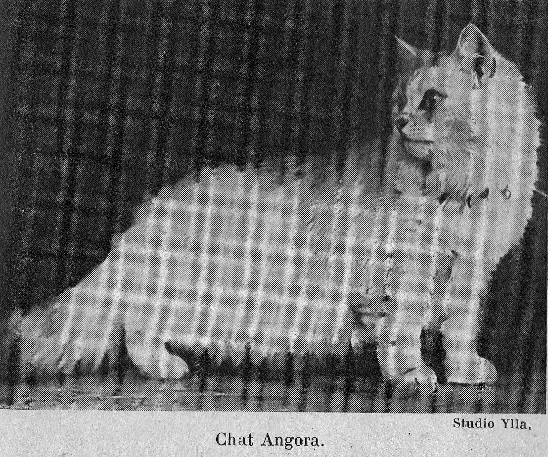
INFECTIOUS DISEASES
This chapter is undoubtedly the most important in feline pathology. The conditions and injuries studied above are more or less serious and although they can be fatal; they are nonetheless isolated incidents whose importance does not go beyond the scope of the individual. With infectious diseases, the situation is different; the seriousness of an individual case is secondary to the key factor of contagion and speed of spread. From this point of view, all the infectious diseases which will now be discussed are not of the same importance, but there are diseases like distemper or typhus, which spread so quickly that it is not uncommon to see them wipe out a cattery in only a few weeks and annihilate an entire breeding programme of several years.
Infectious angina. - Characterized by inflammation of the throat, this strikes cats of all ages, but is especially common and severe in young cats who often succumb.
It begins with general symptoms: depression, decreased appetite and fever, followed by a dry, nagging cough. Soon swallowing becomes difficult and the passage of food requires the cat to extending the head on the neckline when swallowing. Even swallowing saliva is difficult and the cat drools. The throat is tender, painful on pressure, and there is nasal discharge.
On direct examination of the throat, by opening the mouth wide, the whole area is red and congested. Swollen tonsils are often covered with false membranes that may be partially expelled during coughing fits.
In severe cases, complications of conjunctivitis, keratitis and bronchopneumonia appear, and these take a heavy toll on young cats.
Treatment. —- Intervene as soon as possible. Isolate the patient, feed them and support them with easily swallowed liquid foods, particularly lukewarm milk.
Make a diversion ["draw" the infection] under the throat by rubbing it with iodine tincture morning and evening, followed by the application of a cotton bandage. Locally, brush the back of the throat with the following mouthwash:
Methylene blue - 0.5 grammes.
Glycerine - 5 grammes.
Distilled water - 5 grammes.
removing the false membranes if possible. Clean the nostrils and instil a few drops of gomenolated oil into them twice daily.
As an anti-infective and antitoxic, administer morning, noon and evening half a teaspoonful of:
Uroformine - 4 grammes.
Quinine sulphate - 4 grammes.
Foxglove tincture - XX drops.
Cinchona syrup - 50 grammes.
Distilled water - 100 grammes.
Disinfection of contaminated premises is essential in all cases of infectious disease and will be described in some detail at the end of this chapter.
Contagious coryza. – A very contagious infectious disease which is particularly prevalent in spring and autumn among cats of all ages.
Chills and prolonged humidity seem to favour its outbreak, but when it does appear, it spreads quickly from cat to cat, regardless of the housing conditions. All it takes is the introduction of an infected cat into a cattery for all the cats there to be affected within a few days.
It begins with frequent sneezing, which does not significantly alter the cat’s general condition, the affected cat is cheerful, eats well, and does not have a fever; at most we may note a little dryness at the tip of the nose.
Quite quickly, in two to three days on average, a discharge is clearly visible, serous at first, then mucous and purulent. It dries up at the entrance to the nostrils in the form of greyish, cracked crusts which hamper respiration. At this stage, the appetite is greatly reduced if not completely lost; there is fever in the region of 40 C. The cat can no longer breathe through its blocked nostrils and has to breathe through a half-open mouth which often drools foamy saliva.
The inflammation most often spreads, first to the mucosa of the eyes, which become congested, red, swollen and soiled with pus, then to the pharynx, causing pharyngitis. Finally, in some cases, bronchopneumonia may occur.
When the coryza is mild and the inflammation is localized to the nasal mucosa, healing generally takes ten to fifteen days. But there are serious forms marked by deep depression, high fever and intense dyspnoea; these are the ones that develop complications and often end in death.
Treatment. — This is mainly symptomatic. To aid breathing, give eucalyptus inhalations morning and evening followed by instillation of a few drops of gomenolated oil at the entrance of the nostrils. Instillations of a few drops of silver nitrate solution at 1 p 450, at the entrance of the nostrils and between the eyelids, from where it flows through the tear duct to the nasal cavities, generally give good results. Hygiene and cleanliness of the nostrils facilitate breathing and greatly contribute to healing. Finally, support the patient by spoon-feeding with liquid food, particularly hot milk.
Isolating the sick is absolutely necessary. From a prophylactic point of view, one must think about infection during shows and any cat that has just been exhibited must not be returned to the common room until after a one week quarantine.
Infectious gastroenteritis. Typhus. - An extremely contagious infectious disease particularly prevalent in winter and autumn and which results in serious disorders of the general condition and digestive symptoms. Its contagiousness is such that when it appears in a neighbourhood or village, it takes many victims. This is often the price of exhibiting cats and many catteries have been decimated after becoming infected this way.
In the acute form, which is the most frequent and medium intensity form; the disease appears suddenly in the form of severe dejection and depression. The prostrate patient remains lying down and no longer responds to his master's calls. Stringy, foamy saliva drools from the mouth and soils the jaws and face. There is high fever which often reaches 41 C, but in rare cases the temperature may remain normal. The limbs, head, and ears are cold.
These serious changes in general condition are accompanied by particularly serious digestive disorders which fix the diagnosis: the appetite is suppressed though sometimes the patient tries to drink, but either does not have the strength or immediately vomits the liquid. The same is true of anything that can be administered by force. The stomach is sensitive and painful, we note either obstinate constipation or profuse, foetid, sometimes bloody diarrhoea. Uncontrollable vomiting coinciding with either constipation or diarrhoea, along with the extremely serious general condition, are the symptoms characteristic of typhus.
The disease progresses rapidly; the patients succumb in two, three or four days, during which time they lose weight in an extraordinary way. Death is often preceded by a drop in temperature to 36 C, 35 C which heralds the fatal outcome.
During cattery, neighbourhood or village epidemics we may observe slightly different forms. In particular, cases with an acute course which kill in twelve, twenty-four or thirty-six hours, with rapid coma and without time for the digestive symptoms to appear. We also see slower forms with less marked symptoms in patients who resist for eight to ten days, many of which can be cured.
Treatment. - As soon as suspicious symptoms appear, vigorously treat the isolated patients in a room heated at a regular temperature. Various treatments have been recommended, particularly intravenous injections of stovarsol at a daily dosage of 2 to 3 centigrams and the administration of lactic acid bacteria in liquid culture; half a teaspoonful in a little sugar water three to four times a day.
Intramuscular injections of colloidal palladium in at a dose of 1 to 2 cc combined with subcutaneous injections of calcium gluconate 2 cc in 10% solution, a double injection every twelve hours on the first day, every twelve hours from the second, have given encouraging results.
Septazine at a dose of 4 tablets of 0.5 grammes spaced evenly throughout the day gave results clearly superior to anything that had been obtained previously. This is currently the preferred treatment. If vomiting makes it inapplicable, you must resort to soluseptazine in solution, at 6%, 5 cc injected subcutaneously morning and evening.
Whatever treatment is employed, it is important to support the patient with daily subcutaneous injections of 15 to 20 cc glucose serum or caffeine.
If improvement occurs, as soon as possible spoon-feed small amounts of hot sweetened milk. Despite all this care, treatment is frequently insufficient and many patients die.
Vaccination. - A cat that recovers from this disease is immune; hence the idea of ??performing vaccination. The vaccine consists of a splenic emulsion taken from a patient during the feverish period. This is treated with 2 cc formalin water.
The vaccination consists of two subcutaneous injections of 2 cc of the vaccine 5 days apart. Immunity is acquired about ten days after the last injection and the vaccinated cats are therefore immune to the disease. This vaccination should be used systematically in all large catteries and also in all cases of severe epidemics, and these occur frequently.
Distemper ["Disease of young age"]. - A very serious disease, infectious, contagious, inoculable, which strikes young cats aged three to eighteen months, especially the selectively bred improved breeds. The contagion is, here again, extremely subtle; exposures are very often the starting point for serious epidemics caused by infected subjects during exhibitions. It bears more of an analogy to canine distemper, depending on the author, most consider these two different diseases, and attempts to transmit it from one species to the other are generally unsuccessful. But it might be unwise to conclude that the two diseases do not have the same origin and the characteristics that differentiate the two viruses are perhaps only the result of modifications imprinted on a single virus by the different physiologies of the cat and dog. Distemper is different from typhus in that individuals cured of typhus remain susceptible to distemper.
In the acute form, which is again the most common here, the condition begins with a few sneezes, which are often overlooked. But soon the appetite diminishes, the sneezing becomes more frequent and discharge appears. At this time the appetite almost completely disappears and the temperature is around 40 C. The situation worsens rapidly, the patient is dejected, depressed, and prostrated. The conjunctiva are inflamed, swollen, and soiled with mucous pus, this inflammation is sometimes so intense that the mucous membrane forms two swollen bulges that meet and completely cover the eye. Foamy saliva stains the face. This picture is frequently complicated by the appearance of respiratory and digestive disorders. The former progresses in the form of bronchopneumonia with frequent cough, intense dyspnoea, labial murmurs with discharge, and pleurisy with discordant breathing and signs of asphyxia. As for the digestive complications, these manifest as gastroenteritis with vomiting and especially profuse diarrhoea. Death usually results when one or other of these complications occurs, in other cases recovery is possible, but rare.
Alongside these medium intensity cases, we may observe super-acute cases with death in twenty-four hours and no symptoms except extreme prostration and fever of around 41 C. There are also cases with slow progression which are mainly observed in older patients, between 12 and 18 months, who already show some resistance. The symptoms do not differ significantly from those reported in the acute form, but they are less pronounced, and recovery is frequent. But, and this is an important remark to note, these survivors remain carriers of germs for at least a year and, while appearing perfectly healthy, they can infect susceptible subjects brought into contact with them. This shows how dangerous animals can be from a cattery whose health status has not been known for at least a year.
Treatment. — As soon as possible, isolate the patient in a warm and ventilated room. Combat the general health disorders with tonics: glucose serum, caffeine in subcutaneous injections, 10 to 15 cc each day. To support the heart, use 1 to 2 cc camphor oil daily under the skin. In severe cases, create a fixation abscess. Combat general infection and intoxication by administering the potion:
Uroformin - 2 grammes.
Quinine Sulphate - 2 grammes.
Foxglove tincture - X drops.
Cinchona syrup - 25 grammes.
Distilled water - 100 grammes.
Eucalyptus inhalations in the morning and evening followed by nasal instillations of gomenolate oil facilitate pus evacuation and breathing. Wash the eyes two to three times a day with a lukewarm 3% boric solution and instil a few drops of 1.5% zinc sulphate eye drops or better 1 p 450 silver nitrate eye drops. For respiratory complications, use counter-irritant poultices on both sides of the chest renewed daily. To combat vomiting, administer two to three times daily a teaspoonful of:
Soda citrate - 1 gramme.
Distilled water - 100 grammes.
Against diarrhoea, give two or three teaspoons of:
Bismuth sub-nitrate - 4 grammes.
Lactic acid - 4 grammes.
Laudanum - XX drops
Quince syrup - 50 grammes.
Distilled water - 100 grammes.
It is also important to support the patient; when the appetite is not completely suppressed, take the opportunity to administer raw meat and boiled fish. Otherwise, add a spoonful of meat juice and a beaten egg in a little sweetened milk.
Finally, there is a specific treatment that involves the subcutaneous injection of serum from cats hyper-immune to the disease. This serum is curative, it gives excellent results at the onset of the disease, and is also preventative, and cats who receive it under the skin are immune to infection for some time. Unfortunately, this serum is not available in France, but in England it is widely used in cats sent to shows where they could become infected.
Rabies. – An infectious illness, progressing rapidly, considered invariably fatal in all cases.
It is inoculable and caused by a virus which, after entering the body, becomes confined to the nervous centres. Rabies, which has become rare in dogs in France, is even more unusual in cats, because of their living conditions. In all cases, it is caused by inoculation from the bite of a cat or a mad dog. The spontaneous rage which was once believed to exist as a result of deprivation, neglect, etc., does not exist.
In both cats and dogs, the saliva is the virulent element, because the virus that swarms in the nerve centres is eliminated by the salivary glands and is injected directly into the wound by the bite.
Saliva can be virulent and able to spread rabies two weeks before the visible onset of the disease; in other words, the bite of a healthy-looking cat or dog can cause rabies.
The incubation period, that is, the time between the bite and the onset of disease, is very variable; never less than a week, in some cases it can reach a year. In other words, a cat bitten by a rabid animal may show nothing at all abnormal for a year and only then show the first signs of rabies. As a general rule, the incubation period is shorter the closer the bite is to the nerve centres, which explains the urgency of treatment for bites to the head.
Rabies develops as two forms:
1. Furious rabies. This starts with a little restlessness; the cat isolates itself and hides in dark places. Soon signs of excitement appear, the patient is agitated, constantly on the move. The appetite disappears at the same time as we observe the perversion of taste: eating straw, rags, sometimes stones. It is then that stringy drools of saliva flow from each side of the mouth. The voice changes, the cat utters plaintive meows, swallowing is often also difficult, there are signs of pharyngitis which sometimes suggest the presence of a foreign body in the throat.
During this period, the patient becomes aggressive; from his corner he ambushes people, dogs and cats that pass within reach, bites them deeply, lashes out at his victims and not letting go, insensitive to the most violent blows. Death occurs after a few days, either during a furious fit or more often by progressive paralysis which begins with the hindquarters.
2. Dumb rabies. – This is rare and progresses more quickly. After 24 or 36 hours of depression and prostration, paralysis appears. It most often begins with the lower jaw, which hangs inert and death occurs from paralysis.
There is no treatment for rabies, the disease is inevitably fatal in all cases.
Preventive immunization by the Pasteur method is not applicable to bitten cats, because the law states that "dogs and cats bitten or rolled by a rabid animal, or having previously been in contact with it, are immediately slaughtered, by order of the mayor ".
New methods of immunization have been developed in recent years; they have been given to dogs, and in many countries one no longer systematically slaughters contaminated animals which were previously vaccinated, and which are given a new vaccination in the days following their contamination. These methods could be applied to cats; they make perfect sense in countries where rabies is common, though the advisability of adopting them is more questionable with us.
What action should you take in the presence of a cat showing suspicious signs of rabies?
Above all, be careful not to kill him and do your best to capture him, to lock him up so that he cannot bite anyone. Leave him for observation for a few days. If he really is infected with rabies, the characteristic symptoms and death will occur in a short time. If, on the contrary, rabies is not involved, the patient survives. Either way, the situation is clear and the person bitten know what to expect.
Another question arises: since rabies can be transmitted by the bite of an apparently healthy cat, should every bite be considered suspicious? It is essential, in fact, to keep any biting cat under observation for at least 15 days. If, after this period, he remains in excellent health, any danger of rabies is eliminated. On the other hand, anti-rabies treatment is compulsory for the person bitten if, during the fortnight of observation, the cat dies, regardless of the cause of death.
Tuberculosis. —- A contagious disease common in humans and all domestic livestock, caused by the proliferation of the tuberculosis bacillus in the body.
While tuberculosis is less common in cats than that in dogs, it is not as rare as believed for a long time. It has been claimed to affect 2% of the overall cat population; without wanting to give this figure a precise value, it nevertheless gives an idea of the prevalence of tuberculosis in cats.
Cats are most often infected through contact with people with tuberculosis, by ingesting sputum which, in the case of open pulmonary tuberculosis, contains large quantities of tuberculosis bacilli.
Tuberculosis first manifests as general disorders; without apparent cause, the patient loses weight although appetite is unchanged, becomes miserable, appears tired, and the hair becomes dull.
These signs are too imprecise to provide a diagnosis, but they do not remain isolated symptoms for long and soon thoracic, abdominal, cutaneous or bone symptoms appear, which constitute signs, while definitive, of probable of tuberculosis.
Thoracic tuberculosis progresses as pneumonia, pleurisy, and pericarditis.
Coughing is the dominant symptom in pneumonia; it is small, nagging, sometimes painful, more or less frequent, it is rarely accompanied by discharge or, at least not by abundant discharge. The respiratory acceleration is not very marked, and the fever hardly exceeds 39.5 C i.e. remains significantly lower than in common bronchial pneumonia. All this evolves insidiously, without sudden bursts; however, in winter and during cold periods, acute outbreaks of tuberculous bronchopneumonia are noted which differ little from simple bronchopneumonia.
Pleurisy is not significantly less common than pneumonia; it almost always appears as chronic pleurisy with effusion. It manifests as a dry, small, painful cough; dyspnoea with discordant breathing and frequent vomiting. Percussion of the chest indicates sloping dullness at an upper horizontal limit, as discussed on page 60. Thoracentesis results in a discharge of haemorrhagic fluid in most cases.
Pericarditis is rarer than the previous forms, it is also less well expressed from a clinical point of view. Respiratory acceleration, depression and dyspnoea remain the dominant signs, but, at the same time, it is observed that the heartbeat can hardly be heard behind the left elbow and, on auscultation, the sounds of the heart are hard to hear, as if distant and muffled. The pulse is small, rapid.
Tuberculous ascites, most often associated with massive damage to the liver, results in the enlargement and distortion of the belly due to the accumulation of fluid in the peritoneum. An exploratory puncture often yields haemorrhagic fluid. It is the most common symptom of abdominal tuberculosis, sometimes with chronic enteritis.
Skin tuberculosis is common in cats; it manifests as ulcerated wounds, resistant to healing, which exude a small quantity of greyish fluid pus, in which the tuberculous bacilli abound. These sores are mostly seen in the throat area, the lower edge of the neckline and the nose area.
Localizations to the bones manifest almost exclusively as chronic arthritis, with enlarged joints, the hock and the stifle being the most common sites.
When one or more of these manifestations coincide with a severe impairment of the general condition as mentioned above, all presenting a chronic course leading to progressive decline with weight loss and muscle wasting, the tuberculosis is not definite, but is highly probable and severe isolation measures are required while awaiting a confirmed diagnosis.
This diagnosis is made by subcutaneous tuberculination, which is carried out as follows: the patient is immobilized for twenty-four hours, without modifying any of his usual living conditions. We record his temperatures morning, noon and evening, and the average gives us the initial temperature. Early the next morning, e.g. 6 a.m. or 7 a.m., 0.5 cc of tuberculin to the tenth is injected under the skin. The temperature is taken at the time of the injection and then every two hours, until at least 4 p.m.. In cases of tuberculosis, there is usually a rise in temperature four, six or eight hours after the injection. This hyperthermia persists for some time, and generally disappears after twenty-four hours. The animal is considered tuberculous when there is a 1.5 C difference between the highest temperature after the injection and the initial temperature.
In addition to fever, a positive reaction manifests as general disorders: depression, chills, inappetence ... and sometimes also by a hot and painful swelling at the site of the tuberculin injection.
Unfortunately, tuberculination by subcutaneous injection in cats does not give as precise results as in some other species; it is also inapplicable when the initial temperature exceeds 39 C. In the latter case, a complementary deviation can be performed with the serum of the suspect animal, but the latter method is not completely reliable. In any case, whatever diagnostic method is used, care must be taken not to rely completely on the results, if the positive reactions are to be considered as proof of tuberculosis infection, the negative reactions should only be accepted as negatives if they do not contradict clinical observation; in doubtful cases one should not hesitate to conclude the presence of tuberculosis. It is less serious to destroy a dangerously ill non-tuberculous patient than to leave a dangerous tuberculous patient in the family environment or in the cattery.
Tuberculous cats are not practicably curable; they are or will be a danger to people close to them, especially children. Keeping them would not only be a mistake, but an extremely serious fault which nothing, absolutely nothing, and excuse. Destroying them, ending their suffering, is all that can be done for them.
Disinfection. - In all cases of infectious or parasitic disease, disinfection is a necessity. It must be applied not only to the contaminated premises, but to all objects as well: cushions, blankets, cleaning instruments, dishes soiled by the patient.
In cases of infectious disease, various objects - cushions, blankets, combs, brushes, dishes – must remain in the contaminated room, separated by airtight closed doors, windows and other exits. A mixture of trioxymethylene and lime chloride at a rate of 6 grammes of the first to 12 of the second per cubic meter to be disinfected is placed in this room, in a sufficiently large container. Add 20 grammes of hot water per cubic meter at the time of use. Formalin vapours are released which do not damage metals or fabrics. The room is left tightly closed for twenty-four hours, then amply ventilated for two days to complete the process.
In cases of parasitic disease, especially scabies, sulphur is the best disinfectant. The room is prepared as before, and in it one installs a sheet of metal 1.5 or 2 meters from the ground; this is covered with lighted charcoals on which one deposits flower of sulphur or roughly crushed sulphur granules, sprayed with a little methylated spirits, at the rate of 50 grammes of sulphur per cubic meter to be disinfected. After twenty-four hours of airtight closure, the room is ventilated for two days and is ready to be occupied again.