THE CAT : BREEDS - BREEDING – DISEASES
By
E. Larieux
Veterinary Doctor, Member of the Central Society of Veterinary Medicine and the Society of Comparative Pathology.
Ph. Jumaud
Veterinary Doctor Laureate of the Lyon Veterinary School Graduated in Physiology from the Faculty of Sciences of Lyon
Secretary General of the Cat-Club of France and Belgium
With 29 Figures
Paris
Vigot Frères, Publishers
Successors of Asselin and Houzeau, 23, Rue de l'École-de-Medecine
[Philippe Jumaud (1880 – 1942) was a French veterinarian and founder of the Cat-club of France and Belgium. He was also the chairman of the Saint-Raphael tourist office, and historian of Provence. He used the pseudonym Ph. De Magneux for his literary works. He was the managing director of the periodical Les Tablettes de la Côte d'Azur. Les Races de Chats (The Breeds of Cats) began as a thesis in 1925 and was later published as a book that same year. Second edition - 1926 (fully revised). 3rd edition - 1930 (fully revised). 4th edition - 1935 (fully revised). There were also reprints in between the revised editions.]
I. THE CAT
THE CAT is a mammal, belonging to the order of digitigrade carnivores, the felid family, which contains the lion, panther, leopard, serval, cat, jaguar, cougar, lynx, cheetah, etc. The word ‘cat’ comes from the Latin word ‘cattus’ which appeared very late in the Latin language, a commonplace word which, along with the word felis, was used to designate it; some say it is derived from the word ‘cautus’ meaning cunning, clever, while others view ‘cattus’ as coming from ‘cattare’ because the cat watches for its prey to come within range and then seizes it.
The cat is characterised by a short muzzle, a rounded head, short, strong jaws, five digits on the fore limbs armed with fully retractile claws, four digits on the hind limbs and, generally, a well-developed tail.
MONCRIF recounts, in his History of Cats, the following legend which he heard from a Mulla, a minister of the Muslim religion, who accompanied the ambassador of the Porte in France: « During the first days that the animals spent shut up in the Ark, astonished by the ship’s movements and by the new home they found themselves in, they all remained in their separate quarters... The Monkey was the first to get bored of this sedentary life; he proceeded to tease a young Lioness... The affair between the Monkey and the Lioness resulted in the birth of two Cats, one male and one female."
Fossil cat bones have been discovered in Germany, England and France. The naturalist de Blainville thinks that, in the middle tertiary period, fourteen species of cat, whose size could vary from that of our present cat to that of a pony, inhabited the vast forests which covered the globe; most are still found today, but some seem to have disappeared completely from the face of the earth.
THE CAT IN EGYPT AND IN THE ANTIQUITY. - According to Mr. Pictet, the cat was considered a domestic animal by the Egyptians in the 12th dynasty (around 2,200 years BC), and was held in great veneration, if one is to believe Herodotus, who was the first Greek historian who mentioned it in the 5th century BC. It was introduced into Egypt, according to Ruppel, during the conquest of Ethiopia where it originated, by Ousirtasen I, of the 12th dynasty and quickly multiplied. It was deified from ancient times and the god of music was represented by a human body surmounted by a cat’s head, holding a sistrum in its hand, and the goddess of love was represented by a cat’s head resting on a woman's body; moreover the beauty of women was even more appreciated in Memphis as it more closely resembled the cat’s figure.
[Footnote – The sistrum is a sort of Egyptian instrument, oval in shape, consisting of a curved metal blade, fitted with a handle on the inside, and crossed by movable sticks, also made of metal, which resounded when the device was shaken. The upper part of the blade was decorated with three figures: in the middle is a cat with a human face, on its right is Isis, and on its left is Nephtis. The cat-headed goddess Beset (Bastet), a variant of the goddess Pacht, is often depicted holding a sistrum in the left hand.]
All the Egyptian temples housed a family of cats, each temple having its particular species and, if we refer to Diodorus of Sicily, a large proportion of the children were dedicated to the cat. It was, moreover, a huge source of income for the priests who sold small medals representing the head of the cat of the temple where the vow had been made, medals which were hung around the children’s necks.
The he-cat was the emblem of the Sun and Osiris and, according to M. de Caylus, the she-cat was the emblem of the Moon and Isis; it was even claimed that the Moon had given birth to the cat and that Diana took the form of this animal when the Gods, according to the poets, turned themselves into animals to escape the persecution of the giants. In the Delta at Bubastis, there was even a temple where the goddess Isis was worshipped in the image of a cat and in the name of Aelurus.
In the time of Herodotus (430 BC), when a cat died in an Egyptian house, all the inhabitants shaved their eyebrows as a sign of mourning. The body, carefully embalmed with spices, was placed in a small coffin reproducing the animal’s image in bronze or painted wood adorned with rich colours, bearing enamel eyes, and often inscribed with gold; then, followed by the foremost magistrates, the body was taken and buried in a special cemetery, reserved exclusively for cats. Thus in 1890, in a hypogeum called the Cave of Diana near Beni Hassan, 180,000 cat mummies were found and were taken to London; this cemetery was located near a chapel dug into the rock and consecrated by the kings of the 18th dynasty (1703 to 1462 BC) and of the 19th dynasty (1462 to 1288 BC) to a local goddess with a woman’s body and a cat’s head, named Pakhit, Pacht or Bast. Among these mummies, some were simply surrounded by bandages, others, on the contrary, were buried in coffins. Underground cat cemeteries were found at Bubastis, which was the most sought-after necropolis because of the proximity of the goddess whose emblem they were, at Sakkarah, at Stabl-Antar, near Thebes, etc.; these mummies belonged to three species: felis caligulatus, felis bubastis and felis chaus.
If anyone killed a cat, even accidentally, the people would fall on the murderer and kill him in a most cruel manner. Thus Diodorus of Sicily wrote that when King Ptolemy sought the friendship of the Romans, he could not prevent the people from killing a Roman citizen who had accidentally killed a cat. The Egyptians were so afraid of hurting cats that around 525 BC when Cambyses, king of Persia, wanted to seize the city of Peluse (on the ruins of which now stands Port Said) whose garrison was Egyptian, he made a large number of cats march in front of his troops and officers and soldiers each carried one as a shield. The Egyptians were afraid of harming the cats and surrendered without fighting.
The cat was granted the gift of chasing snakes, and the veneration shown to him was, for some writers, merely recognition of the services he rendered by destroying the rodents which invaded Egypt after each flood of the Nile. The people perfumed it, let it sleep in the sumptuous beds and, at feasts, gave it the place of honour. The Louvre Museum in Paris has mummies and statuettes of sacred cats from Egypt in its collections.
The Assyrians and Babylonians did not know the cat and the Bible does not mention it; but a Sanskrit work dating back two thousand years already spoke of the domestic cat.
From Egypt the cat made its way, albeit very late, into Syria and Arabia, where it became the favourite animal of the Prophet Muhammad. Moreover, according to Muslim, believers trace the origin of the cat’s ability to always land on its feet to the Prophet. His cat Muezza, lying one day on the sleeve of his coat, seemed to meditate there deeply. In a hurry to go to prayer but not daring to rouse the animal from her ecstasy, her master cut the sleeve of his coat so as not to disturb her. The creature was grateful to him and when the Prophet returned, Muezza came to thank him for his marked attention by bowing to him. Muhammad then assured his cat a place in his paradise and, passing his hand three times along her back, he gave her whole race the virtue of always falling on their feet. Long before Muhammad, Pliny wrote that the Arabs worshipped a Golden Cat; and the Turks regard the cat as a pure animal, pampering it in their house at the same time as proscribing the dog as an unclean animal; even now cats are still in great honour among all Muslims.
Homer speaks of cats only with the greatest respect, and Corinth owned a colossal bronze statue of a crouching cat. The Alains, the Suives, and the Vandals adopted the cat to represent freedom because it could neither be tamed nor submissive; according to Favyn, their emblem was "silver with a sable cat". In heraldic terms, the cat is said to be herissoné [with its back up] when the hindquarters are higher than the head, and effarouché [startled] when it seems to be crawling. Among the ancient Germanic peoples it was a symbol representing adultery and, at the same time, independence; the Scandinavians chose him as the god of love while, in his sermons, Saint Dominic represented the demon in the form of the cat and the witches were supposed to take the form of a black cat.
Leboux, from Lincy, in his Book of French Proverbs, recounts the curious legend of the Beaugency cats. An architect could not manage to finish the bridge he was building at Beaugency because the last arch always fell as soon as it was finished. After three or four unsuccessful attempts, he called the devil to aid him. The devil was kind enough to undertake the task, but only on the condition that he would have the first soul to pass under this arch. The deal was made, but as soon as the arch was built the thought of sending a cat through it. The enraged devil unsuccessfully tried to destroy his work by kicking it, and, in his fury, he tried to seize the cat. The cat scratched his hands and face and thus escaped, immediately running off to take refuge a league away, in Sologne, in a place which has since become known as Chaffin (cat end).
CAT LOVERS. - Nowadays, humans often consider the cat as a companion and friend. Thus, near the Victory Gate (Babel Nazz) in Cairo there was a hospital where sick or homeless cats exclusively were taken in and cared for. Currently, the League for the Defence of Animals has kennels in Paris where lost, stray or abandoned cats are collected and cared for. A dozen years ago, a restaurant for animals was created near Westminster in London, where rooms furnished with tables, on which were placed bowls containing various provisions, were reserved for cats; the residents could be recognized by a medallion they wore around their necks.
In a pretty sonnet, when Tasso had no candle to write by during the night, in his misery he begged his cat to lend him the light of its eyes. Petrarch, when he had retired to Arca, near Padua, after the death of Laure de Noves, fell in love with a cat whose skeleton is kept in the Museum of Padua. Joachim du Bellay wrote an epitaph of two hundred and two lines to celebrate the virtues of his cat Belaud:
Belaud, my small grey cat, deceased.
Belaud – nature’s finest masterpiece,
Wrought in flesh, fur and form of cat;
Belaud, the deadly scourge of rats
Whose beauty was such, ‘tis true to tell,
He was worthy of being immortal.
Richelieu almost always had his cat Rita near his worktable; he surrounded himself in the morning with a dozen cats with which he played, and he set up a cattery near his room, entrusted to the care of two attendants, Abel and Teyssandier, who were responsible for distributing, morning and evening, the chicken breast paté that formed their food. Montaigne admitted that his cat's games were, for him, as much a recreation as a subject of study. Colbert always had a bunch of young cats in his office. Fontenelle always adored cats, but had a special affection for one of them which he placed in an armchair to make speeches to her in order to practice, but one fine day this cat ran away never to return. The English philosopher Locke always had a cat on his table. Abbot Galiani, the Italian philosopher, wrote to Mme d'Epinay from Naples: "Your life in Paris is less insipid than mine in Naples, where nothing is attached to me except for the two cats I have with me, one of which was led astray yesterday through the fault of my people. This made me mad; I dismissed everyone from me. Fortunately, he was found this morning otherwise I would have hanged myself in despair."
Hoffmann owned a superb cat that he loved very much. Bernardin de Saint-Pierre, Jean-Jacques Rousseau, Chateaubriand who had inherited a reddish gray cat he called Micetto from Leon XII, Baudelaire, Victor Hugo, Prosper Mérimée, Théophile Gautier who had a remarkable collection of cats, Guy de Maupassant, Sainte-Beuve, Flaubert, Champfleury, Alexandre Dumas, Théodore Barrière, Jules Lemaitre, Taine, Paul de Kock, Loti, etc. etc., had a very strong taste for cats; Henri Rochefort owned a cat named Kroumir, which he was very attached to and which died of grief very shortly after its master’s death. Mme de la Sablière got rid of her dogs and replaced them with black cats; the Duchess of Maine composed a rondeau to sing the merits of her cat Marlamain; Madame de Lesdiguières had the following quatrain engraved on a white marble mausoleum, which she had erected in her cat:
"Here is a pretty Cat:
His Mistress, who loved nothing,
Loved him madly;
Why would you ask? It’s plain to see."
Mme Deshoulières said when talking about her cat: "When my husband is away, Grisette is enough for me."
According to an article published in the "Annales" under the signature of Mr. Gabriel Mourey, while at the Elysee Palace Mr. Poincaré enjoyed observing his Siamese cat Gri-Gri which he was particularly fond of. "This cat has a strange and complex personality. Voluptuous and greedy, treacherous and cunning, stubborn and ferocious, authoritarian to the point of tyranny, he has, you see, only flaws, but those flaws, just half of which would make any other cat unbearable, become, by the way he uses them, qualities. Indeed, he is witty, full of talk and verve, offbeat and facetious; he knows the value of a well-placed joke. He pulls himself out of the most difficult situations with a pirouette. He could teach useful lessons to so many timid and hesitant people who are uncertain and slow to reach a decision!"
Mr. Clemenceau during a quick trip where he was going to negotiations on the final conclusion of the peace protocols with the King of England and Lloyd George, bought, in a store on Bond Street, a little Persian cat which he called Prudence and made his mascot.
The following quatrain, found in the report of the Academy of Reims of 1924, whose author is unknown, brings to mind that of Mme de Lesdiguières:
In my garden, without noise or glory
I piously place in the earth
My pretty black cat, Suzie,
Alas! I find it hard to believe it.
[Dans mon jardin, sans bruit, sans gloire
J’ai mis en terre, avec piété
Suzie, ma jolie chatte noire ;
Hélas ! que j’ai de mal d’y croire.]
We also note in the review Les Chats, of April 1, 1914, the following piece by Paul Nagour, taken from his catophile poems.
The Old Cat
With his dishevelled fur and hollow belly,
The homeless old cat wanders on unsteady legs,
Bruised, from fools and wicked humans he has fled,
A flash of vengeance in his green eyes swelling.
He is dirty, he is ugly; - in his barbarous pride
The cruel shopkeeper insults him and drives him away,
When, dying of hunger, weary of war, he arrives
To crouch on his doorstep, pitiful and resigned.
Servile dogs are sent to chase the damned,
When, anxious, he drinks black water from the streams,
Once, long ago, this outcast knew the good times,
Knew comfort, free of malice, free of wicked hands.
Once upon a time, fingers fine and charming
Caressed him, while he rode the tides of dreams,
His eyes - the colour of seaweed from the beach -
Deep and pure, and eloquent as speech.
Oh yes, he was a superb cat, loved by his masters,
And in winter, a silky eiderdown became his bed,
A cat that people loved to see, in happy laughter,
Climb the window curtains or steal thread.
He frolicked like a clown upon the carpet,
And, with shining eyes played hide and seek,
Fading away, showing only a hint of whiskers,
Like a big cat, vanished in his carpet forest.
Alas! the master died, and then the mistress,
And although their pious and devoted friends
Would take him elsewhere, he liked it better,
Close to his old home, in his grief and distress.
When the people are no longer passing,
He sleeps on his old familiar doorstep,
A sad old cat lost in sweet dreams can forget
The lonely day and see again dead masters.
[Le Vieux Chat
Avec son ventre creux et ses poils en broussailles,
Ill erre, le vieux chat sans gite – trébuchant,
Tout meurtri, fuyant l’homme imbécile et méchant,
Ayant dans ses yeux verts l’éclair des représailles.
Il est sale, il est laid ; - en son barbare orgueil
Le boutiquier cruel l’injurie et le chasse,
Lorsque, mourant de faim, il vient, de guerre lasse,
Piteux et resigné, s’accroupir à son seuil.
On lance contre le maudit, les chiens serviles,
Lorsqu’il boit, anxieux, l’eau noire des ruisseaux,
Pourtant il a jadis connu, les bons morceaux,
Le confort, à l’abri des méchancetés viles.
Des mains fines, des mains charmantes, autrefois
L’ont caressé tandis que le vague des rêves
Emplissait ses grands yeux, couleur d’algue des grèves,
Ses yeux profonds et purs, parlants comme des voix.
Alors c’était un chat superbe, aimé des maîtres,
Ayant, l’hiver, pour lit un édredon soyeux,
Un chat qu’on aimait voir, dans les rires joyeux,
Voler le fil, grimper aux rideaux des fenêtres,
Cabrioler ainsi qu’un clown sur les tapis,
Et, dans ses yeux bruyants et fous de cache-cache,
S’effacer, ne montrant qu’un soupçon de moustache,
Comme les grands félins dans les jungles tapis.
|Hélas ! le maître est mort, puis la chère maîtresse,
Et, bien que des amis dévoués et pieux
Aient voulu l’emmener ailleurs, il aima mieux,
Près de l’ancien logis, le deuil et la détresse.
Et lorsque nul passant ne marche plus dehors,
Qu’il peut dormir au seuil de son ancienne porte,
Dans le songe très doux que le sommeil apporte,
Le vieux chat désolé rêve des maîtres morts !]
ENEMIES OF CATS. – At times, however, cats were the innocent victims of ignorance, prejudice and even human superstition. Thus, up until the end of the eighteenth century, a ceremony was celebrated in Metz every year during which, in front of magistrates and festively clothed clergy, an iron cage filled with cats was placed on top of a pyre that was then lit. This ceremony was not abolished until around 1750, on the intervention of the Marshal of Armentieres. In Paris, the Saint-Jean’s fire was lit around a flagpole raised in the Place de Greve, and cats, held in a basket, were released when the flames were already rising around them; their only possible retreat was to climb to the top of the mast from which they then fell, suffocated by the smoke, to the great joy of the spectators. At that time, cats were considered wizards who, on Midsummer's Eve, went to a general Sabbath. An author wrote the following quatrain on this subject in 1619:
A cat, who, with a brief run,
Went up the fire of Saint-Jean-en-Grève;
But the fire did not spare it,
And made it jump up and down.
Alfred de Musset's maternal grandfather, Guyot Desherbiers, in a poem entitled Les Chats, speaks of the wizard cat in the following terms, reproduced by Landrin:
Often, the mere multitude
Surpasses one’s instinct
In the combinations studied.
There are indistinct meanings
In the Great Book of Nature,
Which is not always without deletions
For Pliny or Buffon.
The curious mind of man
Wants everything to be straightforward;
We need this, sensible or not,
We need some sense. And that's how
The few physicist people
Created the magician Cat.
In what seems unbelievable,
He grips the claw of the devil,
If he does not see the finger of God.
The dogma of witchcraft
Introduced in due course
After that of idolatry.
Not that I pretend to make you impious
And believe in the fact of magic,
Doubtless he is a sorcerer,
And the Cat is one of his familiars.
In theology, we teach
That hell must have no orgy,
And that the Saint John sabbath
Must have Master Cat preside.
When in the Holy Office at Goa,
Was in conflagration
For our edification,
Some makers of witchcraft,
Saw the lights of an auto da fe
Monseigneur took his coffee,
We know that the devout ladies
The misbelievers saw souls -
Inasmuch as a soul can actually be seen -
Pass through the infernal manor
In the guise of a Black Cat.
From the great Baldus of Bartole who
Maintained and even increased the school,
I will tell you a sad story.
Knowing everything apart from magic,
With the greatest scientist of Cats,
He learned astrology!
One day they were in discord
(And it was Baldus who was wrong),
And to warn him his cat bit him,
And ink smeared the edge
Of his inaccurate tablature;
We do not know if the cat
With Belzebub had a pact,
But Baldus firmly believed it;
He saw his salvation in danger.
The supernatural criticism
Stormed his grave brain.
In the depths of his veins he felt,
With the poisons of the tooth,
The infernal aftermath,
And he became obsessed
With the spirit named Legion.
Now he reads without method
Digest, News and Code.
From this known syllogism,
Whose master was recognized,
He can no longer find the trail;
Finally, the doctor,to have
Incurred disgrace from a cat,
Lost meaning and knowledge.
Therefore the Cat, the world over,
Is sometimes god, sometimes necromancer,
Constantly able to maintain
His haughtiness of character.
One point of universal faith
(The illustrious traveller Pythia
Attested, at the seven hundredth chandelier),
It is that always the Cat that held
The seals of destiny over oneself,
That Fortune has chained itself,
To the talisman of his favour
And that by a victorious charm,
All its supporters attract
All the magnetism in the heart.
As proof that I speak without laughing,
Let us go no further, dear sister,
I find the effect on yourself
And that sweet and sacred power
That made you, by force or by will,
That heard, saw and loved you,
Can it not be said
That the knot of this supreme love
Unites us with the Cats?
We know that Henry III fainted as soon as he saw a cat. Toussenel, in his 'Spirit of the Beasts' and Honore Schoeffer both accuse this animal of destroying game. According to them, the 6 million cats that existed in France destroyed, each year, 24 million young rabbits and leverets, 72 million partridges, 2,190 million small edible birds (larks, skylarks, ortolan buntings, etc.) figures that seem very exaggerated. In any case, it is easy to keep a poaching cat away from the nests by placing nearby, in the tree or bush, a cloth soaked in empyreumatic oil [medicinal creosote or similar], for which the cat has an invincible repulsion.
Lenz summarizes the question of the usefulness of the cat by giving the following advice, reported by Landrin: "If you have a cat that scratches and bites children, that constantly breaks pots and pans, steals sausages, butter and meat, that strangles chickens and small birds, but never tries to catch rats or mice, then the best thing is to is drown it or give it the coup de grace one way or another. But if you have a nice kitty who is the favourite toy of children, who does not cause the slightest disorder in the house, and who keeps busy day and night hunting rats and mice, then you would do very well to take great care of him like a benefactor."
Finally, although the cat, like most domestic animals, can transmit certain diseases to humans such as mange, ringworm, favus, diphtheria, tuberculosis, rabies, etc., on the other hand it destroys mice and rats that are covered with fleas that can inoculate us with infectious diseases, including plague, sweating sickness [suette miliaire] and pneumonia.
[Translator’s Note: Suette miliaire, or "Picardy Sweat," was probably hantavirus, which is linked to rodents. There were serious outbreaks of "Sweating Sickness" in England between 1485 and 1551, with average mortality rates between 30% and 50%. There were serious outbreaks of Picardy sweat in France between 1718 and 1861.]
THE ORIGINS AND VARIETIES OF THE DOMESTIC CAT
WILD CATS - Today, there are still three different species of cats found in the wild.
[Footnote: We owe thanks to Mr. Tisserant, the director of L’Acclimatation, for allowing us to reproduce the four magnificent drawings by Mr. Malier inserted in the first part of this book; the other figures were provided by our collaborator M. Ph. Jumaud. We sincerely thank them for their kindness.]
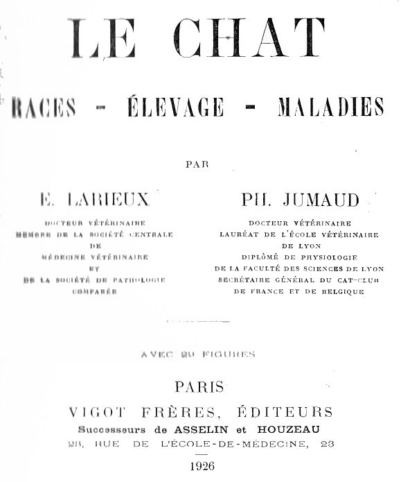
Firstly, the Wild Cat (felis catus ferus) which inhabits Europe and western Asia; we still see it in wooded regions of the Jura, the Alps and the Pyrenees, in Switzerland and in the Thuringian forest, often inhabiting caves or burrows, and keeping watch from trees while concealing themselves along the big branches, mainly at night, to hunt rodents and birds. Its fur, especially on the jowls, is long and bushy; its coat, fawn-grey in colour, has a blackish line along the middle of the back from which parallel blackish bands, more or less distinct, descend down the sides, shoulders and thighs; it is whitish grey on the underside of the body and at the corners of the mouth; the forehead and the upper part of the head have four parallel bands; the lips and soles of the feet are a beautiful black; the ears are straight and stiff; the tail is very bushy and cylindrical with black rings along its length and ends in a black point. The animal is about 35 centimetres in height, and its length varies from 60 to 70 centimetres, of which 25 to 30 centimetres is the tail.
Secondly, the gloved cat (Felis catus maniculatus) seems to be the white-eared caracal described by Buffon; it has also been called the Lydian cat, or Nubian cat, and is found in north-eastern Africa, the Nile valley, Arabia, Asia Minor and Sardinia. It is assumed that it was brought from Meroë to Egypt by the priests and that, from there, it was introduced into Arabia, Syria, then Greece and the Roman Empire. The upper part of its body is a reddish tawny yellow that becomes lighter on the flanks and turns to pure white under the belly; a long black stripe extends along the back, from which emerge narrow, brown stripes across the trunk; the head has 7 or 8 narrow and arched black stripes, and the thighs are also marked on their outer face, with 5 to 6 stripes of the same colour; the long tail is fawn yellow and ends in a black point preceded by a few rings of the same colour; the soles of the feet are black. Standing taller on the legs than the previous wildcat, its hues are very variable and some of its varieties greatly resemble the striped and mottled variety of domestic cat. Its length is 78 to 80 centimetres including 25 centimetres for the tail; it is about the size of our domestic cat.
[Translator’s note : Meroë was on the east bank of the Nile, in the Kingdom of Kush in Nubia, now northern Sudan.]
Thirdly, the Manul (felis manul), which is found on the central plateau of Asia which goes from Tibet to the Amur valley, in the steppes of Tartary and Mongolia; it has a thick pale grey, somewhat reddish, coat with indistinct spots.
Among the wild species derived from the previous ones we find:
Temminck's felis minuta, encompassing cats from Sumatra and Java, which inhabit the Himalayas and are found on the high mountains of Malacca and the Sunda Islands (Sumatra and Java); it is reddish-grey above, white below with four lines of brownish spots on the back, a transverse stripe below the throat, two or three below the neck, and round spots on the flanks. Felis badia of Borneo is also reddish but without spots.
The swimming cat (felis undata), also found in Java and described by Boitard, has a dull grey coat striped with small brown bands and has webbed feet, the toes being united by a membrane extending to the tip of the third phalanx.
Diard's cat (felis Diardis), also inhabiting Java, is 90 centimetres long, 70 of which is the tail; yellowish grey with black spots on the back, neck and legs, it has black rings with a grey centre on the shoulders, thighs and flanks and dark grey rings on the tail. [Clouded Leopard]
Felis Eyra, which is found in the forests of Brazil and Paraguay, is very easy to tame; it is a light reddish colour, with white lower jaw and whiskers; it has a white spot on each side of the nose; its length is about 55 centimetres including 30 centimetres for the tail.
The Chinese felis tristis, described by Milne Edward, has a slate grey coat dotted with dark spots arranged in broad, loosely shaped rosettes; its rather bushy tail has transverse bands giving it a ringed appearance. [Chinese mountain cat (Felis bieti)]
The paler felis scripta is tawny grey with pink spots, the centres of which are a rather vivid tawny colour; its less bushy tail is also ringed, but the individuals are smaller than the previous species; it is found in western China where it inhabits the Moupin mountains. [also Chinese mountain cat (Felis bieti)]
DOMESTICATION OF THE CAT. – According to Ruppert and Ehrenberg, the Egyptian cat derives from the gloved cat; this is also the conclusion of Temminck's work; for Brehm, the domestic cat of Yemen and the western Red Sea coast is similar to the gloved cat. The Egyptians used it for hunting in the marshes and trained it to bring back birds injured by boomerangs. Relying on the fact that the Greeks and Romans did not have cats in their homes and that they used weasels and martens for destroying mice, Link believes that the Egyptian domestic cat was not imported into Europe and Asia until the Middle Ages, at the start of the Crusades, when the Tarentines and Etruscans brought them back as luxury animals (it was not until the 2nd century A.D. that the cat was known to the Romans), but it was still very rare in Western Europe in the tenth century A.D. because, at least in England, it was regarded as a very valuable animal.
However, this data does not seem to agree with the discovery made at Moosseedorf, in the canton of Bern, at Vaunyl, in the canton of Lucerne and at Robenhausson by Professor Rütimeyer, where the remains of ancient lake settlements appearing to date back to the Stone Age were uncovered, in which cat bones were found amid dog, ox, horse, pig, goat, and sheep bones. The wild cat was also found by Professor Steenstrup, in Halvelse (Denmark), in the skjôkkenmôddings (from "kjôkken," cooking and "môdding," heaps of refuse [midden]), heaps of bones and shells that primitive man accumulated around tents or the huts he lived in and which are still very numerous in Denmark, then in Scotland, in England in the counties of Cornwall and Devon, at the mouth of the Somme, in Saint-Valéry, in Australia, in Malaysia, in Tierra del Fuego; these kjôkkenmôddings date from the Paleolithic period or stone age. Finally, Mr. Lartel also found felis catus in the south of France, in Aurignac, in a cave dating from the same period. Moreover, at the time of the Greeks and Latins, the common cat or felis catus already lived in the wild in the forests of Europe and Asia and, if our ancestors the Aryas did not have cats, it is probable that they knew the wild species; this is what made Cuvier believe that our current cat must descend from felis catus rather than felis maniculatus.
It also appears that the manul, which Blyth says freely crossbreeds with the Indian domestic cat, was domesticated by the Chinese and the Tartars, and Pallas believes it to be the source of our so-called long-haired Angora breed which, coming from Persia, was imported into Italy in 1551 by Pietro del Lavale. A century later Ménard, arriving in France from Rome, smuggled an Angora cat and introduced it to France. However, for Dr. H. Beauregard, it was not introduced to us until 1720 by de Peiresc, adviser to the Parliament of Aix.
According to Mr. Pictet, the domestication of the cat in India must go back, as in Egypt, to ancient times and the Indian cat is, moreover, quite different from the Egyptian cat. All in all, and this is Darwin's opinion, it is not yet known whether domestic cats descend from several distinct wild species or from a single species modified by accidental crossbreeding, but it is likely that our current breeds come from several ancestral wild species. Cats have been transported by Europeans to all parts of the earth, but the diversity of climates has had little influence on them.
DOMESTIC CATS. - The domestic cat is distinguished from the wild cat by its smaller size, its slender, longer and more tapered tail, its larger and longer intestine (according to Daubenton, it is five times the length of the body instead of three times, as in the wild cat); this fact would be due, according to Isidore Geoffroy Saint-Hilaire, to the less carnivorous diet of the domestic cat. The main domestic breeds are:
The common cat (felis catus domesticus) or tabby domestic cat has a supple and graceful body, well proportioned, a medium size head, a long and rather broad nose, small erect ears that are hairy on the outside and hairless inside, blue, orange or green eyes, a long and slender tail, short hair, white skin and a coat of varied colours - most often tabby, with tawny, white, grey and black stripes, bands and markings; the lips and soles of the feet are black. This variability of coat colour is due to the inability to monitor and control matings and, as soon as the domestic cat returns to life in the woods, it reverts to the variegated grey tint of the wild cat. It has an average length of 40 to 50 centimetres including a 32 centimetre tail, a height of 27 centimetres and an average weight of 4 kilograms. Fitzenger believes that the striped cat is the product of crossing the Spanish cat with the European wildcat (felis catus); moreover, in Europe and in La Plata, cats that have become wild again are regularly striped, and although in a few cases their size has increased considerably, they do not differ in any other respect from the domestic cat.
The Antiguan Creole cat is small with an elongated head.
The Ceylon cat, also small in size, has flat-lying fur; its head is small, with a receding forehead, wide and thin ears.
The Paraguayan cat is smaller than the European cat; its body is thinner, its fur is short, shiny, sparse, very close-lying especially on the tail.
The Spanish cat (felis catus hispanicus), with rather short and shiny hair, has a coat marked with irregular patches of white, black and orange on the upper and lateral parts of the body, fawn under the belly; the lips and the soles of the feet are flesh coloured. In this species, only the female shows all three colours together, the male, in the vast majority of cases, shows only two colours, either white and black sometimes with a tawny tint, or white and orange, but never has black and orange at the same time. The Spanish cat, according to Fitzenger, is the closest match to the Egyptian cat. This cat is generally gentle, affectionate and very loyal; it is significantly smaller than the common cat.
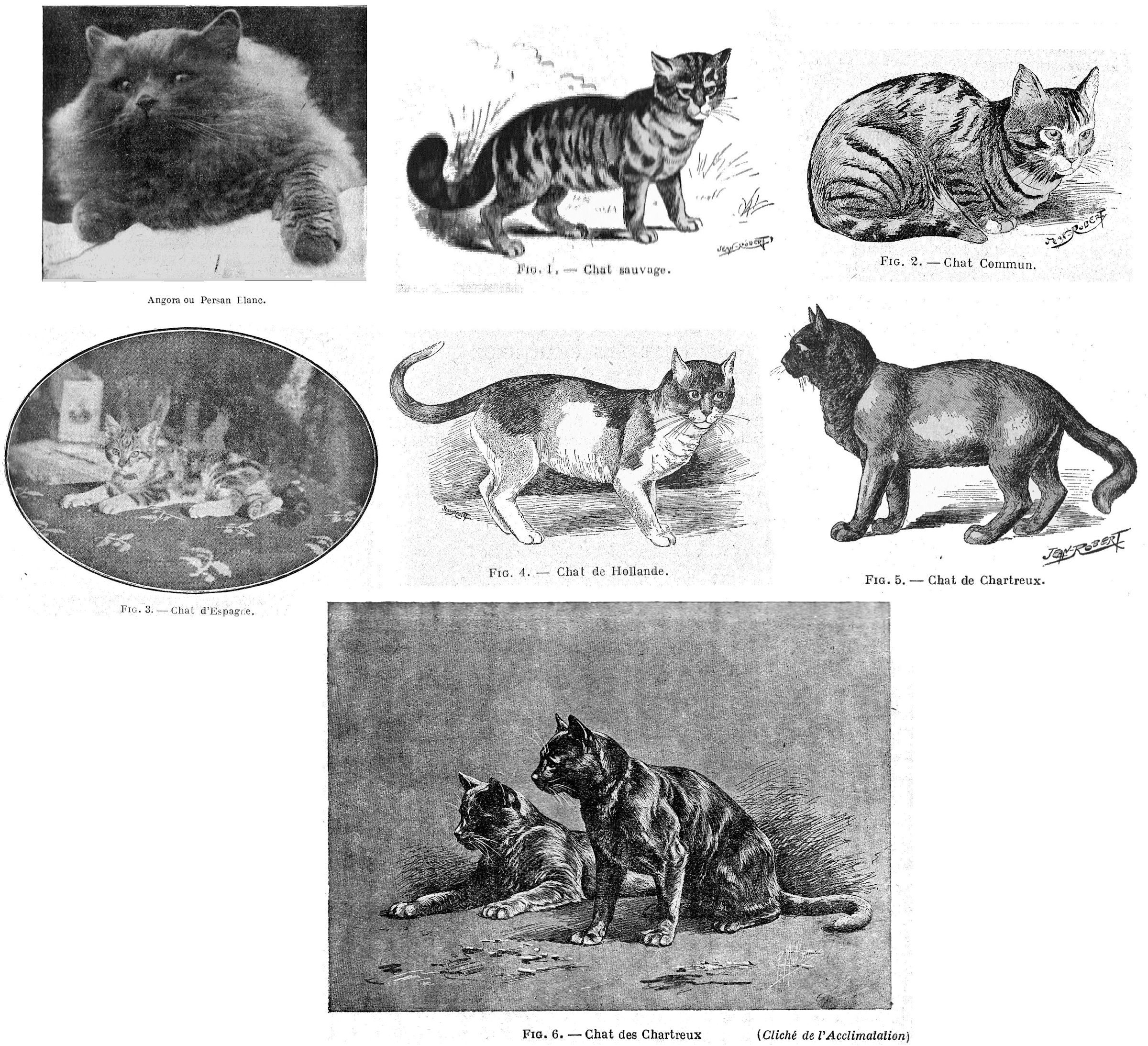
[Footnote - This peculiarity is explained by Mendelian rules and by bringing in the X chromosomes. When the sex cell, before its maturity, is going to divide, the nucleus produces small rods which strongly take up dye (hence their name "chromosome"), in a number determined for each individual, animal or plant; these first meet at the equator of the nucleus, each splitting into two segments in the direction of their thickness and then half moving to each of the two poles of the nucleus. In addition to these normal chromosomes there is a chromosome of variable shape, larger than the others, possessing special properties and which is precisely the X chromosome.
Most individuals, and this is the case with cats, have two kinds of sperm, one containing an X chromosome, the other without, while each egg always contains an X chromosome. Moreover, on the one hand the male is only formed in the presence of one X chromosome and on the other hand the female is only formed in the presence of two X chromosomes so we always find one of these elements in the ovum. The sperm without an X chromosome will always produce a male while the sperm with an X chromosome will produce a female; the formula of the male therefore becomes XO (O representing the absent chromosome), and that of the female XX. (Translator’s note: the Y chromosome was not discovered at that time.)
According to Mendelian law these chromosomes are responsible for transmitting the special characteristics of individuals. While a few (dominant characteristics) can momentarily hide others (recessive characteristics), they can also walk side by side, mingling with each other, as is the case with the pigmentation of the Spanish cat. Note that white, which is a discoloration, is not involved with this pigmentation and that we have only two colours, black and orange. Let us represent for example the orange or red colour by R, the black colour by N, and the the absence of an X chromosome by O, and cross an orange female (whose formula is RR) with a black male (formula NO); if we dissociate the gametes and recombine them then we get NR, female (since there are two chromosomes) having both black (N) and orange (R), and RO male since there is no only one chromosome and orange (R).
On the contrary, let's cross a black female (NN) with an orange male (RO); we still have an orange and black RN female but a black male NO. In both cases the male has only one colour while the female has two.]
The Cypriot cat is light grey, and black on the underside of the legs; it is usually short-haired and white-skinned, it has small, erect ears. It does however have some long-haired sub-varieties.
The Icelandic cat has a grey-blue coat.
The red cat of the Cape of Good Hope (felis domesticus ruber) in which the red colour can be limited to a simple dorsal stripe going from the head to the tail.
The Carolinian archipelago cat, with a reddish yellow coat, has very long legs.
In the Cape of Good Hope we also meet a slate or blue-grey cat with short hair, small, erect ears, a tail of medium length and reddish colour; they are usually red and blue, some subjects may show a reddish stripe on the back.
The Carthusian cat (felis catus coeruleus or felis catüs carthusianorum) was reportedly, according to Fitzenger, a crossbreed of the Egyptian cat and the Manul. It is found in the United States under the name of Maltese cat. Strongly built and well proportioned, its tail has no nodules and is uniform in size. It has a strongly built head, large eyes, small, erect ears and a short nose. Its hair is very fine, woolly, a little long, displaying everywhere a beautiful uniform slate grey colour with bluish highlights; the lips and soles of the feet are black. This rather big cat is very elegant, but lazy.
Related to this variety are the cats from Romania (Caucasus) and Khorassan (Persia) which closely resemble it and have a brownish blue coat; the Tobolsk cat (Siberia) whose size is a little larger than the ordinary cat, and which has a strongly built head, large eyes, a rather short nose, small, erect ears, a very long coat with a woolly consistency and uniformly reddish colour.
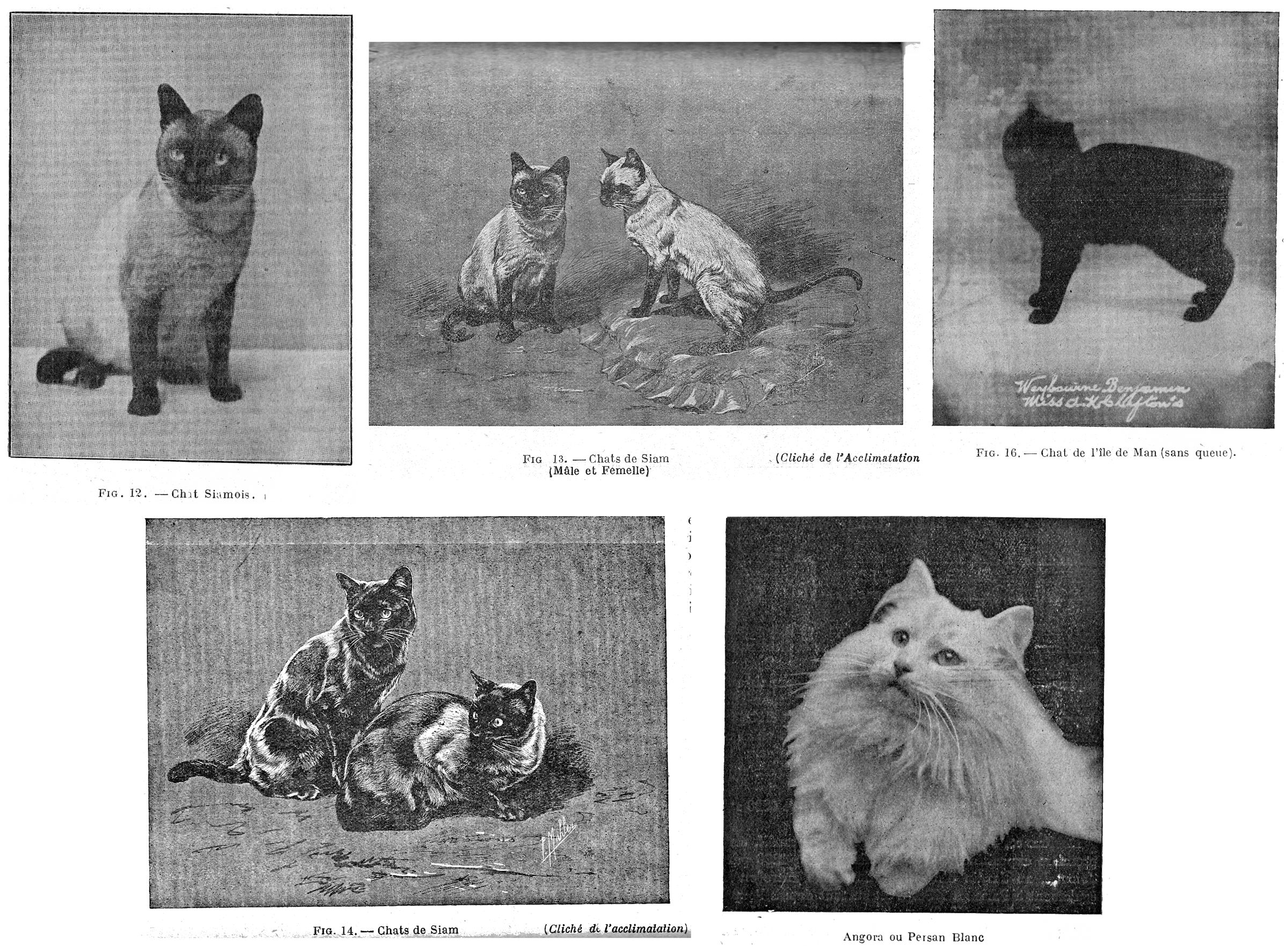
The Angora, or Persian, cat (felis catus angoransis) has fine, silky hair, very long, especially on the neck, under the belly and on the tail; that of the head and legs is short and its coat, usually of a uniform shade, is white, black, grey, blue, orange, cream, silvery, rarely striped or mixed with all these shades in irregular patches; the lips and the soles of the feet are flesh coloured. It is medium size and white individuals sometimes have depigmented eyes that appear to have a red tint. It is indolent, lazy, but very intelligent.
The Gambian cat or negro cat (felis catus nigritia) is found in Gambia, Guinea and on the west coast of Africa; it has black, wrinkled skin, short, blue-grey fur, somewhat bare ears, long legs, a body similar to that of the common cat, and a tapered tail without breaks or knots.
The Chinese cat (felis catus sinensis, or felis catus auriculosa), which is mainly found in China, Pet-chili and Manchuria, is reminiscent of the Chartreux; it is very large, with a rather strongly built head with ears falling to the sides; its fur is semi-long, silky, black or yellow with markings on the side; the tail is thick, medium length, without knots or breaks. It is consumed by the Chinese who fatten it for this purpose, mainly in the province of De Chy Li, where the Manchus exchange their small male cats for sable skins with the inhabitants of the land of Kiliaque.
The Siamese cat and the Malayan cat (felis catus torquata), a little smaller than the European cat, have an elegant and graceful profile, a short and smooth coat, small head, rather large ears lined inside white hair, uniform dark brown tail and legs, a very light isabelline grey coat, paler under the belly, darkening more towards the extremities; the lips and soles of the feet are black and the iris most often shows a blue-grey tint; the whiskers are pure white and the muzzle a beautiful dark pink. The tail has a break towards its end; it is sometimes twisted to the side, and most often ends in a knot.
The Birman cat, small in size and low on its legs, has an elongated body, a rounded forehead, very long whiskers and, on the eyebrows, long tufts of hair. The coat, cream like the Siamese, shows light bronze tints along the spine; the coat is quite long and silky. The tail is furnished with long otter-brown hairs above, grey or beige below.
On the Isle of Man, a small island in the Irish Sea, there is a tailless race (felis catus anura) which is also found on the coasts of Japan; of fairly large size, with hind limbs longer than the forelimbs, a broad, fine head, large round eyes, orange or brown, with semi-long hair, they are tabby or uniformly black, although the latter colour is either rare; the tail is reduced to 5 or 6 caudal vertebrae and is not visible.
In England, where they were introduced and mated with long-tailed cats, they produced fertile offspring which had tails, either in the first generation or in subsequent generations; by crossing an Isle of Man cat with regular she-cats, Dr Wilson obtained 23 small cats, 17 of which were tailless, while crossing an Isle of Man she-cat with regular cats consistently gave him offspring with short, imperfect tails.
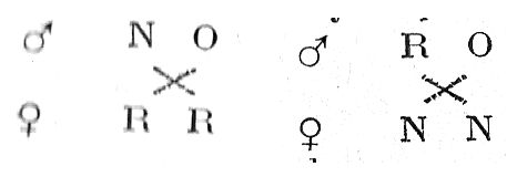
With Professor Cornevin we can classify the different varieties of cats as follows:
According to Trouessart, "The domestic cat crosses quite readily with wild species of the same size inhabiting the country where it is introduced, especially when it returns to the semi-wild state, and these unions, generally fertile, give birth to new varieties which can multiply ad infinitum." These crosses have been observed in Scotland by Sir W. Jardine and in England by Blyth; the latter even thinks that when the domestic cat was introduced to England it, being very rare, interbred frequently with the wild cat or felis sylvestris. Isidore-Geoffroy Saint-Hilaire relates the crossing of a domestic she-cat with a local wild cat (felis lybicus) and Jeitteles reports the same fact in Hungary. Sir W. Elliot met, near Madras, half-breeds resulting from the crossing of the domestic cat with felis chaus, characterized by a broad brown stripe on the inner side of the forearm, and he often found this stripe on the forelimbs of domestic cats of India. According to Dr D. Short, in Hansi, in India, a large number of domestic cats, can no longer be distinguished from the wild cat (felis torquatus).
The same facts were also observed by E. Layard in the Indies and in Paraguay. According to Darwin, in Europe and La Plata cats that return to the wild life are regularly striped; and although their size is sometimes larger, they show no other peculiarities compared to the domestic cat. In cats, we do not meet with such clearly defined breeds as in other species because, due to its nocturnal and wandering habits, it is difficult, not to say impossible, to make selections and monitor the crosses and mixtures.
ECONOMIC VALUE OF CATS
The public's justified infatuation with dogs might suggest that the same is true of cats here. It is not so; this domestic animal is generally not the object of any solicitude, despite the services it renders and the value of certain individuals.
In England, and in Belgium, it is not so. Luxury cat breeding is practiced by a number of breeders, generally women, who derive a significant income from it. Owing to the number of special clubs, to exhibitions, to public taste, the prices attained are often considerable. Animals with good pedigrees, 10 to 12 months old, commonly sell for £30 - £40 and some champions fetch prices of £100 and even £200.
There are Cat-Farms in America, and also Kitten Raising Farms where special attention is paid to the scientific breeding of expensive kittens. In France, enthusiasts who have taken an interest in cat breeding are very satisfied with the results.
Mme Brassart, whose Maritza cattery (Blue Persians) groups together several award-winning females and studs, easily sells her youngsters for 300 and 400 francs at weaning, 500 and 600 francs for cats from 5 to 10 months depending on the beauty of the individual; the animals awarded prizes at shows immediately acquire real added value and several of Mme Brassart’s cats would not be sold for less than 3,000 or 4,000 francs. 6,000 francs were paid for some animals imported from England. In 1923, Mme Brassart acquired an adult white Persian for 114 pounds which, together with the 20% customs, transport costs and luxury goods tax, represents about 12,000 francs. In September 1924, she acquired a blue cat which cost her 10,000 francs.
The prices of Siamese and other short-haired cats are generally lower. At weaning, ordinary subjects sell for 80 to 150 francs; from 5 to 10 months, prices stabilize between 300 and 500 francs. As for the Blue Persian, the award-winning females and studs, employed in breeding, fetch high prices: 1,500 to 3,500 francs. Some rare cats are rated very highly, they are orange, cream, tortoiseshell cats or the extremely rare Birman cats.
The breeding of pedigree cats is practiced in France by around 1,500 breeders, who, in 1924, bred an average of ten animals; if in 1925 the number of breeders increases in proportion to the last three years, it will exceed 2,000 by the end of the year.
Cat breeding is also of economic interest because, for several years, couturiers have launched "fancy" furs in which cat skins figure, not only from the tabby cat, but also from various colours of long-haired Persian and Angora, and from the Royal [Seal-point] Siamese, etc. Cat fur is used for adornment and as a warm lining, but its use is not limited to this; several large novelty stores recently exhibited car blankets made from 15 or 20 almost identical tabby-and-white cat skins, so closely matched it can be assumed that it took several hundred to find this number of matching skins.
The use of these furs has provoked protests from cat lovers, a protest currently legitimate, as it is very likely that the animals which provided the furs were stolen and slaughtered since no utilitarian breeding exists in France. But, by generalizing and protesting, the cat lovers miss the point, and since fashion, a tyrannical god to whom we must bow, calls for cat skins, we can envisage their rational production in France without misplaced sentimentality. In this we will follow America, where successful ranching farms deliver sizable financial results. There is no reason not to breed these animals for fur if they are given good care during their lives and humanely sacrificed; by breeding cats for this purpose, many small investors could increase their income as some already do by exploiting rabbits in this way.
The foregoing considerations and figures indicate that cat breeding is an element of wealth likely to be developed with the aim of reducing the importation of pedigree cats, to organize their export to countries where the pedigree cat is particularly appreciated and, finally, to use their fur.
STANDARDS
Because of the economic value of cats, the detailed study of feline breeds, especially luxury breeds, in the most extreme detail of their characters, expressed by standards, is therefore useful.
Their establishment is generally the consequence of the work of clubs trying to codify the beauty and the purity of the breeds which interest them. Until 1912, French authors describing the breeds of cats contented themselves with mentioning, without much detail, certain typical characteristics without considering all parts of the body. In England, Miss Simpson, in "The Book of the Cat," published in 1900, published a series of standards mainly based on colour and fur type. Since the foundation of the Cat Club de France, the drafting of standards has been attempted for the main breeds; some, which were drafted by serious breeders, have hardly been modified; others, published by leaders of clubs wishing to create races in order to govern them, are generally of little value.
The standards published here are those that have been drawn up by specialist registries in France or Belgium, but it is mainly in England that the breeds in this study have been described. Unfortunately, their wording is far from precise, practical and scientific; in the interests of breeding, they should be reviewed, refined and supplemented by a competent official committee, both technical and scientific, to which we could add long-serving breeders and show judges.
The standards should describe:
Firstly, the general appearance of the breed.
Secondly, the fundamental characteristics of the breed, mentioned by main parts;
Thirdly, the size, shape and weight of the animals with maximum and minimum figures, taking into account the sex of the subjects: male, female or neuter;
Fourthly, a description of the skin and coat with a precise indication of the colours, their distribution and the fixed characteristics of the coat.
With very exact standards, it will be possible to eliminate from the breeding population, or at least eliminate them from being registered in stud books, those cats not meeting the minimum standard according to its scale of points.
These official standards should be approved by the Superior Committee of Genealogical Books appointed by the Ministry of Agriculture; they would then be truly adopted by breeders in all countries who demand precise and scientific guidelines.
DUTCH CAT
Colour. – A variety of the common cat that we have managed to fix; it can be black and white, blue and white, orange and white, or cream and white, each colour being well defined and not showing marks of another colour.
Markings. - The same colour (i.e. black, blue, cream or orange) starts immediately behind the shoulders, all around the body and continues on the tail and hind legs. The tips of the hind legs are white. The ears and the mask have the same colouring as the hindquarters.
White is found on shoulders, neck, forelegs, feet, chin and lips. An extended blaze between the ears connects at the back of the head to the white at the nape of the neck and divides the head exactly into two parts. The markings sought are those seen in the variety of rabbit known as the Dutch rabbit.
The eyes are copper, orange or amber for black and white cats and for blue and white subjects; brown, orange or amber for orange and white and for cream and white.
Body and tail. - The animal is set low on the legs, well-muscled, the chest broad and open, the tail almost continuing as the extension of the dorsal line, thick at the root, curving slightly upwards, ending in a point, long rather than short.
Legs and feet. - The legs are shorter rather than long, well-muscled, the feet are rounded.
Head and neck. - The head is broad between the ears, the cheeks are well developed, the face and nose are short, the head is well set on a short, thick neck.
Ears. - Small and round at the tip, they are carried a little forward and not too large at the base.
The coat is always very short.
Scale of points. - Colour 20; markings 25; eyes 5; body and tail 15; legs, feet 5; head and neck 10; ears 5; hair 5; overall impression 10. Total 100.
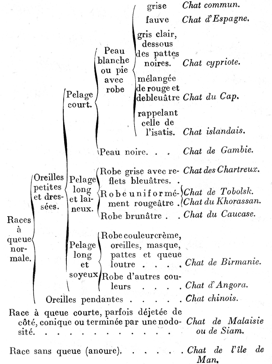
CHARTREUX CAT
Rather rare in England and France, it is quite common in many other countries, especially in the United States where it is known as the Maltese cat. This beautiful animal is widespread in mining districts and in agricultural regions, far from inhabited centres. It is also found in Russia where it constitutes the sub-races of Tobolsk, Khorassan and the Caucasus.
It is one of the races which can live in the low temperatures of the higher mountainous districts; moreover, this is where they are taken from before taking them into Russian territory. Their lives go from one extreme to the other; during the brief Russian summer, they roam the forests infected with poisonous insects; in the winter, they are imprisoned within the four walls of a snow-covered cabin, and forced into domestic service, until the thaw drives them out. Many of the beautiful furs that come to us from Russia are actually the skins of those cats, there is a large and prosperous industry in preparing these for the market.
Behaviour. - The Chartreux has a general tendency to be lazy, however, it is a good hunter of mice and small birds.
Size. – Strongly built, handsome, with well-proportioned limbs that blend well with the rest of the body.
Tail normal, of equal size along its length, without knots.
Head strongly built, with large full eyes, small erect ears, short nose.
Coat. - Semi-long, woolly coat, this woolly consistency forms the main characteristic of the breed; it is even found in individuals whose hair is not very long.
Colour. - Uniform grey colour with bluish reflections in the Chartreux breed proper, reddish in the Tobolsk variety.
Scale of points. - Size 20; tail 5; head 10; coat (woolly texture) 25; colour 25; overall impression 15. Total 100.
BIRMAN CAT
Like the Siamese, this is native to the Far East. Raised in temples, the Birman cats are strictly guarded, and their transfer is prohibited. However, a few years ago, Mr. Vanderbilt was able to acquire a couple of them from which the subjects that currently exist have been bred.
Behaviour. - These cats are very sociable, intelligent, cheerful and cuddly, following their masters in the same way as dogs; there are exceptions, however, and some subjects have been particularly savage.
Size. - These animals have elongated bodies, slender legs and are well proportioned. Adult weight varies between 3 and 4 kilograms.
The head is long with large, erect ears covered with felted hairs; the forehead is rounded. The males have a bushy, cream-white forelock between their eyes, which covers them oddly like a griffin. The whiskers are thick and long. The eyes are intense royal blue, very mobile.
Coat. - The fur is, the same length as the fur of the half-Angoras [domestic longhairs/half-bred Persians] and separates along the back as though by a comb. The tail fur is very bushy and forms a plume.
The colour is that of a creamy white Siamese, with perhaps, more golden tones. The mask, tail, ears and legs are dark otter-brown, the four legs are terminated by a white glove which ends below the wrist.
The tail has no knots or nodules; it is long, covered with fur to form a plume, and carried aloft in the manner of a squirrel.
Reproduction and breeding. - Breeding these animals has always been particularly difficult, and you shouldn't expect to raise more than one in ten kittens. Regarding food’ these animals find good boiled fish and cooked salad very good; others will only accept raw meat.
Scale of points. - Coat (length and texture of hair) 20; colour and markings 20; head 15; eyes 15; tail 20; body 10. Total 100.
ANGORA
The angora [small "a" meaning "longhair"] race or more precisely the Angora breed originates from the Angora region of Anatolia. This Persian region of Angora has been recognized as a remarkably promoting the fur and the fleece of all breeds born in its climate, and the name "Angora" has become attached to any cat with a long, thick silky coat. These cats are quite common in Turkey and Armenia, and there are some fine samples in England and France.
Behaviour. - The Angora cat has an indolent, frivolous character; he is lazy, sleepy and lacks the carnivorous instincts of felines. Rarely does it hunt mice, rats and other small mammals. His intelligence is however highly developed.
Realizing that it is a subject of luxury, the Angora is fond of soft cushions, armchairs and, resting motionless for hours in living rooms, it seems to be part of the objects of art collected to indulge the privileges of wealth.
Owing to his indolence, he moves slowly, grows fat easily, meows little and sleeps much more than other breeds of cat.
Colour. - The permitted colours are white, black, blue, or fawn. The fur should be uniformly single-coloured without any intermingling.
The coat should be silky, the hairs long and abundant, especially in the region of the neck, stomach and tail.
Size and shape. - The cat must be large without being too coarse, but it must be fleshy and have short legs.
The head should be long and broad, the nose short, the ears large and pointed with a tuft of hair at the top, the lips flesh pink.
The large, expressive eyes should be blue or orange with the white and fawn colour varieties, orange with the black and the blue varieties.
Scale of points. - Colour 25; coat 25; size and shape 20; head 10; eyes 10; beauty, overall impression 10. Total 100.
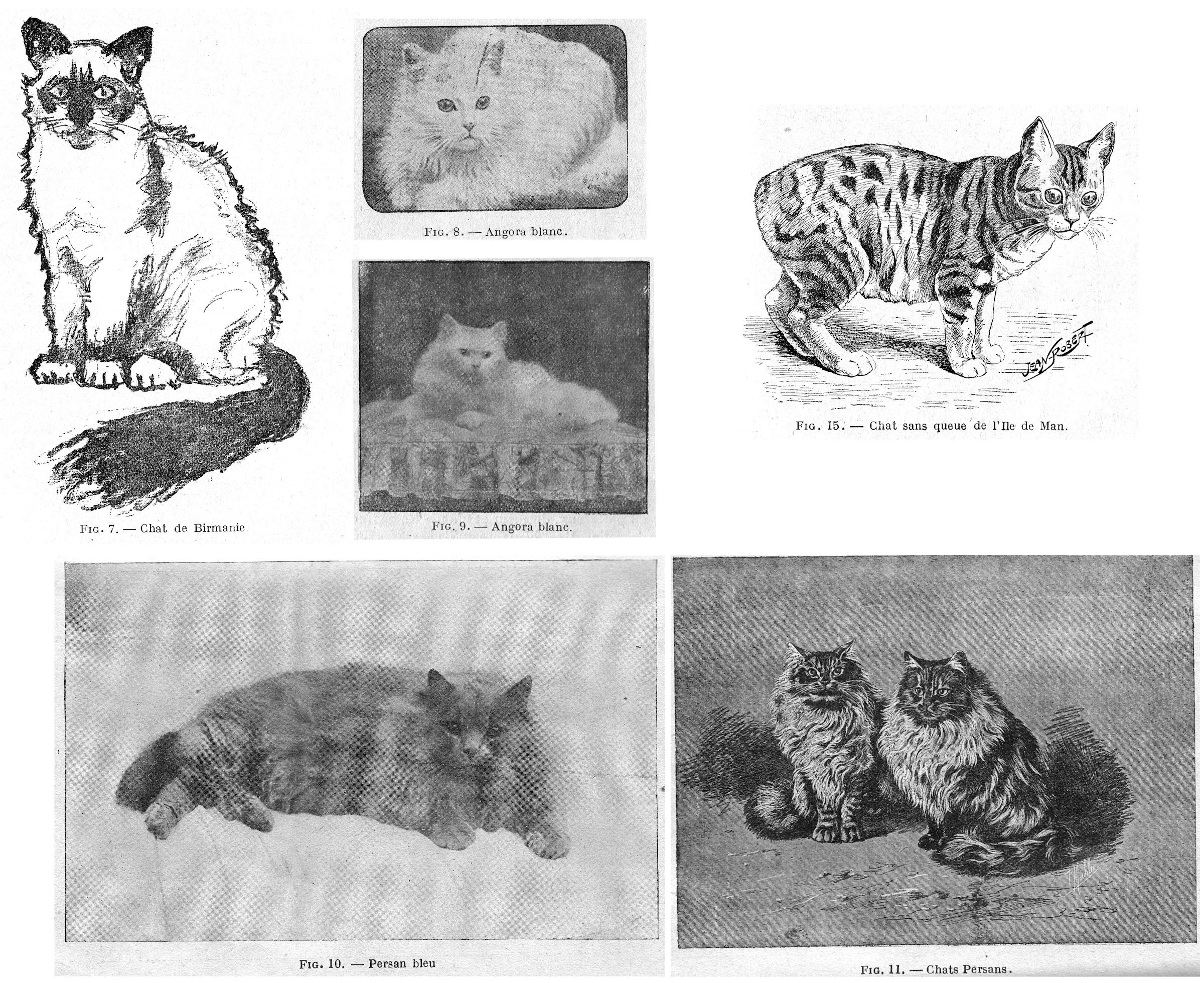
BLUE PERSIAN CAT
The Blue Persian cat is a variety of the Angora characterized by the shade of its coat. It is native to Asia Minor where it is nevertheless quite rare. The specimens imported into France, and especially into England, come from the caravans which, twice a year, cross all of Persia and go to Bombay to sell horses and other commodities.
Behaviour. - These cats are quite sociable. The castrated specimens become fat and very indolent.
The size is generally larger than the white Angoras.
The head is large with slender, erect ears covered with felted hair. The face and nose are short. The head is surrounded by a very thick ruff. The tail is normal, without knots, not tapered, of medium length and covered with long fur.
The coat is long and silky, and long fur covers the legs and even the feet.
The colour is blue, but there are many varieties and by selective breeding you can get just about any shade of blue. All shades are allowed on condition that there are no white hairs and that the shade is uniform.
In French shows, the judges of the Cat Club de France have adopted the following standard:
Coat. - All shades of blue are allowed: solid and well-defined colour, without markings, shading, or white hairs. The coat must be long, thick and soft to the touch, the ruff very thick.
Large, round head with space between the ears. The face and nose are short. Ears thin and covered with close-lying fur. Well-developed cheeks.
Eyes large and round, orange in colour.
Body cobby and set low on the legs.
Short, full tail, not tapering.
Point scale: Coat 30; head 25; eyes 20 body 15; tail 10. Total 100.
TORTOISESHELL CAT
This breed is a variety of long-haired cat that originated in England as a result of selections made by cat lovers and breeders in the United Kingdom.
Behaviour. - Luxury animals, specimens of this breed are generally quite indolent and not well suited to chasing mice.
Size and shape. - Large in size, robust without being fat, se low on the legs, all of which constitutes a graceful and elegant whole.
Large, round head, short nose, small, well erect ears. The eyes are large, round, full, brown or bright orange in colour.
Colours and markings. - Three colours are allowed: black, orange and yellow. These colours should be as vivid as possible, free of any stripes or white markings, the latter of which distinguishes the tortoiseshell cat from the Spanish cat.
Long tail, thicker at the base.
Coat. - The coat is silky, soft and very long.
Scale of points: Colour 30; coat 20; size and shape 20; head 15; eyes 5; overall impression 10. Total 100.
The term tortoiseshell is also given to short-haired cats exhibiting the characteristics described above. In this variety we sometimes find specimens that have white forelegs and breast, and a white patch between the eyes, extending like a blaze down to the muzzle.
SIAMESE CAT
Native to the Far East, this breed is found in Siam, the English Indies and the Malay Archipelago. In Europe, we now find many specimens imported from, or born in, England or France.
Behaviour. - These cats are very intelligent, and training is easy; they can be taught to open doors and wardrobes as well as acrobatic tricks. Very attached to their master, they follow him like little dogs, but it should be noted that they are greater thieves and more independent than other domestic cats. Siamese cats, very cautious, seek warmth; some of them can nevertheless face the cold to hunt, although, in winter, the Siamese fall into a state of dullness which is not always very noticeable. They eat mainly fish and boiled rice, but they are great hunters and devour their prey: birds, mice and rats which they are very fond of. They have a wide vocal range, and they use it with different and modulated intonations, especially during the period preceding the mating: at this time the females make cries that resemble those of wildcats.
Size and shape. - They are generally smaller than our European cats; the male is noticeably larger than the female. The profile is a rather long, but elegant and graceful. The legs and neck are thin.
The head is always small, wide between the two eyes, tapering between the ears. The forehead is flat and receding, the nose is long and rather broad, the lips are rounded and full, the ears are quite large and broad at the base with sparse downy hair inside. The eyes are almond-shaped slanting towards the nose, the iris is a greenish blue; these eyes take on almost red hues when the animal is irritated or frightened.
Tail. - Shorter than other European cats, it is either straight and slender like that of a pointer dog, or short, curved, broken and even twisted like that of the pig. At the base, there is almost always a knot, which is one of the characteristics of the breed.
Coat. - The hairs are short, soft and silky to the touch. On the face, legs and tail, they are shiny and glossy. The coat should lie flat on the body, showing the hard and firm muscles, because the Siamese cat should not be too fat.
Colour. - Adults have a solid background colour; light café au lait, pale silvery grey, light orange, or bright fawn are the preferred colours, but the lighter colours are most popular; the belly and the underside of the animal are always lighter, the back being darker. The head (in whole or in part), tail, and all four legs are dark brown, and the blue eyes stand out against the dark mask. The hairs lining the inside of the ears are white as down. There is often a white patch on the front between the neck and chest; any other white spot is a sign of degeneration and, consequently, of disqualification. The hairs of the long whiskers and those of the eyebrows are very light. Adults have a brush of hair, similar to whiskers, on the forearm. Usually, in the umbilicus region, there is a blackish patch that stands out well on the very pale coat in that area.
Reproduction and breeding. - For a long time, the number of kittens born in Europe was very small because the King of Siam, very jealous of his breed of cats, does not allow entire males to be exported. Females cannot mate without extreme danger, as Siamese kittens are born much smaller than those of other breeds. It should be noted that the female gives birth about a week later than the female cats and the gestation period is generally sixty-five to sixty-six days. The young are born white with a small dark line on the edge of the ears. If, at birth, there is the slightest stain on the immaculate coat, they should not be kept because they are not purebred. Be careful not to overdo raw meat: it is good to mix it with bread or with the cooked rice that they prefer; give plenty of milk as a drink and keep clean water available for the kittens.
The standard adopted by the judges of the Cat Club de France is as follows:
Body colour. - The hue of the body should be as light as possible and preferably cream, but the tawny shade is also allowed without stripes, spots or other markings on the body. The mask, ears, legs and tail are dark, clearly marked, otter-brown in colour. The mask, separated by a faint line as in the small cats, has no spots or undefined lines. The general appearance depends a lot on a good mask which should maintain the marten face as much as possible.
The eyes should be blue, bright and vivid.
The coat is shiny and flat, close-lying on the body.
Form. - The body should be rather elongated, the legs thin and well proportioned.
The head should be long and pointed.
The general appearance should have the following peculiarities: the gaze should be somewhat curious and striking; it is important that the subject is not too big, which would detract from the slender type which is so appreciated. In summary, in each of its peculiarities, the Siamese cat must be the opposite type of the domestic short-haired cat, retaining the colour contrasts along with a knot on the tail that is a particularly distinguishing characteristic.
The Cat Club de France recognizes as Siamese cats only those which meet the above standard and have a cream or chocolate colour; it eliminates blue, black, white, tabby or other coloured cats. The points of the chocolate coloured Siamese cat are the same as those above, except for the body, which has a particular colour.
Scale of points. - Body color 20; size 10; dress 10; head 10; eyes 20; mask 15; point report 15. Total 100.
Any cat which does not obtain, in each show, a minimum of 75 points, cannot aspire to the Championship of the Cat Club of France.
TAILLESS CAT
This breed of cat currently originates from the Isle of Man in the Irish Sea; but it is very likely that it was imported from the Far East, Japan or China, or neighbouring countries of Malaysia, the home of short-tailed cats. Japanese or Chinese paintings, in which tailless cats are often seen, confirm this opinion.
Currently, this breed is especially widespread in England, but very rare in France.
Behaviour. - These cats are very friendly, attached to their master, and very good at hunting mice; they feed preferably on meat.
Size and shape. - A little above average size, the body is as short as possible. The hind legs are higher and stronger than the front ones, so that the animal no longer runs like a cat, but like a rabbit.
The head is large and fine, not pointed; the eyes are large, round and full, like common cats, the medium ears are slightly rounded at the top. The eye colour varies: it is orange, golden yellow or brown, depending on the colour of the coat; always orange with a black coat.
The Isle of Man cat does not have a tail, and this is the main characteristic of the breed; there should not even be the semblance of a tail stump.
The colour is very variable, however, no whites are known and blacks are very rare; it is generally spotted or silvery.
Coat. - The fur is semi-long, especially the top coat; the undercoat is slightly cottony and soft to the touch like that of a rabbit.
Scale of points. - Body and legs 25; head ears and eyes 10; coat, consistency to the touch 25; no tail 25; general appearance 15. Total 100.
CAT CLUBS AND SHOWS
Like lovers of other animal breeds, cat lovers have come together in Societies, these being especially numerous in England. Below is the list of official clubs which we have had the opportunity to contact.
ENGLAND
The National Cat Club of London, founded in 1887.
The Cat Club of London (1898).
The Nothern Counties Cat Club (1900).
The Silver and Smoke Persian Cat Society (1900).
Black and White Club.
The Blue Persian Cat Society (1901).
The Siamese Club (1900).
The Orange, Cream, Fawn and Tortoise-shell Society (1900).
The Chinchilla Cat Club (1901).
The Short Haired Cat Club (1901).
The Scottish Cat Club (1894).
The Midland Counties Cat Club (1901).
The British Cat Club (1901).
The Man Cat Club (1901).
AMERICA
The Beresford Cat Club of Chicago (1899).
The Chicago Cat Club (1899).
The Louisville Cat Club (1900).
The Pacific Cat Club of San Francisco.
The Atlantic Club of New York.
FRANCE
The Cat Club of France (Secretariat in Saint-Raphaël, Var).
The French Siamese Cat Club.
The French Blue Persian Club.
BELGIUM
Friends of the cat, in Antwerp.
Feline Society of Flanders, in Ghent.
Brabant Cat Club, in Brussels.
All these societies regulate the breeds of cats to improve their purity and to keep them in conformity with the adopted standards. To this end, they organize cat shows, the oldest of which, to our knowledge, is the one which took place at the Crystal Palace in London in 1870; Since that date, the National Cat Club and the special clubs have organized annual events in London and other British cities which have always been very successful, both in terms of the number and the quality of the specimens exhibited.
In Holland, the first cat show dates from 1890 and in Belgium from 1891.
In France, the feline movement came later because the first exhibition was not organized until 1896 by the Journal, at the Jardin d'Acclimatation. Another took place in Paris in 1898. It is mainly since the Bordeaux show (1910) that cat shows have become widespread. Let us mention those of Nice (1912), Cannes (1912), Nice (1913), Lyon (1914), Aix-les-Bains (1914), Cannes (1922, 1923 and 1924), Marseille (1924 and 1925), Lille (1924 and 1925), Angers (1925); in all of them, alone or with Miss Simpson and Veterinarian Dr. Hasse, veterinarian Dr. Ph. Jumaud was appointed as judge. At the beginning of 1926, the Cat Club of France and Belgium organized an international cat show in Paris which, according to the promises [entry forms] received, should bring together a series of remarkable specimens. It is hoped that this exhibition will become an annual one, and will each year group together the most beautiful specimens of cats.
ANATOMY
SKELETON
The cat has 285 to 290 bones distributed as follows: 50 to 54 vertebrae, 30 in the head, 8 for the sternum, 26 ribs, 40 in each front limb, 41 in each of the hind limbs, 1 hyoid, 1 penile bone and 4 ossicles in each ear.
Spine. - The vertebrae break down into 7 cervical, 13 dorsal, 7 lumbar, 3 sacral and 20 to 24 coccygeal.
The cervical vertebrae are long and thick; their head is almost flat and, consequently, their posterior surface is slightly excavated; the vertebral laminae are broad and overlap one another; the posterior and anterior articular processes are united by a continuous bony lamina and the spinal process is higher the closer it is to the dorsal vertebrae. The atlas has relatively wide wings, transverse processes extending outward and a little at the back, and a very faint process at the insertion of the extensions of the neck; it has no tubercles on its underside. The body of the axis is quite elongated and its odontoid process, cylindroid and curved from top to bottom, is constricted at its base; the spinal process, very thin, is simple at the back, and the vertebral foramen, seen from behind, is higher than it is wide. The 3rd vertebra is wider than it is long, and the following ones gradually decrease in thickness. The 4th and 5th have a pointed spinal process and the transverse processes are simple or divided into two or three lobes. The spinal process of the 6th is more than five millimetres high and that of the 7th reaches one centimetre; in the latter, the transverse processes are not perforated.
There is no inferior ridge below the vertebral body of the dorsal vertebrae; the anterior articular surfaces of the arch are close together or united to each other; the spinal processes at the withers are pointed and triangular, especially that of the 10th vertebra which is vertical, and the transverse processes decrease as they go from front to back; they even disappear completely in the last three, where they are replaced by a tuberosity or accessory process.
The lumbar vertebrae, long and thick, have a voluminous body which increases from the 1st to the 5th to decrease in the last two; the spinal process descends and becomes acute in the last vertebrae to the point of being pointed and vertical in the 7th; the transverse processes tilt sharply forward and downward, and the first five vertebrae puts out an accessory process; the underside of the body of the vertebra is almost flat; the articular head and the posterior surface are flat.
The sacrum, most generally formed by the fusion of three sacral vertebrae, is short, surmounted by a poorly developed supersacral spine forming a thin, sharp ridge, and lacks a median ridge on its underside; it has an almost square shape and is extended by two posterior horns formed by the transverse processes of the last sacral vertebra.
The coccygeal or caudal vertebrae are well developed and lack a spiny process; the first four or five, which articulate like true vertebrae, still have well-developed transverse processes directed obliquely backwards, but these diminish as one moves away from the sacrum, and the vertebral bodies transform into cylinders whose anterior end is larger than the posterior, extending to the Xth vertebra and then shortening, so that the latter forms only a conical ossicle with an obtuse apex. The increasingly narrow spinal canal extends as far as the 6th vertebra.
Head. - The head consists of the skull, formed of 9 flat bones, 7 of which are unpaired and only one, the temporal is paired. The face, including the upper jaw, is made up of 19 broad bones of which only one, the vomer, is unpaired, and of the lower jaw.
The bones of the skull are: The occipital, in which the cervical tuberosity of the external occipital protuberance is poorly developed, the styloid processes short, the basilar process wide, long and thick. The parietal, very convex, shows very faint ridges and, on the other hand, two large bony laminae coming to separate the cerebellar cavity from the cerebral cavity. The almost flat frontal bone has fairly long orbital processes and an incomplete orbital arch, completed by a ligament which can sometimes ossify; we do not see an eyebrow hole, the frontal sinuses communicate with the nasal cavities by a narrow slit. There is a very deep pit in the ethmoid and well-developed, but few, volutes. The anterior sphenoid is very short and carries two broad wings on the sides, going up in the temporal fossa; on the contrary, the posterior sphenoid, which is very narrow, has very small lateral extensions; there is neither a sub-sphenoidal duct nor carotid notch, the latter uniting with a similar notch in the temporal bone to form the carotid foramen. The temporals, lacking any hyoid extension, present a very curved zygomatic process, an external auditory canal, a well-developed mastoid protuberance and very small mastoid processes; on the other hand, one sees neither a carotid canal, nor a temporal hole, nor a mastoid hole; there is a gutter across the promontory, providing passage to the trigeminal nerve. The temporal fossae are well developed, the external auditory hiatus is very wide.
The face is made up of the following bones. The upper jaw or large maxilla, very short, lacks a maxillary spine. The intermaxillary bone is very small and the palatal bones, although very extensive, do not enter into the formation of the sphenoid sinuses; their horizontal portion forms almost half of the vault of the palate and the vertical portion shows a very large nasal hole, joined to the large sphenoidal slit by a well-marked fissure. The pterygoid, very extensive, has the general shape of a quadrilateral. The zygomatic only articulates with the upper jaw at its base, which has no indentation. The lacrimal, very small, forms a small elongated, quadrangular lamella, lacking a lacrimal facet. The nasal bone is poorly developed, wider below than above, without a nasal extension. The vomer is short and thick, has broad wings and is only a dependency of the ethmoid. The turbinates present numerous folds which do not form part of the frontal sinus or the maxillary sinus; we can distinguish on each side a well developed upper turbinate and a much smaller lower turbinate. The lower jaw shows a well-marked triangular fossa at the point of insertion of the masseter, a very strong, very high and very broad coronoid process; its branches are short and wide apart, its condyle very elongated.
In general, the head, which is very short, has a convex profile on its upper face; the parietal region is rounded, the frontal is flat, the nasal is very short; the cerebellar compartment is very spacious.
Hyoid. - The hyoid body is made up of three parts that remain isolated in adult animals, the thyroid horns or uro hyal not being attached to the middle part of the body or basi hyal; it is accompanied by its three branches, the upper (stylo hyal) a little curved outwards, the middle (cerato hyal) as long as the previous one and the lower (hypo hyal) short, but strong and irregularly prismatic.
Thorax. - The sternum, flat and elongated, is made up of eight sternebrae, rarely seven, elongated from front to back, hollowed out in their middle part and swollen at their ends. Its underside is slightly convex; the first sternebra is pointed at its free end, the last terminates posteriorly with a thin xyphoid cartilaginous appendage which can oscillate vertically.
There are 13 pairs of ribs including 4 asternals which, with the exception of the first, are thick and narrow; they increase in length from the first to the seventh and decrease from the tenth; curved over their entire length, they present a fairly strong tuberosity, the articular facet of which remains isolated from the posterior facet of the head, except in the last three ribs where it is lacking. The costal cartilages get longer and longer the further back you go; the cartilages of the true ribs articulate with the sternum; the cartilages of the four false ribs are attached to each other and form costal arches; The postero-internal vasculo-nervous groove is almost obliterated.
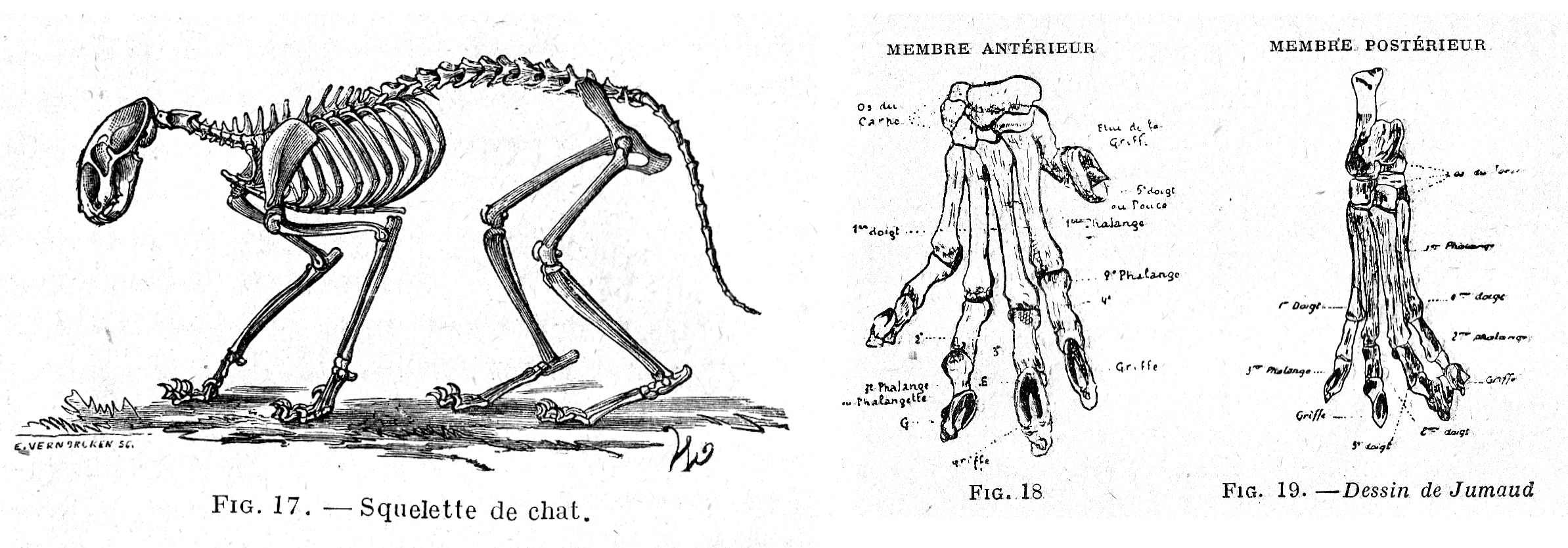
Forelimb. - The scapulum or shoulder blade is short, broad, lacking a cartilaginous extension; its anterior border is rounded and the upper and infraspinous fossae are about the same width. There is a clavicle, a small styloid bone that connects the acromion to the sternum through two fibrous cords. The humerus shows a simple trochlea without condyle at its lower end, a little protruding deltoid impression and, on the inner side of the lower extremity, a particular hole (epitrochlear hole) which forms a vascular arcade; the coronoid and olecranon fossae do not communicate with each other. The radius and ulna, almost equal in volume, are slightly curved over their length and meet at their ends, crossing slightly; the anterior surface of the ulna is roughly flat and the upper articular surface of the radius is cup-shaped; the upper end is larger than the lower in the ulna, smaller in the radius; these two bones remain independent throughout the life of the animal.
The carpus has eight bones; in the first row we find the pisiform or suscarpal bone, elongated and prismatic; the highly developed pyramidal, which articulates with the ulna, the hamate bone and the first metacarpal; the scapholunate, resulting from the fusion of the semilunar bone and the scaphoid; the second row, where the size of the bones decreases from the first to the fourth, consists of the hamate bone, which corresponds to the first two metacarpals, the large bone which articulates with the third metacarpal, the trapezoid in the fourth metacarpal and the trapezius in the metacarpal of the thumb; there is also a small bone, fulfilling the role of a sesamoid, located above the trapezius and articulated at the posterior part of the scapholunar, being only a few millimetres in diameter, which has been given the name of phacoid bone. The third and fourth metacarpals on the one hand, and the second and fifth metacarpals on the other hand are the same length, but the first two are longer than the others; the second metacarpal extends a lateral process to the third, which also extends one to the fourth; the metacarpal of the thumb is also strong, but is shorter than the others. There are five fingers, the inner (thumb) with two phalanges, the others with three phalanges; the first phalange is accompanied by two large sesamoids at the level of the metacarpo-phalangeal joint, the internal being smaller than the external, except at the level of the thumb where there is only one; the third phalanx is conical, curved downwards and lacks a small sesamoid.
Hind limb. - The pelvis is longer, behind than in front. The ilium, almost vertical, has a depressed outer face; the oval holes are spacious and regularly elliptical. The thick pubis does not have a groove on its underside. The length of the ischium is two-thirds that of the ilium; the notch forming the ischial arch involves only the medial half, the posterior border of the ischium, which is regularly convex from outside to inside; the supra-acetabular [supra-cotyloid] crest, also convex, ends imperceptibly behind; the rear of the acetabulum is widely open and does not have a precotyloid tubercle.
The femur is elongated, rectilinear and cylindroid; it has neither a third trochanter nor a supracondylar fossa and the top of the greater trochanter is lower than the head; the lesser trochanter is shaped like a nipple; the neck is long and narrowed, the digital fossa deep; the condyles are surmounted posteriorly by a small facet joint corresponding to two small, sesamoid bones found in the thickness of the posterior tibial-femoral ligament. The patella, which is quite large, is flattened from front to back. The tibia, long and thin, has a prominent anterior crest; at its upper end it lacks a tendinous slide between the anterior tuberosity and the external tuberosity, but there are two grooves at its lower end, the external one completed by the lower end of the fibula, the internal one bordered by a prominent malleolus. The fibula, of the same length as the tibia, is completely independent, almost rectilinear, sharp in front, rounded behind and on the outside.
The tarsus is formed of 7 bones, due to the presence of three cuneiforms; the talus has a relatively short head by means of which it articulates with the scaphoid, and the calcaneus has an infero-external process and a shallow beak; the cuboid, which has a narrow groove through which the long peroneus slides, articulates with the fourth and fifth metatarsals; the scaphoid has no posterior slide; the cuneiforms articulate with the corresponding metatarsals; three cartilaginous and sometimes even bony granules can also be found in the tarsus, fulfilling the role of sesamoids, which are applied against the posterior surface of the head of the three outer metatarsals and serve as points of attachment for ligaments or tendons. There are four well-developed metatarsals and a rudimentary fifth, articulating with the internal cuneiform or first cuneiform and which is never accompanied by a phalanx, so that there is no thumb. There are four fingers each having three phalanges, the first phalange being accompanied by two sesamoid bones, but the last lacks small sesamoid.
LIGAMENTS
The cat lacks a cervical ligament and the interspinal ligaments are replaced by small muscle bundles, which explains the animal's extreme flexibility. The odontoid ligament of the atloid-axoid joint is also replaced by two lateral cords (starting from the odontoid process and inserting inside the condyle of the occipital) and a transverse ligament; the atloid-occipital joint has only one synovial capsule. The maxillary condyle fits exactly into the temporal cavity, preventing lateral and horizontal sliding movements. The humero-radial joint is provided with a very thick external lateral ligament and the internal lateral ligament is inserted on the ulna by a very short bundle, on the internal side of the head of the radius by another equally short ligament, finally on the posterior surface of the radius by a longer and deeper bundle. The two bones of the forearm are united by an interosseous ligament completed at the top by an annular ligament, at the bottom by a new interosseous ligament.
Each of the metacarpophalangeal joints has an intersesamoid ligament, an inferior sesamoid ligament consisting of two crossed bands, two small lateral sesamoid ligaments, two lateral metacarpophalangeal ligaments and finally a capsular ligament. The last two phalanges are united by two lateral ligaments and a third, very strong, located in front of the joint, made up of elastic tissue, which mechanically produces the retraction of the nail when the flexor muscles stop contracting; in addition, the articular pulleys of the last interphalangeal joint have a sufficiently strong obliquity to allow the nail to come to be lodged, by rising, between the two fingers; the articular surface of the third phalanx is completed by a fibro-cartilage serving as a pulley for returning the tendon of the perforator. There is no femoral-patellar capsule on the hind limb and only one tibio-rotular ligament is found; the other two (external and internal ligaments) are represented by two strong fibrous bundles coming from the tendons of the external and internal twins. The two bones of the leg are united in their middle part by an interosseous ligament.
MUSCLES
The muscles of the upper cervical region are large; the rhomboid is bifid at its origin; the angle of the shoulder merges with the large serrated edge; the splenius does not have a cervical portion; the large complexus is divided into two portions, the obliques and straight posteriors of the head are very thick. In the lower cervical region, the platysma splits into two parts; the two portions of the humeral mastoid are clearly separated, the superficial or cleidomastoid portion going to the ridge taking the place of the mastoid process, the deep or trapezoidal portion joining the side of the occipital protuberance; the omotrachelion has two upper insertions; the sternohyoid and the sternothyroid are thick; we find a trachelo-atloid, a scalene from the first rib and a supra-costal scalene attaching at the top on the axis, at the bottom by two attachment points, the superior on the fourth rib, the inferior on the ninth ; it has no omoplathyoid.
The spinal region of the back and lumbar region shows a well-developed trapezius and a voluminous latissimus dorsi; the lower branch of the iliospinal has two kinds of bundles; the small anterior serratus, very thick, attaches to the ribs, from the third to the tenth, while the small posterior serratus attaches to the last three ribs; the common intercostal and the spinal transverse of the back and lumbar are very developed; the longissimus dorsi is thick, the longissimus spinal is extended to the axis and the interspinous are very strong. In the region of the tail, the lateral sacrococcygeus is divided into two parts, upper and lower; the upper sacrococcygeal arises from two branches. In the lower lumbar region, the iliac psoas almost merges with the large, poorly developed psoas; on the other hand the lesser psoas is as voluminous as the large psoas, darker in colour and covered on its posterior half with a beautiful pearly aponeurosis; the lumbar quadratus is very developed.
The region of the face has a labial or orbicular muscle of the lips which is quite rudimentary; the alveolo-labial or buccinator is formed of only one muscular plane; the zygomato-labial continues above with the zygomato-auricular, and ends below at the corner of the lips; the supra-nasolabialis does not divide at the bottom and attaches to the side of the upper lip; the supramaxillo-labial and nasal supramaxillus are a single muscle terminating both in the outer wing of the nose and in the upper lip; neither the small nasal supra-maxillo, nor the transverse nasal passages are present, and the point of the chin suspensor muscle is, like the latter, barely developed. On the other hand, in the temporomaxillary region, the masseter and the temporal are very thick, the digastric is formed of a single belly; the stylo-hyoid is very thin, the kerato-hyoid, on the contrary, is well developed; the external pterygoid and the transverse hyoid are absent.
In the costal region, the anterior serratus merges with the angle of the shoulder. The abdominal tunic is thin and opaque, the rectus muscles form a real thickening on each side of the linea alba which appears like a spindle. The very thin crural arch is formed by an aponeurosis continuing the external oblique muscle of the abdomen.
The diaphragm is relatively thick and attaches to the very edge of the base of the thorax; the supports are quite short, which pushes the muscle back.
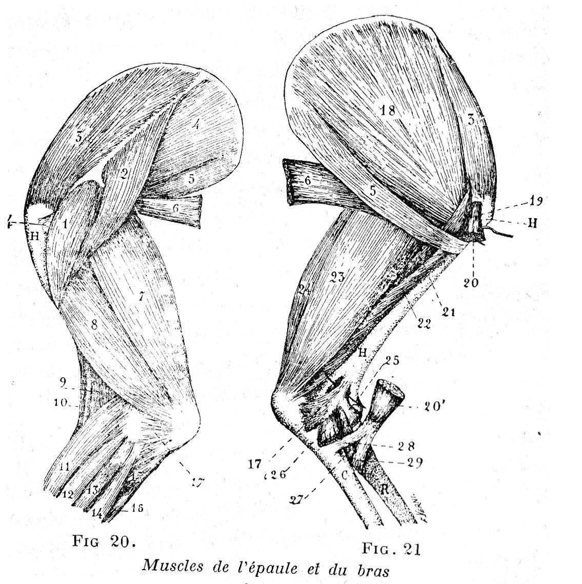
Muscles of the shoulder and arm.
Legend: 1, 2, the two portions of the deltoid; 3, supraspinatus; 4, infraspinatus; 5, teres major;
6, latissimus dorsi; 7, long anconeus, without an accessory head; 8, external anconeus; 9, brachialis anterior; 10, brachioradial [long supinator]; 11, anterior metacarpal extensor; 12, anterior phalangeal extensor (common digits extensor); 13, lateral phalangeal extensor (extensor of three outer digits); 14, external ulnar [external cubital]; 15, 16, both heads of the internal ulnar [internal cubital]; 17, small external anconeus; 17', small internal anconeus; 18, subscapularis; 19, coraco-brachialis; 20, origin of biceps; 20, termination of biceps; 21, deep anconeus; 22, internal anconeus; 23, accessory anconeus of latissimus dorsi; 24, long anconeus; 25, origin of the pronator teres; 26, joint insertion of the epitrochlear muscles; 27, internal ligament of the elbow joint; 28, ulnar [cubital] tendon of the biceps; 29, radial tendon of the ulnar. - H, humerus; R, radius; G, ulna [cubitus]; an arrow crosses through the epitrochlear foramen.
Among the muscles of the shoulder, the abductor longus of the arm is very developed, as well as the supraspinatus which ends in a single branch leading to the trichiter; the infraspinatus does not have a deep branch at its posterior end and the coraco-brachialis, which is very short, is formed of a single muscular bundle which may, however, present a slender bundle descending to the bottom of the humerus. At the level of the arm, the coraco-radial or long flexor of the forearm provides at its lower end, and after being attached to the bicipital tuberosity of the radius, a small tendinous branch sliding on the internal side of this bone to fix itself towards the base of the olecranon, medial to the ulna; the flexor digitorum brevis or anterior brachialis terminates in a small tendon that also attaches to the ulna; the medial extensor of the forearm or internal anconeus is highly developed; the large forearm extensor or long anconeus is simple; there is a small internal anconeus or epitrochlear ulna.
The muscles of the forearm are very similar to those of a man; the anterior extensor of the metacarpus divides at its lower part into two inserted branches, one at the metacarpal of the index finger, the other at that of the middle finger; the oblique extensor attaches to the metacarpal of the thumb which it moves away from the other digits and gives a small slippery branch, with the help of the phacoid bone, on the third bone of the lower row of the carpal (trapezoid) and is then merged with the posterior carpal ligament; the anterior phalangeal extensor is called the common extensor digitorum, and its tendon ends in four branches, each leading to one of the digits; the lateral extensor of the phalanges is continued by a tendon with three terminal branches inserting into the anterior face of the three outer fingers; we also find the analogue of the oblique flexor (internal ulnar [internal cubitus]), external flexor (internal ulnar/cubitus) and medial flexor (flexor carpi radialis) of the metacarpus of solipeds; the perforator leaves above the knee a small muscle bundle terminated by a very slender tendon constituting the small palmar muscle, and its radial portion forms the long flexor of the thumb; the perforated, separated from the perforator by the internal ulnar, terminates in a tendon dividing into four branches which each attach to the second phalange of the four main fingers; we also find, in the forearm of the cat, five muscles peculiar to predators which are: the extensor proper of the thumb and index finger, a small muscle located under the lateral extensor; the long supinator, a small band of muscle placed anteriorly and medial to the anterior extensor of the metacarpal; the supinator brevis, a triangular muscle hidden under the extensor anterior of the metacarpal and the common extensor digitorum of the digits; the pronator teres, a thick, short muscle located above and medial to the forearm, between the flexor digitorum metacarpal and extensor digitorum anterior in the same region; the pronator quadratus, placed behind the forearm bone.
The muscles of the forefoot are: abductor pollicis brevis, an atrophied muscle; the opposing muscle of the thumb, a small, pale muscle that is almost completely fleshy; the flexor digitorum brevis, a very small dark muscle; the abductor of the index finger, elongated and prismatic; the cutaneous palmar formed by a musculo-fatty nucleus forming the base of the outer tubercle which is located behind the carpus; the abductor of the little finger which, coming from the axis-carpal bone, is placed outside and behind the external metacarpal and ends in a very elongated tendon which ends outside the upper end of the first phalange of the small finger; the flexor digitorum brevis, located medial to the abductor; the opposing muscle of the little finger, stretched up and down and flattened back and forth; the three lumbrical muscles, occupying the intervals between the four terminal branches of the perforator tendon; the four interosseous metacarpals, bifid muscular bundles at their lower end, placed in parallel, in front of the flexor tendons and behind the four large metacarpals, which replace the suspensory ligament of the horse's fetlock.
In the hind limb we find, in the region of the croup, an underdeveloped superficial buttock muscle [gluteus maximus], a medial buttock muscle [gluteus media] united very closely to the deep buttock muscle [gluteus minimus] which, for its part, is very developed; the scansorius or accessory of the gluteus minimus shows bundles arranged in a fan; finally, there is a short, but rather large, pyramidal muscle of the pelvis. In the thigh, the fascia lata, presents, anteriorly, a supernumerary bundle extending from the external angle of the ilium to the patella; the semitendonosus goes under the ischial tuberosity; the semimembranosus is bifid at its lower extremity; the adductor brevis of the leg or internal rectus is thin and narrow; the adductor longus of the leg or sartorius is formed by a very distinct fleshy band running from the external angle of the ilium to the internal face of the tibia, which forms its posterior portion, and also has an anterior portion ending in the kneecap; the great adductor of the thigh is very developed; the twin-headed gastrocnemius of the pelvis consist of two small but clearly isolated bundles; there is also a parameral muscle, a small bundle starting from the origin of the coccyx and continuing with a short tendon to the kneecap, and a lumbricoid muscle detaching from the transverse process of the second caudal vertebra and losing itself in the infero-posterior part of the femoral biceps.
We find in the leg a fairly large anterior tibialis muscle; an extensor proper of the thumb, very atrophied, a long common extensor of the digits, similar to that of the foreleg, a long lateral peroneus, a short lateral peroneus, and a small piniform abductor muscle of the foot; an extensor proper of the external finger, and a soleus; on the other hand, neither a lateral phalangeal extensor nor a metatarsal flexor can be seen; the perforated one merges with the external twin in its upper two thirds and its tendon ends in four branches; the perforator's tendon also gives a branch for each finger; the posterior tibialis, completely distinct, is formed of a fleshy, well-developed body, continued by a tendon which merges with the posterior tarso-metatarsal ligament. The hind foot shows a foot muscle formed by three bundles starting from the lower end of the calcaneus and ending in the second, third and fourth fingers; a few muscular tabs are attached to the perforated tendon; there is an accessory muscle of the perforator, small and atrophied; two or three strips, pale and rudimentary located near the thumb; a thin and elongated external digit adductor; four interosseous metatarsals and three lumbricals.
DIGESTIVE SYSTEM
The lips are thin and mobile; the upper one carries two lateral tufts formed by very large and very mobile tactile hairs constituting the whiskers. The mouth is widely slitted, the mucous membrane of the cheeks and palate are often marked with black spots; the palate is traversed in its width by five or six ridges divided by a median groove running its whole length. The tongue is thin and very mobile, and its anterior two-thirds are lined with backward pointing filiform papillae, and covered with a thick horny sheath making it hard and rough; at its base, inside the anterior pillars of the soft palate, there are two tonsils formed by an agglomeration of closed follicles. The soft palate is very short and the isthmus of the throat is widely open.
The dental system of the cat has 26 teeth inthe first dentition with the following dental formula:
Upper: incisors 3: canines 1: molars 3
Lower: incisors 3: canines 1: molars 2
these last include in the upper jaw a pre-carnassial, a carnassial, a tuberculous and in the lower jaw a pre-carnassial and a carnassial. The second dentition consists of 30 teeth, the dental formula becoming:
Upper: incisors 3; canines 1; molars 4
Lower: incisors 3: canines 1: molars 3
the molars are divided into 2 pre-carnassial, 1 carnassial and 1 tuberculous at the top, 2 pre-carnassial and 1 carnassial at the bottom. We therefore find, on each jaw and on each side:
Firstly - Three incisors, spaced apart from each other at the top, closer together at the bottom, the sizes of which increase from the centre to the periphery, which are distinguishable by pincer, division and corner, and in which we can recognize a root, a neck and a crown. These are more developed in the upper jaw than the lower jaw. The exposed portion of the virgin tooth has three lobes, the median being stronger than the laterals and the first to be attacked by wear; the root, very well developed and flattened from side to side, is separated from the crown by a very pronounced neck. The appearance of the corner teeth is similar to that of the canine. Milk incisors are smaller and sharper than adult incisors.
Secondly, a canine or hook tooth, separated from the incisors at the top by a fairly large gap, and at the bottom by a smaller gap, and from the molars by a gap generally even larger; a very strong pointed tooth, elongated, conical in shape, curved backwards and outwards, deeply ridged on its outer face. The upper canine is larger than the lower and the milk canine is more slender and more elongated than the adult one.
Thirdly, there are four molars at the top and three at the bottom, the first two on each side of the jaw constituting the premolars and being shed and replaced by the second dentition teeth; sharp and serrated like a saw, they do not meet at their crowns, but at their sides, the lower jaw being narrower than the upper jaw. The upper molars are subdivided into the relatively very small fore-molar; the larger main molar with a triangular crown and almost central apex, with two anterior roots and a posterior root (these two teeth constitute the pre-carnassials); the first rear molar or superior carnassial, very large, formed of a two-lobed sharp, pointed central part, the anterior lobe being conical and rather small, the posterior lobe being much more extensive, forming a sort of sharp wing, so that the tooth has three points; the second rear molar or tuberculous tooth, small and quite rudimentary, terminating in two blunt lobes, equipped with a single flattened root, often bifid at its end. The lower molars are distinguished as the main molar, slightly triangular with a basilar heel in front and a much larger, almost bi-lobed heel behind; the first rear molar is similar to the upper one but a little stronger (these two teeth forming the pre-carnassials), second rear molar or lower carnassial, elongated behind and depressed on its lateral parts, having only two points, having only two sharp lobes separated by a deep notch, and equipped with two roots; there is no tuberculous tooth. When the jaw is closed, the lower teeth cross the upper ones on the inside to make it easier for the animal to cut meat.
The parotid is not very bulky (it weighs about 6 grams) and the Stenon canal crosses the masseter; the sublingual, carried back, is narrow and elongated; it is not united with the maxilla and does not have a posterior part; the submandibular, reddish, weighs about 4 grams; the labial, lingual and staphylin lower molar glands are also very small; there is no upper molar gland.
The very large pharynx dilates with extreme ease.
The oesophagus, also very expandable, enters the stomach via an enlarged funnel-shaped cardia; its fleshy, reddish layer sags on itself. The stomach is shaped like a pear curved at its top (which corresponds to the pylorus) and forms a single sac whose mucous membrane, in the empty state, is lined with many regular folds: it contains about 30 at 32 centilitres.
The intestine, very short (about 2 meters, including 35 centimetres for the large intestine), has an average capacity of 01.114 for the small intestine and 01.154 for the large intestine. The small intestine, which rests on the lower abdominal wall, is supported by a long mesentery; it expands in its posterior half and has thick walls in which we find 5 or 6 Peyer's patches; the caecum is reduced to a small appendix of about 2 centimetres, curved into a hook, showing a true Peyer's patch at the bottom of the mucosa; the colon, held by a short mesentery, is much larger than the small intestine, especially at its origin, and has no indentations or longitudinal bands; Brünner's glands are only seen near the pylorus, while Lieberkühn's glands are found throughout the intestine. There are two omentums, a large and a small; the mesentery forms a continuous fold, inserting itself under the spine and enveloping the various parts of the intestine. On each side of the anus, on its free edge and towards its lower part, there is an orifice leading, through a very short canal, to the anal glands.
The very large liver weighs 150 to 180 grams; it is deeply indented and divided into five lobes; the gall bladder, curved onto itself, follows a bypassed cystic duct and is lodged in the large notch of the right intermediate lobe, which is much more developed than the left intermediate lobe; Spiegel's lobule is clearly detached from the right lobe; the common bile duct is inserted 3 to 4 centimetres from the pyloric orifice, right next to the pancreatic duct; the liver is partially accessible to external palpation (behind the 10th rib) which makes it possible to recognize its hypertrophy or the presence of tumours: it then spills over the circle of the hypochondrium behind it. The pancreas is very elongated with its anterior end curving behind the stomach to come forward again; it is enclosed between the laminae of the mesenteric layer supporting the duodenum. The spleen, slightly pointed at its anterior end, is narrow, elongated and curls like a billhook at its lower end: palpation can also recognize its hypertrophy.
RESPIRATORY SYSTEM
The tip of the cat's nose is made up of a shagreen-like protrusion, bare, wet and cool, most often pink, sometimes divided by a median groove, and has two comma-shaped nostrils whose convex parts ae opposite each other. The nasal passages are very short; each superior much more developed than the inferior; the olfactory mucosa is thick and dark in colour. There is only a single, very small maxillary sinus, and a slightly more developed frontal sinus, which opens into the nasal cavity through a small slit located near the middle septum of the two frontal sinuses.
The larynx has a large and strong cricoid, a thyroid with narrow wings and a large hyoid horn, a long, flexible, very sharp, triangular epiglottis; the lateral ventricles are shallow; the upper vocal cords, 4 to 5 millimetres in length, are very thin, soft, widely spread anteriorly and meet at the bottom of the sub-epiglottic sinus; the lower vocal cords, thick and closer together, are separated from the preceding by an excavation, located on a more internal plane and in contact at their thyroid end; there is no arytenoid Wrisberg cartilage or anterior thyro-arytenoid muscle; according to A. Landrin the upper vocal cords determine the purr and the growl of anger, the lower cords the meow. The tracheal rings, about 40 in number, overlap at the back, forming a ridge inside the trachea and on its posterior surface.
The left lung has three lobes (sternal, cardiac and diaphragmatic lobes, the latter being by far the largest), the right lung has four, having in addition the mediastinal lobe; all are separated from each other by deep grooves extending to the root of the lung; the azygos lobe is not very elongated and triangular in shape; there is no sharp indentation in the heart, not to the right or the left. The posterior mediastinum is imperforate.
The thyroid bodies are flattened, thinned internally, located below the larynx and on the sides of the trachea; they are each accompanied by an external parathyroid housed in their superior outer border and an internal parathyroid, located towards the upper third of their internal surface.
GENITO-URINARY SYSTEM
The kidneys weigh on average 15 to 20 grams each and have very apparent subcapsular veins, radiating from the hilum on each face; they are simple and the pelvis has at its bottom a large elongated tubercle having at its base a few very short supports; often floating, they then become noticeable on palpation and can be mistaken for tumours or foreign bodies. The adrenal capsules are made up of two yellowish-white discs, the size of a bean. The ureters open through two oblique openings at the base of the bladder, which is retracted and provided with a thick wall on which the bundles of the muscular tunic can be seen very clearly.
The rounded testicles are located in the perineal region. The cat does not have a seminal vesicle and the elongated bladder neck is almost entirely surrounded by the prostate, which is yellowish in colour, concave on its upper surface, divided into two lateral lobes on its lower surface; the two Cowper's glands are well developed. The urethral canal shows, at the level of the ischial arch, a bulge under the bulb and the bulbo-cavernosus muscle extends it quite far around this duct. The penis, short and directed backwards in the resting state, on the contrary moves forward, when erect, for mating. The free part of the penis is conical and its top, near which the urethral orifice is pierced, is based on a small incomplete penile bone surrounded by a layer of erectile tissue; this free part is covered with an integument bristling with small, somewhat harsh papillae, backwards-pointing, but likely to straighten during erection.
The ovaries are housed in a fold of the broad ligament; oval in shape, they have a lobular appearance due to the protrusion of the de Graaf's vesicles; the oviduct is not very flexuous and the body of the uterus is short, the uterine horns on the contrary are elongated and straight; the vagina, relatively long, does not have Gartner ducts. The rounded lower commissure of the vulva houses a small clitoris with a core of cartilage. There are 3 or 4 mammary glands on each side, each with a very short nipple, distinguished as inguinal, abdominal and pectoral mammary glands.
CIRCULATORY SYSTEM
Heart. - The heart, ovoid, almost globular, weighs about 20 grams and lies almost entirely on the upper surface of the sternum, its anterior surface thus becoming inferior; its point, directed backwards, rests on the anterior surface of the diaphragm while the pericardium is attached to the aponeurotic centre, being united to the sternum only through the mediastinum. The inner surface of the ventricles and atria have many thin fleshy columns.
Arteries. - The pulmonary artery, part of the right ventricle, gives rise to two branches above the left atrium, one for each lung; each one enters this organ with the bronchi where it ramifies ad infinitum.
As the aorta does not form an anterior branch, the arteries of the head and thoracic limb arise directly from the arch. The thoracic aorta gives rise to: the arteries of the heart, the right smaller than the left, both providing a horizontal branch and a vertical branch; the posterior intercostal arteries from the fourth intercostal space (the anterior intercostal arteries coming from the superficial intercostal artery); the oesophageal arteries, 4 or 5, which descend into the mediastinum and then distribute to the oesophagus; to the first two pairs of lumbar arteries.
The abdominal aorta leaves the third lumbar pair between the two pillars of the diaphragm, the fourth, fifth and sixth lumbar pairs before the origin of the femoral and the seventh pair after the origin of this vessel. The coeliac trunk is detached between the pillars of the diaphragm and gives rise first, after a very short distance, to the right branch or hepatic artery which supplies the duodenum with the pyloric artery and then continues by the gastro-duodenal artery dividing, at the level of the pylorus, into the pancreatico-duodenal artery which goes to the intestine, and the right gastro-omental artery which goes into the pancreas before going to anastomose with the left omental artery. The coeliac trunk continues with a very short trunk from which a central branch or gastric artery emerges which supplies a few small branches to the cardia, sends a branch to the pancreas and ends in ramifications on both sides of the stomach; then a left branch or splenic artery giving pancreatic branches, a gastro-splenic artery dividing into gastric artery, splenic artery and left gastro-omental artery. Very close to and behind the celiac trunk, the great mesenteric arises, which irrigates almost the entire intestinal mass; it gives rise to a branch from which are born successively the median colonic artery, the right colonic artery and the ileo-caecal artery, then ends with the artery of the jejunum which sends numerous branches to the small intestine, one of which, among others, anastomoses with the pancreatic-duodenal branch of the gastroduodenal artery. Right next to the great mesenteric artery, the aorta provides a diaphragmatic branch and a sub-lumbar branch.
The phrenic, renal and spermatic arteries present nothing unusual. The small mesenteric brings blood to the floating colon and rectum; it begins near the end of the aorta and divides into an anterior branch or left colonic artery going there towards the diaphragm to anastomose with the ipedian colonic artery and a posterior branch or inferior hemorrhoidal artery which goes to the anus. The iliac circumflex arises directly from the abdominal aorta.
Towards its termination, the latter gives rise to the primary iliac arteries or crural trunks which supply:
Firstly, the external iliac artery proper;
Secondly, the femoral artery, first detaching voluminous muscular branches, then two large muscular arteries, the posterior of which provides the prepubic artery which itself gives rise to the external pudendal artery; the latter sends a branch through the thickness of the mammaries which ends up with the mammary branch of the internal thoracic, then ends in the lips of the vulva where it anastomoses with the vulvar branches of the internal pudendal artery;
Thirdly, the saphenous artery gives rise to two branches, one small, accompanying in front of the hock the nerve and the internal saphenous vein, which unites with a branch of the anterior tibial to supply the dorsal metatarsal interossei; the other, more voluminous, engages under the flexors of the phalanges, giving cutaneous divisions and is divided into two branches, one of which ends at the level of the hock by very fine arterioles, while the other sends a few branches to the tarsus then emits, in the tarsal sheath, two very slender branches coming to form the plantar arch and finally ending with three very small branches going to flow into the interosseous metatarsal arteries;
Fourthly, the popliteal artery supplies a few rudimentary arterioles constituting the posterior tibial artery which will anastomose with the saphenous vein, then a femoro-popliteal branch continuing through the anterior tibial artery; the latter provides, in front of the hock, the tarsal artery which anastomoses with the anterior branch of the saphenous vein, continues under the name of dorsalis pedis artery, and ends, at the level of the metatarsals, terminating with the arch of the plantar arteries which gives rise to three plantar metatarsal interossei; these interossei communicate with the dorsal interossei and bifurcate above the interdigital spaces to give the digit collaterals, except however the outlying collaterals of the extreme digits which come from the anterior tibial or dorsal branch of the saphenous vein.
The internal iliac arteries result from the bifurcation of a pelvic trunk extending the aorta to the level of the first intersacral joint; this trunk gives rise to first the umbilical artery, going towards the bladder, then the internal iliac artery which divides into two branches towards the entrance of the pelvis; one, the visceral, provides the internal pudendal artery terminating in the cavernous [deep artery of the penis], bulbous and dorsal arteries of the penis in the male, and to the uterine artery in the female; the other, parietal, or sub-sacral artery, will be lost in the muscles of the thigh after supplying the ilio-lumbar artery, the anterior gluteus and the lateral coccygeal. The middle sacral artery gives rise to the coccygeal arteries.
The axillary arteries arise directly from the aortic trunk and supply successively:
Firstly, a bulky trunk giving rise to the dorsal artery passing between the first two ribs, to the superior cervical, very small, passing in front of the first rib and to the superior intercostal which gives descending and ascending branches;
Secondly, the vertebral artery, going to anastomose with the retrograde branch of the occipital artery;
Thirdly, the inferior cervical artery, which produces pectoral branches and a suprascapular branch;
Fourthly, the internal thoracic artery, very voluminous, providing a superficial division intended for the mammaries going to mouth with a branch of the external pudendal;
Fifth, the external thoracic artery, reminiscent of the suprascapular artery;
Sixth, the subscapular artery.
The axillary artery is then termed the humeral artery which, arriving at the level of the upper end of the radius, divides into two branches, the ulnar artery and the radial artery. The ulnar, smaller than the radial, unites with the posterior interosseous and provides branches anastomosing with the internal collateral of the elbow and with the divisions of the radial artery; near its origin it gives rise to the interosseous artery of the forearm which extends to the lower third of this region and divides into two branches; the anterior descends towards the carpus forming the dorsal interosseous artery; the posterior, which continues the original blood vessel, forms the palmar interosseous; it produces an internal branch which anastomoses with the radio-palmar then, arriving at the supra-carpal bone, it receives the ulnar artery and then divides into two branches. The smallest branch anastomoses with the superficial palmar artery, the larger one opens with a small fibre from the radio-palmar artery to form the deep palmar arch from which eight interosseous metacarpal arteries originate: four posterior or palmar meeting with the collaterals of the fingers, four anterior or dorsal joining the branches of the anterior interosseous of the forearm and coming to meet, at the level of the metacarpophalangeal joints, at the collateral arteries of the fingers. The radial artery gives rise to the volar artery of the forearm, then the radio-palmar artery; reinforced by the ulnar, it forms the superficial palmar arch from which four palmar metacarpal or collateral branches of the digits break away, which soon receive the metacarpal interossei; the internal runs along the thumb and at the same time gives the outlying collateral of the index finger; the second provides the collaterals of the adjacent edges of the second and third fingers; the third, the larger one, also divides into two branches joining the adjacent edges of the third and fourth fingers; the last sends branches to the corresponding edges of the fourth and fifth fingers.
The primary carotids arise in isolation from the right axillary artery; they give a voluminous thyro-laryngeal branch before ending in three branches. One, the occipital artery, supplies a mastoid artery, a retrograde artery anastomosing full-channel with the vertebral artery, and branches off into the occipito-muscular artery and the cerebrospinal artery. The second, or internal carotid, travels through the carotid canal from back to front, then describes a loop that exits the skull through the carotid foramen and re-enters after receiving a branch of the external carotid artery; it then anastomoses with the divisions of the sphenospinous artery and the re-entrant branches of the ophthalmic artery, forming a plexus from which the cerebral arteries originate. The third branch or external carotid continues the original vessel and gives rise successively to a branch running along the side of the larynx and going to meet at the carotid loop; to a superior laryngeal artery providing a few branches to the submaxillary gland; to a large lingual artery; to an external facial or maxillary artery which is divided into two branches, one going into the point of the chin, the other providing on the face, in front of the masseter, ascending and descending branches among which we see the inferior and superior labial arteries and the posterior auricular artery. The external carotid ends with a superficial temporal artery giving a posterior or atrial branch, and an anterior or temporal one, and by the internal maxillary artery which in turn gives rise to the inferior dental artery; the deep temporal artery and its masseterine branch; a very fine tympanic arteriole; the sphenospinous artery which mainly forms the plexus of the cerebral arteries; some pterygoid arteries; the ophthalmic artery, giving rise to the muscular arteries of the eye, the ciliary arteries, the central artery of the retina, the lacrimal artery and a bundle of branches reaching the internal carotid and the sphenospinous artery; the deep anterior temporal artery; the fairly large staphyline artery; a palatal artery; an oral artery and an alveolar artery; finally the superior dental artery, terminus of the internal maxillary artery, gives rise to an orbital branch and an infraorbital branch. Behind the condyle of the lower jaw and at the origin of the medial maxilla is a highly developed impressive network.
Veins. - The pulmonary veins accompany the bronchi in the interior of the lung and come together in four or five trunks leading into the left atrium.
Cardiac veins are made up of insignificant branches (small cardiac veins) coming from the right ventricle to flow into the right atrium and from the large cardiac vein, formed of two roots, which meets with the trunk of the preceding when it arrives in the auricle.
The anterior vena cava results from the union of the two brachiocephalic trunks, each formed by the meeting of the jugular vein and the corresponding axillary vein; it flows into the right atrium after receiving the internal thoracic vein; the vertebral vein, whose trunk anastomoses with the occipital vein and the internal jugular and then ends in a common trunk with the superior cervical vein; the dorsal or hemi-azygos vein and finally the large azygos vein which arises in the abdominal cavity from two small trunks collecting in part the blood of the lumbar and abdominal muscles and which receives the first lumbar veins and the intercostal veins.
The external jugular, the first branch of the anterior vena cava, is formed by the union of the external and internal maxillary veins; it receives the internal jugular and flows into the axillary vein in front of the first rib. The external maxilla receives the veins of the face (alveolar vein where the veins of the eye, upper dental vein and nasal veins end), the labial and sublingual veins; the buccal vein does not exist and the voluminous sublingual vein replaces the lingual. The superficial temporal with its two roots, the anterior auricular vein and the subzygomatic vein, the lower dental, the trunk of the deep temporal veins and the pterygoid veins, flow into the internal maxilla.
The internal jugular, which comes from the lower cerebral vein, receives the occipital vein, the upper thyroid, and several tracheal and oesophageal venules. The veins of the brain unite into two main trunks united with the veins of the spinal cord, the vertebral vein, the internal and external maxillary veins, and the internal jugular. The veins of the dura mater take the name of sinuses and form the false sinus of the brain, the cavernous or sub-sphenoid sinuses and the occipitosphenoid sinus; the interior veins of the brain unite in a large trunk or Galen's vein which empties into the sinus of the falx cerebri; we also find two veins lodged in the tentorium cerebelli, which form the petrosal sinuses. We also find, along the floor of the spinal canal, two series of venous reservoirs constituting the spinal sinuses.
The axillary vein, the second branch of the anterior vena cava, receives blood from two kinds of vessels: superficial vessels, bringing blood from the veins of the metacarpus, leg and digits, which communicate with the deep veins and the internal jugular; from deep vessels, following the course of the arteries of the limb; these veins are distinguished into anterior or dorsal, and posterior or palmar. The dorsal collateral veins of the digits unite into three dorsal metacarpal veins which continue in the forearm through the anterior subcutaneous vein; this receives, at the bend of the elbow, a branch of the radial veins and then forms the cephalic vein. The palmar collateral veins of the digits also unite in three posterior or palmar metacarpal veins going to form a venous palmar arch under the carpus from which a median subcutaneous of the forearm goes to join the anterior subcutaneous and branches continuing through the deep veins of the forearm: This is how the ulnar [cubital] vein and the interosseous communicate with or originate from the palmar arch. Joining with the anterior and posterior radial veins, the ulnar forms the humeral vein which receives the thoracic subcutaneous vein and the suprascapular vein, after which it takes the name of axillary vein.
The posterior vena cava is formed by the union of the primitive iliac veins and receives the iliac circumflex vein, the lumbar veins, the spermatic veins, the renal veins, the portal vein formed by the meeting of the splenic veins, the common trunk of the two mesenterics and the gastro-omental vein, and finally the diaphragmatic veins.
The original iliac veins consist of the external and internal iliac veins. The external iliac, rather long, arises from the dorsal collateral veins of the toes which give rise to three anterior or dorsal metatarsal veins continuing by the very voluminous external saphenous, which receives a branch of the internal saphenous vein and then goes into the femoropopliteal vein; the plantar collateral veins unite in three posterior metatarsals, covered by the interosseous muscles and form a plantar arch under the tarsus; from there two branches leave, one constituting the origin of the saphenous vein, the other flowing into the external saphenous vein of the same side; the femoro-popliteal vein joins the popliteal vein, formed by the union of the deep veins of the leg (anterior and posterior tibial veins) and it then takes the name of femoral vein; the latter receives the prepubic vein, formed by the posterior abdominal and external pudendal veins, and then becomes the external iliac. The internal iliac vein, which begins with the ischial, receives the lateral coccygeal vein, gluteal, umbilical, ileo-lumbar vein and empties into the external iliac. Superficial veins carry blood from the skin of the thigh and areas below, while deep veins collect blood from the area served by the femoral artery.
Lymphatics. - We find in the head and on each side an elongated and very small parotid ganglion, two submaxillary ganglia, a retro-pharyngeal or superior cervical ganglion. There is an elongated inferior prescapular or cervical lymph node, a small axillary lymph node, a tiny brachial and popliteal lymph node, and almost imperceptible superficial inguinal lymph nodes. In the thoracic cavity, we see generally three bronchial nodes at the bifurcation of the trachea, two mediastinal nodes, one of which is against the arch of the aorta, and a small lymph node group against the internal mammary vessels. In the abdominal cavity and the pelvic cavity we find, on the side of the external iliac arteries, two iliac ganglia; in the aortic plexus, small lumbar ganglia, a group of ganglia at the origin of the colon, anastomosed with Aselli's pancreas, two ganglia in the cavity of the caecum, three or four mesenteric ganglia accompanying the mesaraic vein, a ganglion above the small curvature of the stomach, finally an enormous thick ganglion, the pancreas of Aselli, formed by the fusion of the ganglia of the stomach, the spleen, the liver and most of those of the omentum.
All the lymphatics of the thorax terminate either at the thoracic duct or at the tracheal trunks. The thoracic duct begins with a fairly large dilation or Pecquet's reservoir, continues through a duct located to the right of the aorta and empties at the level of the second intercostal space into the terminal part of the left axillary vein after receiving the jugular. The two tracheal trunks receive the lymph from the corresponding side of the head, neck, part of the chest wall and the forelimb; each of them arises from the submandibular and retro-pharyngeal ganglia, accompanies the internal jugular, receives the vessels coming from the cervical ganglia and opens, after having received the lymphatic vessels, on the right in the terminal part of the axillary vein, on the left most often in the thoracic duct.
The superficial lymphatics of the head almost all arrive in the submandibular lymph nodes; the deep lymphatics, coming from the tongue, the eye, the brain, the oesophagus and the larynx, end up in the retro-pharyngeal ganglia. The vessels from the neck, shoulders and back open into the cervical lymph nodes, the lower one joined to the axillary lymph node by anastomotic branches, which supply several vessels to the tracheal trunk. The forelimb has superficial lymphatic vessels which collect the lymph from the plantar tubercles, the skin of the paw and the lower part of the forearm and open into the lower cervical nodes and deep lymphatics, accompanying the arteries, which communicate with the superficial lymphatics. The lymphatics of the shoulder accompany the subscapular artery and vein and form a main trunk, running along the humeral artery and vein, which forms an axillary lymphatic plexus, before emptying into the axillary lymph node; the latter provides a large vessel which accompanies the axillary vein and unites with the tracheal trunk; a few isolated branches may directly join the thoracic duct.
The superficial lymphatic network of the hind limb collects lymph from the foot, which arrives in the popliteal lymph node; from there begin two or three trunks reaching the deep lymphatics and a vessel joining, in the pelvic cavity, the hypogastric plexus. Deep lymphatics come from the muscles and bones of the leg and thigh; the vessels coming from the popliteal ganglion drain into the main lymphatics which, located in the abdominal cavity, receive vessels from the inguinal ganglion and thus form a femoral plexus in the abdominal cavity. Lymphatics from the skin of the leg and from the outer side of the thigh open into the inguinal lymph nodes; those coming from the pelvis enter the abdominal cavity to empty into the femoral plexus or the iliac nodes.
Some lymphatics from the chest wall end up in a vessel coming from the diaphragm, others directly into the thoracic duct. Those of the diaphragm flow partly into the thoracic duct, while the others form small vessels in which lymphatics from the axillary ganglion end. A number of lymphatics from the abdominal wall go to the inguinal lymph node, while the rest go to either the femoral plexus or the iliac lymph node. The lymphatics of the pelvic cavity form the hypogastric plexus, the contents of which are poured into the iliac ganglion; those of the penis and uterus, which are very numerous, communicate with the inguinal lymph nodes.
The lymphatics of the abdominal viscera unite in a much branched aortic plexus, surrounding the posterior vena cava and the abdominal aorta, which also receive lymphatics from the iliac ganglion; this plexus, which encompasses the lumbar ganglia, runs from the origin of the femoral arteries to the space between the pillars of the diaphragm, where its branches terminate in the cisterna of Pecquet. Lymphatics from the testis give three to five trunks crossing the vas deferens to the femoral plexus, iliac ganglion, and aortic plexus. The lymphatics of the stomach and omentum unite in several trunks; those of the large curvature communicate with the lymphatics of the spleen to flow into a large mesenteric node anastomosing with Aselli's pancreas; the lymphatics of the small curvature will flow either into this ganglion or directly into Aselli's pancreas which also receives lymphatics from the small intestine, caecum and part of the colon; the rest of the lymphatics in the colon join the aortic plexus. The vessels from Aselli's pancreas also go to the aortic plexus or to the cisterna of Pecquet. Lymphatics from the liver accompany the portal vein, anastomose with those from the gall bladder, and flow into Aselli's pancreas. The lymphatics of the kidneys join the aortic plexus, those of the other urinary organs join the hypogastric plexus. The lymphatics from the spleen accompany the blood vessels of this organ and flow into the large ganglion which receives the vessels of the large curvature of the stomach.
Lymphatics from the lungs reach one of the bronchial nodes which drains the lymph directly into the thoracic duct. Those of the heart unite in several trunks and also end in the bronchial ganglia; those from the mediastinum go to the small mediastinal ganglia.
NERVOUS SYSTEM
The cat's brain weighs about 30 grams; the ratio of the weight of the medulla to that of the brain is about 1 to 3.75, and that of the cerebellum to that of the brain from 1 to 6.
Envelopes. — The very strong dura mater has several folds: the falx cerebri, separating the two cerebral hemispheres; the tentorium cerebelli, extending between the brain and the cerebellum; the very small falx cerebelli. The pia mater, the arachnoid and the choroid plexuses do not present anything unusual.
Centres. - The isthmus has a large and deep fourth ventricle; the restiform bodies, protruding and sanding out clearly; the protuberance is broad, the cords of the bulb very developed, the pyramids very voluminous, the trapezoid body wide and accentuated, the olivary nuclei well marked, the tubercular testes are larger and more set apart than the nates, the internal geniculate bodies of the optic layers are very prominent; located at the base of the brain, in the sella turcica, the hypophysis or pituitary gland is connected to the underside of the tuber cinereum by the pituitary stem; it consists of a voluminous, reddish-yellow anterior lobe of glandular origin and a smaller, grey posterior lobe of nervous origin, nested in the previous one as in a crescent; between the two is a third or intermediate portion, quite developed, strongly adhering to the posterior lobe which it partly envelops. The cerebellum has a depression at the level of the quadrigeminal tubercles and the posterior vermis, narrow and slightly sinuous, communicating at its upper part with the anterior flexuosity of the lateral vermis. The ovoid-shaped cerebrum is especially well-developed in the direction of its width, presenting few convolutions and a thick layer of grey matter; the olfactory lobes are very developed, the striate body is bulky.
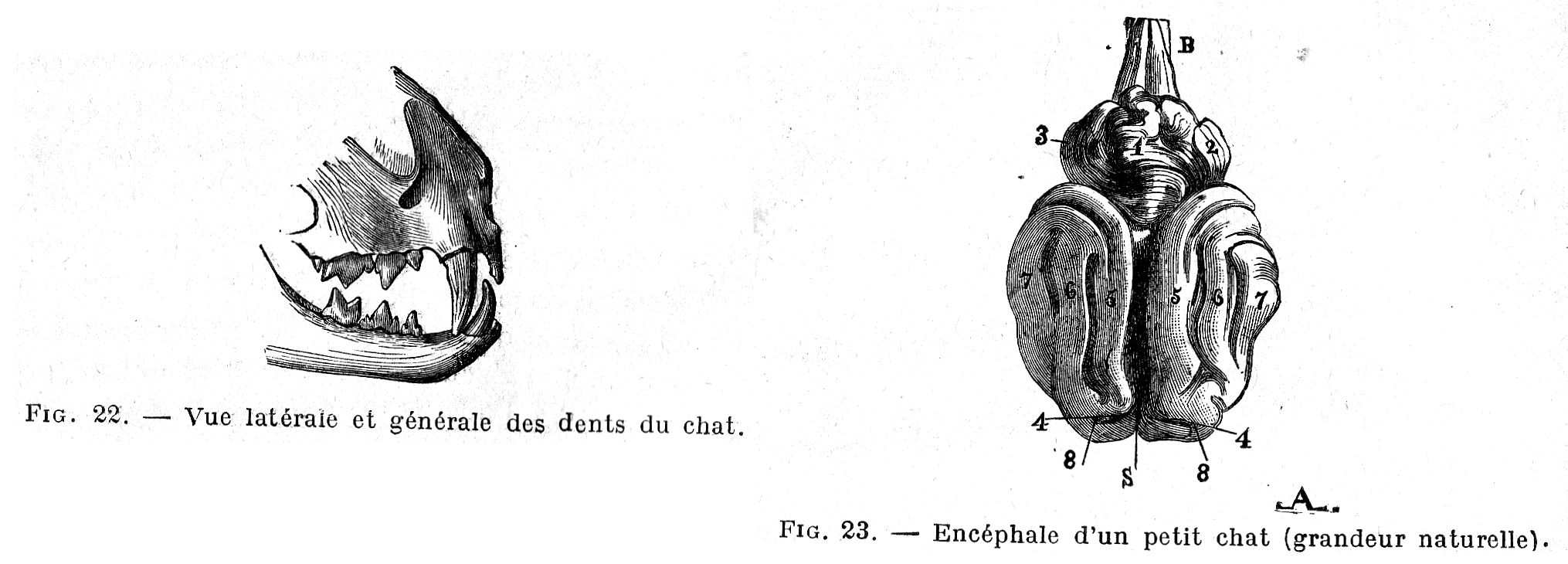
Fig. 23. - Brain of a small cat (life size).
B, medulla oblongata; G, cerebellum; 1, mid-lobe; 2, 3, side lobes; S, inter-hemispherical fissure; 4, 4, crucial sulcus; 5, sagittal gyrus; 6, ectosagittal gyrus; 7, ectosilvian gyrus; 8, sigmoid gyrus.
Cranial nerves. - As in other animals, cats have: firstly, nerves for special sensations (olfactory nerves 1st pair, optic nerves 2nd pair, acoustic nerves 8th pair); secondly mixed nerves with double roots (trigeminal nerves 5th pair, glossopharyngeal nerves 9th pair, pneumogastric nerves 10th pair); thirdly motor nerves with single roots (common oculomotor nerves 3rd pair, internal oculomotor or pathetic nerves 4th pair, external oculomotor 6th pair, facial 7th pair, spinal 11th pair, great hypoglossal 12th pair).
The olfactory (1st pair) and optic (2nd pair) nerves offer no interesting peculiarities to report. The motor nerves of the eye (3rd, 4th and 6th pairs) come together, in the middle of the muscles of the orbit, with threads of the ophthalmic branch of the trigeminal.
The trigeminal nerve (5th pair) has a sensory, bulky root, and a much smaller motor root. The sensory root comes from the annular protuberance, then arrives in the ganglion of Gasser which sends out two large branches, one of which gives rise to the inferior maxillary nerve, while the other branches off after traversing the external fissure of the sphenoid, thus giving two upper branches, the ophthalmic branch and the upper maxillary nerve. The motor root also emerges from the annular protuberance, below the sensory root; it arrives under the inferior surface of the Gasser's ganglion and runs entirely into the inferior maxillary nerve, which is the only branch of the three-branched trigeminal which is both sensitive and motor. The ophthalmic branch ends with three nerves: a frontal branch dividing into two threads, a lacrimal branch and a palpebral nasal branch.
The superior maxillary nerve supplies first an orbital branch, then the greater palatine nerve, the staphylin nerve, the sphenopalatine or nasal nerve, and the superior dental nerves; it ends with the infraorbital branches. From its origin, the inferior maxillary nerve gives rise to the masseter nerve, the buccal nerve, the nerve of the internal pterygoid muscle and the superficial temporal; on arriving at the posterior edge of the jaw, the latter partially joins the middle branch of the facial and sends its other fibres to the anterior auricular nerve; the inferior maxillary nerve then sends out, almost as soon as it leaves the skull, the mylohyoid nerve, which is divided into two branches, one anastomosing to the lower branch of the facial, the other following the mylohyoid muscle, as far as the chin symphysis, then the lingual nerve supplying the vasomotor and excito-secretory nerves of the submaxillary and sublingual glands.
The facial (7th pair) gives rise to, as soon as it leaves the fallopian aqueduct, a cervical branch crossing the parotid, and ending in three branches: the more voluminous upper branch gives rise to an anterior auricular branch and branches to the muscles of the auricle; the middle branch accompanies Steno's canal, anastomoses with fibres from the lower branch and terminates in the upper lip and tip of the nose; the lower one receives, near the maxillary fissure, a sensory branch which joins it from the mylohyoid (5th pair), gives up a few threads to the middle branch, then continues in the lower lip.
The sensory pneumogastric roots (10th pair) start from an irregularly elliptical surface and are about twenty in number, they emerge in three main bundles; the voluminous motor roots, are divided into two series of fibres: the anterior ones, which first merge into their own small ganglion, then flow into the jugular ganglion; the posterior ones which attach to the medullary root of the spinal (11th pair) at the level of the jugular ganglion where they leave it to unite with this ganglion. Below the foramen lacerum the pneumogastric provides the highly developed pharyngeal nerve, which first sends a very thin branch (upper branch) to the pharynx; then another (middle branch) which receives a branch of the superior laryngeal, then sends a division to the thyropharyngeal and finally reaches the cricothyroid; finally a lower branch, innervating the cricopharyngeal and the thyroid gland, reinforced by fibres coming from the plexiform ganglion and sending, at the level of the thyroid body, an anastomosis of the recurrent laryngeal nerve. The pneumogastric then gives rise to a plexiform and elongated ganglion from which the superior laryngeal nerve begins, which in turn gives rise to the external laryngeal. The upper laryngeal sends fibres to the glottis, epiglottis, base of the tongue and the oesophagus; the branch of Galen does not anastomose with the lower laryngeal and exits the larynx a little inside the recurrent laryngeal nerve, then descends along the trachea to reach the thoracic cavity and give rise to two divisions: on the right side, the more important branch receives a large branch from the lower cervical ganglion, then the two divisions unite to join the pneumogastric, a little behind the point of emergence of the lower laryngeal; on the left side the anastomoses are stronger and the preceding branch follows the reverse path of the recurrent laryngeal nerve to join the pneumogastric at the point where the recurrent laryngeal originates on the latter. In the cervical portion, the pneumogastric is intimately attached to the sympathetic. The lower laryngeal arises from two distinct branches on the right side, proceeding from the same point on the left side; the external branch of the nerve (recurrent branch) receives a few fibres from the branch of the superior laryngeal.
The bronchial nerves are numerous and large, the oesophageal plexus is very rich, and the hypoglossus (12th pair) forms a long branch that runs along the sides of the larynx and into the muscles of the anterior aspect of the trachea.
The acoustic (8th pair), glossopharyngeal (9th pair) and spinal (11th pair) nerves do not show any special features.
Spinal nerves. - There are 8 pairs of cervical nerves whose lower branches, united by anastomotic threads, form a very elongated cervical plexus; 13 pairs of dorsal nerves which distribute themselves to the muscles of the back, ribs, abdomen; 7 pairs of lumbar nerves, 3 pairs of sacral nerves and 6-7 pairs of coccygeal nerves.
Brachial plexus. - The brachial plexus is formed by the last four cervical pairs and the first dorsal pair; the fifth cervical pair give rise to an insignificant fibre only.
The anterior or musculo-cutaneous brachialis is formed by a fibre of the 6th cervical pair and by more voluminous branches of the 7th pair; it branches off at the level of the scapulo-humeral joint, one of its branches losing itself in the mass of the biceps, the other adjoining the humeral artery and ending in a branch running out in the anterior brachialis and by a very thin fibre, subcutaneous at the elbow, descending along the internal edge of the forearm and becoming lost at the level of the carpal bone; the anterior brachialis sends a branch slightly below the middle part of the humerus to the median.
The radial nerve formed by the 8th cervical pair receives fibres from the median, ulnar and axillary nerve; the internal branch or muscle branch, which sometimes joins the external branch or cutaneous branch, runs along the internal edge of the pastern [metacarpal], forms a small branch on the dorsal aspect of the thumb and then forms the internal dorsal collateral nerve of the index finger; the cutaneous branch passes in front of the carpal and, at the origin of the third interosseous space, divides into three branches for the second, third and fourth interosseous spaces, each branching off at the root of the fingers to give the dorsal collateral nerves; the very thin outer anastomoses between the first and second fingers with the dorsal branch of the ulna.
The median nerve, formed by the last cervical pair and the last two dorsal pairs, passes through the epitrochlear foramen at the lower end of the humerus and divides, below the carpal arch, into three branches: the internal goes to the rudiment of the thumb and the inner palmar edge of the index finger (second digit); the median descends into the second interosseous space, sends a fibre to the large bulge of the leg, and branches off to form the collateral palmar nerves of the index finger and internal of the middle finger; the external enters the third intermetacarpal space and gives the palmar collaterals external of the middle finger and internal of the ring finger (fourth digit).
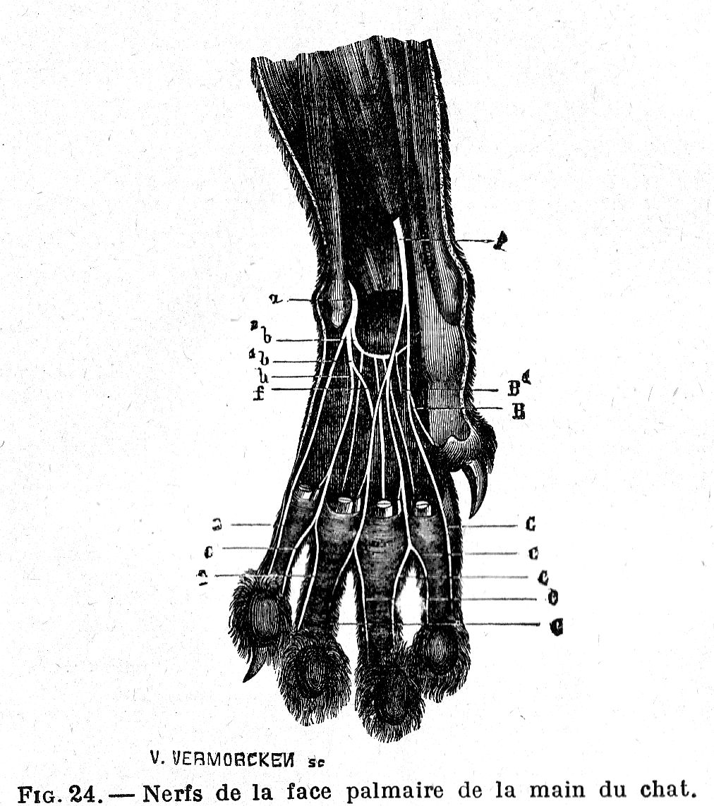
Fig. 24. - Nerves of the palmar face of the hand of the cat.
A, trunk of the median: it is divided into two branches; B, internal branch giving a rudimentary fibre to the thumb; B1, external branch receiving a thread f from the ulnar; C, C, C, C, collaterals provided by the median; a, volar branch of the ulnar; it is divided into three branches; b, internal branch: it provides the thread f at the median; 1b, external branch; 2b, deep branch; C, C, C, collaterals provided by the ulnar.
The ulnar, formed by the first two dorsal pairs, provides two branches, one dorsal, the other palmar; the dorsal branch divides into two filaments at the level of the carpal, one forming the dorsal collateral of the little finger (fifth digit), the other gaining the fourth interosseous space, then receiving a branch from the radial and providing a little further on the internal dorsal collaterals of the little finger, the external of the ring finger; the palmar branch passes inside the pisiform bone and immediately divides into several fibres; a few end in the muscles of the little finger and thumb; another forms, on the outer edge of the little finger, the external palmar collateral nerve; one of the longest, after being lodged in the fourth intermetacarpal space, gives the fibre of the large bulge of the paw and the inner palmar collaterals of the little finger and outer ring finger.
Lumbosacral plexus. - The lumbar plexus is formed by the anastomoses of the lower branches of the 3rd, 4th and 5th lumbar pairs, the sacral plexus by those of the lower branches of the last two lumbar pairs and the first two sacral pairs. The femoral nerve and the obturator nerve originate from the 3rd and 4th lumbar pairs; the saphenous nerve, originating from the first, lodges on the internal face of the tarsus, attaches to the second metatarsal and forms the internal collateral nerve of the second toe; the obturator nerve receives a reinforcing branch from the 5th lumbar pair.
The greater sciatica is large and has two terminal branches, the external popliteal sciatica and internal popliteal sciatica, which separate from each other slightly above the posterior surface of the tibial-femoral joint.
The first branch or external popliteal sciatic nerve passes over the surface of the external gastrocnemius, engages between the flexor digitorum longus and lateral peroneus longus, and divides into two parts, the musculocutaneous nerve and the tibialis anterior. The musculocutaneous branch becomes superficial towards the lower third of the leg, passes in front of the tarsus, provides at the level of the tibiotarsal joint a very thin fibre going to form the external dorsal collateral nerve of the fifth toe and, arriving at the upper part of the metatarsal, gives rise to three branches; each of them runs along an intermetatarsal space and is divided into two fibres at the height of the metatarsophalangeal joints: the external branch gives the dorsal internal collaterals of the fifth toe and external of the fourth; the middle branch supplies the dorsal internal collateral nerves of the fourth toe and external of the third; finally, the internal branch supplies the dorsal internal collateral nerves of the third toe and external of the second.
The second branch of the external popliteal sciatica or the interior tibial nerve accompanies the artery of the same name and gives rise to two terminal branches at the level of the carpus, one going to the tarsal joints and the foot muscle, the other following the second intermetatarsal space and anastomosing, towards the metatarsal phalangeal joint, with the internal branch of the musculocutaneous. The second terminal branch of the greater sciatica or internal popliteal sciatica gives rise to articular, muscular and cutaneous threads (including the external saphenous vein) and continues with the posterior tibial nerve which ends with the two external and internal plantar nerves. The latter is attached to the internal edge of the flexor digitorum superficialis muscle of the phalanges, leaving, when reaching the middle part of the metatarsus, a thin thread forming the internal plantar collateral nerve of the second toe, then passes obliquely in the direction of the last toe or external toe and successively provides three threads (one for each intermetatarsal space) going to anastomose with the terminal branches of the external plantar nerve, at the level of the metatarsophalangeal joints. The external plantar nerve passes between the two flexor tendons of the toes where it leaves a thread constituting the external plantar collateral nerve of the fifth toe; it arrives outside the flexor digitorum profundus, engages under the flexor digitorum brevis to divide into several muscle branches and into three digitate branches; each of these follows an interosseous space, bifurcates at the level of the phalangeal metatarsal joints, receives the fibre supplied by the internal plantar nerve and forms the internal plantar collateral nerves of the fifth toe and external of the fourth, internal of the fourth and external of the third, internal of the third and external of the second toe.
Great sympathetic. — The cord of the great sympathetic, which presides over the vegetative life, includes four regions. A cervical portion, which begins in the superior cervical ganglion and soon merges intimately with the pneumogastric; the two nerves separate at the first intercostal space and the sympathetic gives rise to a very small middle cervical ganglion and an inferior cervical ganglion. In its thoracic portion, the great sympathetic nerve provides a first elongated thoracic ganglion, sometimes welded to the inferior cervical ganglion then, from the fourth intercostal space, a small ganglion at each of the intercostal spaces, finally, at the level of the thirteenth rib, the great splanchnic nerve. The original cord constitutes only a thin thread in its lumbar portion, a thread swelling at each lumbar vertebra in a lumbar ganglion which sends very thin threads to the aorta, to the arteries and to the lumbar veins, and whose union constitutes the aortic plexus. Finally, the sacral portion becomes thinner and thinner, still forming small sacral ganglia and is imperceptibly lost towards the rear.
The cervical portion communicates with the glossopharyngeal, pneumogastric, spinal and hypoglossal nerves and the lower branch of the first cervical pair by means of fibres forming the guttural plexus. The superior cervical ganglion sends superior carotid branches accompanying the internal carotid into the skull and forming the cavernous plexus; a lower carotid bundle reaches the origin of the terminal branches of the primary carotid artery where it forms the carotid plexus; small fibres go into the wall of the pharynx and constitute, with the pneumogastric and the glossopharyngeal, the pharyngeal plexus. The lower cervical ganglion receives the vertebral nerve from the second, third, fourth, fifth and sixth cervical pairs and a branch from the eighth cervical pair; it sends on each side two or three cardiac nerves which go to form the cardiac plexus.
The ganglia of the thoracic portion receive the inferior branches of the dorsal nerves and supply: 1, the large splanchnic nerve, which is formed towards the seventh ganglion; it goes to the adrenal gland and forms part of the adrenal plexus which sends anastomoses to the coeliac plexus, the anterior mesenteric ganglion and branches that unite with fibres of the anterior mesenteric ganglion to form the renal plexus; 2, the small splanchnic nerve formed by two or three fibres emanating from the last thoracic ganglia. The coeliac plexus is made up of short threads uniting the two right and left coeliac nodes and the anterior mesenteric nodes; its branches form the gastric, hepatic and splenic plexuses. The anterior mesenteric plexus contains the anterior mesenteric ganglion, united with the coeliac plexus, the posterior mesenteric plexus, and the anterior mesenteric plexus on the other side. The renal and adrenal plexuses are indistinguishable from each other. The lumbar-aortic plexus is formed by numerous branches originating from the solar plexus and meeting at the posterior mesenteric plexus.
The lumbar portion communicates with the lower branches of the lumbar nerves, sends some branches to the lumbar-aortic plexus, the others going to the origin of the small mesenteric artery where they form the posterior mesenteric plexus. This plexus is formed of fibres coming from the anterior mesenteric plexus, the cord of the lumbar portion of the sympathetic, the aortic plexus and the small posterior mesenteric ganglion; it sends fibres which constitute the spermatic plexus formed by branches accompanying the internal spermatic artery and also coming from the lumbar portion of the great sympathetic; in the she-cat, the spermatic plexus is replaced by the ovarian plexus.
The sacral portion receives the lower branches of three or four sacral nerves and sends fibres to the rectum constituting the hypogastric plexus which is merely the extension of the aortic plexus into the pelvic cavity and encompasses the hypogastric ganglia.
SENSE ORGANS
Taste. - The taste organ has its seat in the corpuscles grouped around the papillae of the tongue; two principal circumvallate papillae are found near the base of this organ, with very small papillae in their vicinity; the filiform papillae that cover the back and the base of the tongue are compound and covered with a thick horny layer; between them, regularly disseminated, are fungiform papillae.
Eye. - The cornea has a circular outline. The choroid shows a light golden yellow or greenish yellow carpet around the papilla, green at the periphery and a dark carpet of intense black, these two carpets being separated by an indigo blue band; the papilla, located in the lighter carpet, is roughly circular and is also surrounded by an indigo blue ring; there are retinal vessels (arteries and veins), supplying the entire internal face of the ocular body. The lens presents an anterior face more convex than the posterior and its volume of 0.5 cc is, compared to that of the eyeball, in the ratio of 1:9.8. The iris is greenish in adults and light blue in young; the pupillary opening is round when dilated, elliptical from top to bottom, sometimes even reduced to a simple vertical slit, when contracted. In the dark, the fundus may appear phosphorescent. The third eyelid is very small, together with the lacrimal gland there is a Harderian gland.
Ear. - The conchinian cartilage of the outer ear is short, pointed, almost triangular, erect and open in front. The middle ear has an extremely developed eardrum, giving the cat very acute hearing; the mastoid cells are found distributed in a special compartment of the tympanic cavity constituting the tympanic bulla; the other compartment contains the ossicular chain. The inner ear with the labyrinth (vestibule, cochlea and semi-circular canals) is unexceptional; the cochlea describes three turns.
Touch. - On the lower face of each limb we find blackish plantar tubercles, six on the front legs and five on the hind legs; the smallest or finger pads are found under each digit (except the thumb) at the joints of the second and third phalanx; the largest, the central, which vaguely resembles a clover leaf, corresponds to the metacarpophalangeal or metatarsophalangeal joints, to which it is joined by very strong fibrous bands; the last or carpal pad is found at the level of the supracarpal or pisiform bone, from which it is separated by a small mucous bursa. They are made up of connective tissue, elastic tissue and fat; there we find very developed sweat glands and tactile corpuscles. The claw, completely retractable especially in the front feet, consists of a horny sheath curved at the bottom; it is protected by a fold of skin and in the normal state is lifted effortlessly by an elastic ligament going, in each digit from the second to third phalange; to extend its claws, the animal is forced to contract its flexor muscles and, as soon as the voluntary contraction ceases, the claws are raised, coming to lodge in the interdigitated spaces. Among the epidermal structures, we also find whisker hairs which are true tactile hairs, remarkable due to the presence of a blood sinus, hollowed out in the thickness of the fibrous follicular sac, showing on its internal wall a veritable tactile ring where the nerve endings are concentrated.
PLACENTA AND FOETAL SACS
The feline placenta has the appearance of a thick girdle circling the middle part of the chorion (simple circular placenta); this chorion forms an elongated sac, the two ends of which come into contact with neighbouring foetal sacs. The allantois presents a poorly adherent internal or amniotic layer and an external or chorial layer closely united to the chorion. The umbilical vesicle constitutes a transversely elongated sac, between the amnion and the internal allantoic layer, provided in its middle part with a narrow pedicle which extends into the umbilical cord; it has extremely vascular walls. The allantoic portion of the umbilical cord is very short and enveloped in a large fold of the allantois.
PHYSIOLOGY
The cat has a rounded head, strong whiskers, a short, stocky neck, an elongated and narrow body, and strong limbs; the male is distinguished from the female by his larger size and a stronger and wider head. The cat’s gait is supple and graceful; it always bends itself, curls up, gets up, lies down and walks silently, making no noise; its legs are wonderfully organized to give it lightness and dampen the reactions of the ground. When pressing down, the fingers extend and move away slightly from each other to disseminate pressure by distributing it over a wider surface; in addition, the cellulo-fibrous tubercles located on the underside of the foot flatten out by gaining the interstice of the phalanges and come return to their shapes thanks to their elasticity as soon as the pressure ceases. Also due to the extreme flexibility of its limbs, the cat can fall from a great height without ever hurting itself because it always lands on its feet and never on its back.
[Footnote - In a dissertation sent in 1700 to the Academy of Sciences, we find the following explanation of this fact. "Cats, when they fall from a height, usually fall on their feet, even when they first have their feet topmost, and should, therefore, fall on their head. It is, of course, not possible for them to turn themselves over while in mid-air, where they have no fixed point to lean against; but they are gripped by a fear which makes them bend their spine in such a way that their entrails are pushed upwards. At the same time, they stretch out their head and their legs towards the place from which they fell, giving these parts greater leverage; thus their centre of gravity moves from the centre of the body, and is placed uppermost. From this it follows that these animals can make a half turn in the air and point their paws downwards which almost always saves their lives.]
In 1895 M. Marey had recourse to chronophotography to know the successive phases of the phenomenon. The cat, held by an assistant with its paws in the air, is let loose in front of a chronophotographic camera capable of shooting sixty images per second.
Examination of the photographs thus obtained shows that the animal "First bent so that his back is strongly convex and directed downwards, straightens his spine and curves in the opposite direction; at the same time, a torsion occurs along the vertebral axis and the torque resulting from muscular action tends to rotate the front and back of the body in opposite directions to each other. But the rotation of the two halves of the body is very unequal. It focuses almost exclusively on the front end; then, when it has turned about 180 degrees, the rear end turns. Inspection of the figures excludes, first of all, the idea that the animal imprints a rotary movement by taking a point of support on the hands of the operator because the first images of each of the two series show that in the first moments of its fall, the animal still had no tendency to turn to one side or the other: its rotation only began with the twisting of the loins. As for the hypothesis of support from the resistance of the air, it is no longer admissible, because, due to the direction of the movements of the animal, if this resistance had appreciable effects, it would produce a rotation reverse of that which is observed."
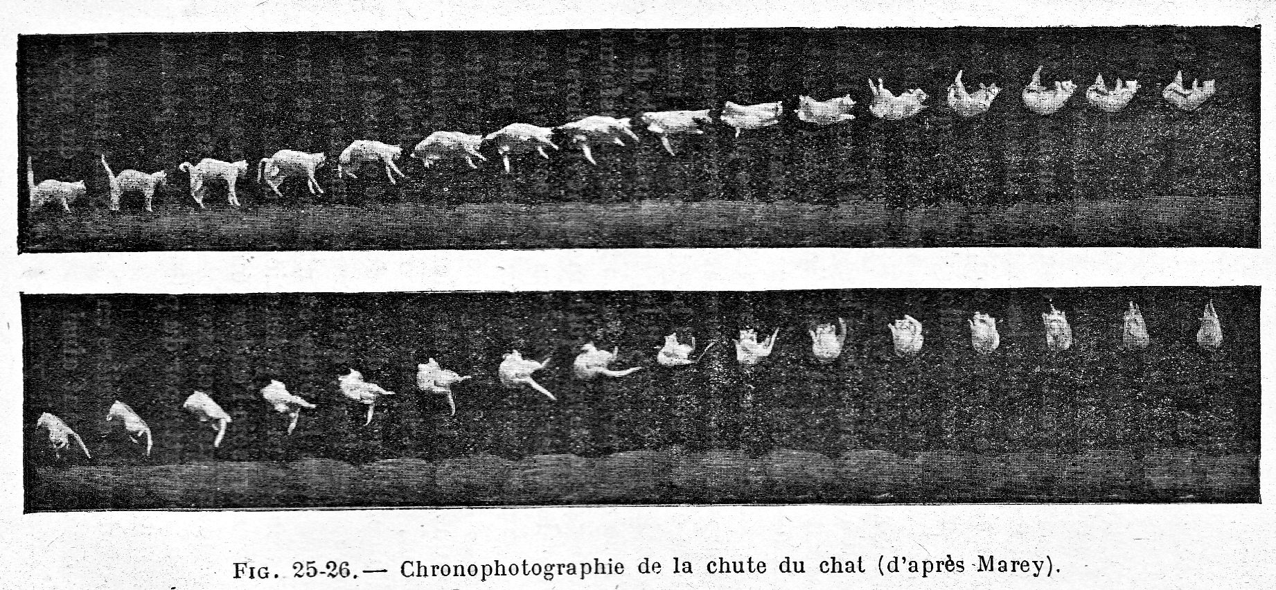
The result is therefore that the cat turns around on its own, without involving any force or any external support. This may be surprising, because it is in fact admitted that a material system subjected only to gravity and its mutual actions and starting from rest, cannot rotate itself around a horizontal axis passing through its centre of gravity, describing a zero sum of areas (area theorem). The fact is, however, correct because a cat is not a homogeneous system like the bodies considered in mechanics. It would take too long to relate here the discussions which took place on this subject; let us content ourselves with saying, with Messrs Marey and Guyon that "it is in the inertia of its own mass that the animal takes successive supports to turn around. The torsion couple which produces the action of the muscles of the vertebra acts at first on the forelegs, which have a very small motion of inertia on account of the front feet being foreshortened and pressed against the neck. The hind legs, however, being stretched out and almost perpendicular to the axis of the body, possesses a moment of inertia which opposes motion in the opposite direction to that which the torsion couple tends to produce. In the second phase of the action, the attitude of the feet is reversed, and it is the inertia of the forepart that furnishes a fulcrum for the rotation of the rear."
The cat can stand on the most shaky and narrow branches and climbs with remarkable agility along trees or walls; he leaps with incredible lightness, but, on the other hand, he is unable to run and when he feels threatened, his dash turns into a series of successive leaps that quickly put him out of reach; unable to force his victims to run, he is content to wait for them, served wonderfully by his patience and the finesse of his ear. It swims easily, its legs move as in walking, giving impetus by hitting the water, symmetrically to go straight, more strongly on one side to go to the opposite side; however, he feels a real antipathy, even fear, for water, avoids the rain and does not like getting his paws wet, taking a thousand precautions on wet ground and choosing carefully where to put his feet. There are, however, a few examples of cats diving into water to catch fish and of fishing for pleasure.
He sits on his hindquarters, leaning on his forelimbs, thus maintaining the attitude of the fabled sphynx. To sleep, he sometimes takes the lateral position, lying on his side, limbs extended or slightly bent; but more often he makes a circle, bending his neck and tucking his head into his belly, greatly bending his spine and bending his limbs towards his body.
He can dream, as evidenced by the cries and movements that are sometimes made during sleep.
He loves and seeks heat, lies in the hottest places of the house, behind ovens, fireplaces and even in front of the fire.
Very clean, the cat spends its time polishing its coat and cleaning itself by licking itself, or by passing its paws, wetted with its saliva, over areas of the body that the tongue cannot reach; this is mentioned by a song, reproduced in "Chants et chanson populaires de l'Ouest" [Chants and Popular Songs of the West] by Jérôme Bujeaud:
Jeannette’s cat is a very pretty beast;
And when he wants to make himself handsome,
He licks his muzzle with his tongue;
And he does his washing with his spittle.
Its fur is easily electrified and at night, in dry weather, can give off small sparks when you run your hand against it. He hides when voiding excrement and, when possible, covers it with earth or ashes.
The mother's instinct is very strong. Not only does she always show herself to be a good mother to her family, constantly sheltering her young from the father's teeth, but she will also adopt other animals and examples are cited of young puppies, rabbits and squirrels being adopted by she-cats; we even find in the "Mercure de France" of 1731, an extract from a letter, dated January 16 of that year, relating that in 1664, in Evreux, a she-cat belonging to Dupuis, living in Rue Trienne, after having exterminated a nest of mice, adopted one which had been hidden beneath her and fed it alongside her kittens.
Regardless of what Buffon says, there are many facts to prove that the cat is capable of attachment and even devotion to its owners. In the Dictionary of Anecdotes of 1820, Vol. II, p. 274, we find the following fact, already reported by Landrin: "An man was murdered in his house by a relative who wanted to enjoy the inheritance. The Justice went to the deceased's house. A big cat sprang from a wardrobe and threw himself into the midst of the crowd and onto a man whose face he clawed furiously. By a presentiment, the surgeon exclaimed: ‘This is surely the murderer, please arrest him.’ At those words the unfortunate man, covered with scratches and blood, tried to escape, but was arrested. Overcome by terror, he threw himself at the feet of the magistrates and publicly acknowledged his crime."
L'Estoile, in the "La Presse" of September 14, 1862, relates that a well-known woman, when she shut herself up in her small living room to paint, always saw an alley cat arrive near her who would snuggle up between the andirons and the screen. She fell ill; the cat ventured into the bedroom, the first few days doing nothing but appearing, then staying there for an hour, then most of the day. He spent the last night under the dying woman's bed and ran away crying as soon as she expired; the next morning he was seen hanging from the forked branch of a chestnut tree.
M. Desherbiers reports that Mme Dupin, daughter of farmer general Samuel Bernard, had a cat, named Bibi, whom she was particularly fond of, which died two days after her at its mistress's grave.
The famous Arabian horse Godolphin, one of the founders of the current English thoroughbred breed, befriended a black cat; when, in 1753, the horse died, the cat remained on its friend's corpse until it was removed and, shortly after, was found dead in a nearby barn.
If, in the wild state, the cat manifests an unsociable ferocious and rapacious temperament, even if, as Cuvier says, it does not show great intelligence, "its wiles consist of little more than silence and mystery," it is no longer the same in domesticity where its intelligence awakens, develops and also increases. The cat is naturally very mistrustful; if he has been corrected after committing some misdeed, we can call him back, present him a piece of meat; he looks and listens, but is careful not to approach, which already indicates a certain degree of intelligence; moreover, if you throw the piece of meat to a familiar cat, it runs up and takes it if you are at some distance away, but does not dare to grab it if the piece has fallen at the feet of its master; on the other hand, it extends its paw hesitantly, pulling the morsel towards it, moving as little as possible if the piece was dropped a short distance. Dugès sees this again as proof of intelligence, because the animal compares the distance which separates it from its master and judges whether it may be within the range of correction. There are many facts to prove the intelligence of the cat.
The Marquis de Nadaillac relates that a secretary to our British Ambassador walking one day in a street in London saw a cat, which gently brushed against his leg; at first he didn't care; but the animal having renewed its merry-go-round several times, he began to look at it; this was what the cat was waiting for, it immediately turned around and, by the expression of his gaze, seemed to invite him to follow him; the secretary stepped forward and, a few steps further, saw the animal stop in front of a house, climb the steps that led to the front door and jump after the doorbell as if to catch it; very intrigued he rang the bell and told the adventure to the servant who opened the door for him: the latter informed him that the cat, being used to running loose, always acted like this when it wanted to return.
Conch claimed to have known a cat that had found a way to open a cupboard in which the supply of milk was kept; the animal would sit on a table next to the cupboard and administer a series of taps on the key ring; the lock being old and playing easily, the key ended up turning and the cat thus got its ends.
Otto relates that a cat belonging to Mr. Parker Bowman, of Parara, was usually locked in a room that had only a hinged window closed with a pivot cross member as an exit; when he wanted to go out he jumped on the support of the window, stretched out as much as possible to reach the crossbar, managed to move it to the upright position and then leaned with all his weight on the window which opened under this pressure.
Romanes had a cat thato, on several occasions, was able to open a door connecting the stable to a courtyard with the windows of his house; the animal walked nonchalantly and unobtrusively towards the door, with a bound grabbed the loop-shaped handle with one foreleg, squeezed the trigger with the other and, at the same time, played with his hind legs against the jamb to push open the door.
Mr. James Stewens, a judge in New Brunswick, while walking in his garden on a winter day when a thick layer of snow covered the ground, saw a robin alighting on a shrub, about a metre above the ground; a cat, which was prowling nearby, saw it, approached gently and stopped a short distance from the bird, but realizing that the snow would prevent him from gaining momentum to reach this prey, he first tried to make the bird fly to a more favourable location. Despite all his efforts, the torpid robin did not move, however, after a while it flew a little further and the cat immediately followed him, making a detour, hiding behind the bushes and skirting a fence, then, with a bound, rushed towards the bird ... which he missed. Dr Klein had a cat that as soon as he saw the window opening after meals and the breadcrumbs thrown out, hid in the clumps to wait for the birds to arrive. Another cat, doing the same thing, was surprised one morning to find snow had fallen overnight and scratching at it furiously. Then, removing the crumbs from the place he had thus cleared, he spread them on the surface of the snow and then went off to watch the birds from behind the clumps. Lastly, Dr Frost had a cat which, when crumbs stopped being thrown outside, scattered them to attract birds and surprise them. These three examples cited by Romanes show that the first cat had noticed that the crumbs attracted birds, the second also highlighted the crumbs to better attract the birds and finally the last one spread the crumbs itself, which denotes an even higher degree of observation and, consequently, of intelligence.
The same author also cites the following example: a cat who had befriended a parrot was left alone in the kitchen; suddenly, the cook, who was on an upper floor, saw him coming to her, excited, meowing, pulling her by her apron to invite her down; she followed him and was greatly surprised to see the parrot calling and flapping its wings, entangled up to its knees in a bowl of dough she had left near the fire to make it rise, and becoming gradually bogged down.
Finally, Bouviller relates the story of a cat raised in a convent, who noticed that when the bell was heard at certain times of the day, the monk who performed the functions of porter and cook, took a portion of meat and carried it to the poor man who rang the bell; one fine day this cat went to hang onto the doorbell rope and took advantage of the cook’s absence to take a piece of meat; the feat was repeated daily and this merry-go-round could have lasted a long time had the greedy cat not set its sights on a chop intended for the head of the community; that was its downfall, the cook having discovered the chop bone left in a corner. Needless to say, the cat was denied entry to the kitchen from that point on.
Brehm, in his book Man and Animals cites the following example reported by Wood. A young woman had a cat called Prettina (pretty) known as Pret. Suffering from a nervous illness the young woman was forced to go to bed. The cat slipped into the room, doing everything possible to amuse her mistress and taking on the role of nurse; she quickly learned to know the hours her patient had to take food or medicine, waking up the actual nurse, if necessary, by biting her lightly in the nose.
Ern. Menault, in his Animal Intelligence, relates that a cat who went for a walk on the roofs every evening would jump at the bell-pull when it wanted to return. He also cites the example of a female cat, who used to respect birds in cages; one of her young was corrected several times as it was about to get hold of a bird; from then on, the cat prevented her kitten from going to the cage by correcting it herself as soon as it seemed to be heading towards the birds.
Another cat used to let a canary ride on her back; one fine day, her master saw her pounce on the bird, take it between her teeth and climb onto a desk; when he made her let it go, he saw that a foreign cat had entered the room and was threatening the canary.
The cat enjoys an excellent memory, especially as regards remembering places; it is excessively difficult to disorientate it when you want to get rid of it and, on several occasions, carrying a bulky cat about twenty kilometres away during the night to get rid of it by losing it in the woods, the animal was back home even before its master arrived back.
Fabre, who incidentally was led to study cats through his experiments on homing ability of certain insects, on two occasions saw adult cats find their way home despite the distance and the completely unknown places they had been taken to.
It is a tradition in the countryside that, to prevent a cat from returning to the home you want to keep it away from, and to make it lose its desire to return home, it suffices to place it in a bag and to rotate the bag – and its therefore its inhabitant – several times. This is a mistake, because experience proves the opposite: the gyration of the cat locked in a bag does not make it lose its taste for home, especially if its old home is not sufficiently far away from the new home.
His physiognomy is very expressive; he knows how to ask people who live with him for caresses by his attitude, his look, his light touches. Likewise, he testifies to his pleasure and satisfaction with the arching of his back, the quivering of his tail and by purring. When he watches a mouse or a bird, everything in his attitude indicates sustained attention; he gets his satisfaction by jumping on the victim he was waiting for and takes joy in letting his bruised prey run away and recapturing it immediately. As soon as he is irritated his gaze becomes threatening, he raises his spine, stands on his feet, bristles his whiskers; he grimaces, and his breath becomes a growl. We even go so far as to give the cat the power to predict the weather like a barometer. If he bends his paw and wipes his head with it the weather will change: if the weather is nice it will rain; if it is raining, the sun will soon come out. If wind or cold are about to arrive, the cat will flatten its hair against its skin to concentrate the heat in its body; if, on the contrary, the temperature rises, it raises and bristles its hairs to increase heat loss.
The cat has a very emphatic voice; sometimes he purrs, as when a hand strokes his back, sometimes when he is irritated, he gives a low growl. As Desmarets rightly points out, we can distinguish, from the tone of meow, the call of females, the cries of distress that summon the attentions of the male, the sweet sounds they make to call their young, the muffled whistles, and the more or less prolonged rumblings that the males make to females in heat. According to Dupont de Nemours "the cat has the advantage of a language in which are found the same vowels as those pronounced by the dog and, moreover, six consonants: m, n, g, h, v and f." On several occasions we have tried to do cat concerts, most often by communicating the keys of a keyboard with a string attached to the paw of a different cat; but we must admit that the effect obtained was never very artistic.
By a remarkable arrangement described in the "Anatomy" chapter, there are in a way two glottis in the cat (an upper, between the membranous vocal ribbons, and a lower between the musculo-membranous ribbons) and two types of cords, each of whose vibrations appear to produce effects of a particular type. M. Segond's experiments tend to show that the superior glottis and the cords that border are responsible for the falsetto voice, and the inferior glottis with its cords as being responsible for the throaty voice. But Longet, having obtained intense and very acute sounds after severing of the upper cords, relates the falsetto and throaty sounds to the lower cords; he even goes so far as to deny that the membranous cords are able to sustain sound vibrations. The role of the cat’s two glottis is, therefore, not perfectly determined although everything suggests that the lower vocal cords, meeting the conditions which belong to the same organs in other animals, play the main role in producing certain sounds, while the higher ones, while still having the ability to vibrate, only play an accessory function.
His sense of touch is extremely developed; the hairs on the lips and around the nostrils, which constitute the whiskers, transmit the impression of the contacts they receive to the bulb in which each hair is implanted; and their sensitivity is such that it is enough to touch one whisker and the animal immediately pulls back. The papillae of the skin, carpal, plantar and finger pads also perceive the impression of contact with external bodies, but to a much lesser degree.
The cat has excellent eyesight, both day and night, thanks to the extreme sensitivity of its iris and the ease with which the pupil transforms into a simple vertical slit in bright light, while in darkness it becomes very large and circular in shape.
Due to the great development of the eardrum, the hearing is very acute; the animal sleeps lightly and the slightest noise awakens it. Numerous observations show, however, that white cats with blue eyes are very often deaf, but they can hear clearly if their iris has even the slightest coloured marking; Dr Sichel observed a cat whose iris began to take on a dark shade at the age of 4 months and which only heard sounds from then on. For Darwin, this fact could be explained "by a slight arrest in the development of the nervous system connected to the sense organs", assuming that the development of the organs of sight and hearing is stopped in the young while the eyelids are not not yet open, the iris, which at birth is always blue, would remain blue for life, and the ears would remain incapable of perceiving the slightest sound.
Its sense of taste is very delicate; if he is offered any food, piece of bread or meat, he turns it over several times, eats the parts that he likes and, if he is not hungry, leaves the rest.
His sense of smell, on the other hand, is relatively undeveloped, however he likes perfumes and readily lets himself be caressed by people who wear them. The smell of valerian (valeriana phu), catnip (nepeta cataria), and teucrium marum [cat thyme] impress him so strongly that he immediately seems transported with pleasure; when he finds these in a garden, the cat will go back and forth over the plant if it is not protected by a fence,destroying it in a short time.
The nose ends in a rather small point, beside and below which the nostrils open. The facial angle is very developed (approximately 41 degrees) and the ratio of the surface of the skull to that of the face is 1 to 0.68. Chewing is slow and difficult because the teeth are arranged to tear, not to grind; the jaws, which are very short, can only perform opening and closing movements. The cat grasps solid food with its jaws and teeth; often, to steady the piece of meat it is tearing, it pins it to the ground with its front paws; its incisors then act like cutting scissors and its canines, long and curved, tear or lacerate the prey. It drinks frequently; to do this it laps, that is to say it pushes his tongue out of its mouth, plunges it into the liquid, then reverses the point backwards and withdraws its tongue abruptly by throwing it towards the teeth, thus throwing small quantities of the drink into the mouth. The cat vomits very easily; besides, for the cat, chewing is very perfunctory and it consumes the most copious meals in an instant as its mouth can open widely, its stomach, which is very dilatable, secretes gastric juice over its entire extent, and the pylorus is narrow, so a large lump can easily enter the stomach and remain there for a long time, until almost dissolved; after twelve hours all the meat in it is gone. The gastric mucosa is very vascular, lacks squamous epithelium and is covered with a thin layer of mucus which is easily detached; under these conditions it presents an enormous absorbing power and the foodstuffs, as a result of their prolonged stay, are sufficiently changed to be, in part, immediately absorbed. One or two bowel movements occur per day.
A 3.5 kg cat absorbs 1,450 litres of air in twenty-four hours; it consumes 69.36 litres of oxygen, burns 27.12 grammes of carbon and releases 50.88 litres of carbonic acid [carbon dioxide]. In the resting state there are 20 to 30 breaths and 120 to 130 heartbeats per minute. The quantity of blood, which varies according to size, can be evaluated, on average, at 1/35th of the body weight, but can vary from 22 to 40 grams per kilogram of animal; circulation is very active and the rectal temperature ranges from 38.5 to 39 degrees centigrade.
The cat can live fifteen years or even longer, but the average lifespan rarely exceeds 10 to 12 years; it reaches its full development between 15 and 18 months. According to Lesbre, the incisors, canines, lower molars and upper second molar of the first dentition appear two to three weeks after birth; the first and third upper milk molars erupt at around 1 and a half months. The replacement teeth erupt from 7 to 9 months in the following order: incisors of both jaws, lower carnassials, upper carnassials, second lower pre-carnassial, second upper pre-carnassial, canines, first lower pre-carnassial, then upper, finally tuberculous.
It is usually twice a year, in the spring and autumn, sometimes three or four times, that mating occurs in cats; Buffon had already noticed "that the female appears more ardent than the male; she invites him, seeks him out, she calls him to her; she announces with loud cries the fury of her desires or rather the excess of her needs and, when the male flees or disdains her, she pursues him, bites him and forces him, so to speak, to satisfy her, although the couplings are always accompanied by great pain." Oestrus lasts from nine to ten days and, from 1 year old, after a gestation of fifty-five to fifty-six days,[1] the domestic cat can give birth to five to six young each time, generally at the end of April and in August. She hides to give birth because the males sometimes devour their offspring. She nurses her young for five to six weeks,[2] then brings them mice and small birds to accustom them to a carnivorous diet.[3] The young, who can already crawl during the first few days, barely start to walk until after a week; their eyelids are closed at birth and do not open for eight to nine days. Between 1 and 5 years, a she-cat generally has little respite between weaning a litter and becoming pregnant again.
Cat urine has a foul and persistent odour; males urinate without squatting, unlike females. The quantity emitted within twenty-four hours varies from 100 to 500 grams depending on size and species; normally yellow, it can turn brownish in summer or with a dry diet. Its specific gravity is approximately 1,030 and it exhibits an acid reaction.
[1. The Siamese cat is most often pregnant for sixty-five to sixty-six days.
2. Essore Mascheroni reports the example of a female cat who nursed one of her offspring for 23 months, at the same time as feeding sometimes other offspring, sometimes very small offspring.
3. When we want to wean the young, we must first start by separating them from the mother for a very short time, just a few minutes, and gradually increase the duration of the separation. When the kittens know how to drink, it is good to transform their liquid food into light broth, then into bread-and-butter soup to which we add a little milk to make it lighter.
BEHAVIOUR
The cat, from an aesthetic point of view, is one of the most harmonious animals that one can find; his mobility and agility are extraordinary, his flexibility proverbial; but often his true character has been overlooked.
All naturalists have described its behaviour and Buffon, in volume VI of his Histoire Naturelle, which dates from 1769, even draws an unflattering portrait of it. [Translator’s note: I use the 1781 English version by William Smellie.]
"The cat is an unfaithful domestic, and kept only from the necessity we find of opposing him to other domestics still more incommodious, and which cannot be hunted; for we make no account of those people, who, being fond of all brutes, foolishly keep cats for their amusement. Though these animals, when young, are frolicksome [sic] and beautiful, they possess, at the same time, an innate malice, and perverse disposition, which increase as they grow up, and which education learns them to conceal, but not to subdue. From determined robbers, the best education can only covert them into flattering thieves; for they have the same address, subtlety, and desire of plunder. Like thieves, they know how to conceal their steps and their designs, to watch opportunities, to catch the proper moment for laying hold of their prey, to fly from punishment, and to remain at a distance till sollicited [sic] to return. They easily assume the habits of society, but never acquire its manners; for they have only the appearance of attachment or friendship. This disingenuity of character is betrayed by the obliquity of their movements, and the duplicity of their eyes. They never look their best benefactor in the face; but, either from distrust or falseness, they approach him by windings, in order to procure caresses, in which they have no other pleasure than what arises from flattering those who bestow them. Very different from that faithful animal the dog, whose sentiments totally centre in the person and happiness of his master, the cat appears to have no feelings which are not interested, to have no affection that is not conditional, and to carry on no intercourse with men, but in the view of turning it to his own advantage. By these dispositions, the cat has a greater relation to man than to the dog, in whom there is not the smallest mark of insincerity or unjustice.
"Though cats live in our houses, they are not entirely domestic. Even the tamest cats are not under the smallest subjection but may rather be said to enjoy perfect liberty; for they act only to please themselves; and it is impossible to retain them a moment after they choose to go off. Besides, most cats are half wild. They know not their masters, and only frequent barns, offices, or kitchens, when pressed with hunger.
"When carried to the distance of a league or two, they return of their own accord, probably because they are acquainted with all the retreats of the mice, and all the passages and outlets of the house, and because the labour of returning is less than that which would be necessary to acquire the same knowledge in a new habitation."
Cuvier, a few years later, judged him much less harshly.
"Although spread over almost the entire surface of the globe, the manners of cats are almost the same everywhere. Endowed with prodigious vigour, and provided with the most powerful weapons, they rarely attack other animals with open force; cunning and daring direct all their movements and are the source of all their actions. Walking noiselessly, they arrive at a place where the hope of finding a prey takes them, crawling near their victim, crouching in silence without any movement betraying them, awaiting the auspicious moment with an unalterable patience, then suddenly rushing forward, they fall on the prey, tearing it with their nails and quenching for a few hours that thirst for blood which consumed them. Satisfied, they retire to the centre of the domain they have chosen for their empire. There, in a deep sleep, they wait for some new desire to urge them to leave. That of love, no less powerful on their senses than that of hunger, comes in turn to snatch them from rest; but the ferocity of their naturalness is not satisfied by this need whose only goal is the conservation of life. The male and female call out with sharp cries, approaching each other with distrust, satisfying their ardour with menaces and parting in fear. Only the mothers show love towards the offspring; male cats are the cruellest enemies of their offspring. Distrust seems to be the most marked feature of the cat’s character; it is never completely erased by domesticity and it presents the greatest obstacle when one wants to tame them. The slightest new circumstance is enough to frighten them, to make them fear some danger, some surprise. It seems that they judge themselves as we judge them ourselves."
Finally, Mr. Boitard seeks to rehabilitate the cat a little in the following terms:
"This animal has a timid nature; he becomes savage by cowardice, suspicious by weakness, cunning by necessity, and thief by need: he is never wicked except when he is angry, and never angry except when feels his life threatened; but then he becomes dangerous because his fury is that of despair, and he fights with all the courage of a coward driven to extremity.
"Forced to live continuously in society with his cruel enemy, the Dog, his natural suspicion increases. It is probably because of this Buffon attributes it with falsehood and insidiousness; it has retained enough of its independence to secure a life in the position we have placed it, and if this position is improved, as in Paris for example, where people love animals, it will give up some of its independence in proportion to the affection given to it."
It must be recognized, however, that while the different climates the cat has been transplanted to have hardly changed it, upbringing, on the contrary, has brought about profound changes in it, especially from a moral point of view; because while some cats remain greedy and suspicious thieves, others never steal anything and become so attached to their masters that they follow them around. We must realize that the cat, from the point of view of domestication, cannot be compared to the dog; the cat is an animal seeking solitude, males and females hardly coming together except to mate, and yet, despite its completely opposed instinct to sociability, it allowed himself to be domesticated. But if it lives in a house, it seeks man’s home rather than man’s company; the caresses and food it receives do not cause its affection or recognition; it remains pretty much what it was in the wild, the mixed diet and plant food it receives have dilated and lengthened its intestine and at the same time weakened its destructive instinct without entirely suppressing it; even if fed enough, it will run through the woods and fields to destroy nests and birds. Man has only been able to domesticate it by using its carnivorous instincts as a defence against mice and rats. Very gentle and very cuddly when young, the cat soon resumes its wild nature and returns to its solitary habits; its affection is selfish and self-serving, feigned rather than sincere, and when it becomes attached to someone, it is always out of self-interest and to get some caresses.
The maternal is highly developed in she-cats. Before giving birth, she usually looks for a secluded place to prepare her nest and hides her retreat, even from the male, until her young are able to walk. As soon as they are born, she abandons the fireside where she loved to sleep and comes home only to eat; she shies away from the caresses she previously sought and hastily returns to nurse, warm and even protect her young. She carefully conceals the place where she has deposited her kittens and the path she takes to get there and becomes anxious and restless when she notices her nest has been discovered. When she thinks they are threatened, she grabs them by the neck with her lips, walks with her head up, carrying them one by one to the new retreat she has chosen for herself and gently lays them on the ground, licking them. As long as she is nursing, she leaves them only to eat and provide theirs if necessary; as soon as they start to walk she accompanies them to watch them, reminding them with a particular meow if they stray too far, correcting them if necessary when they disobey, but in case of danger defending them without worrying about her own security.
Scheitlin describes young cats as follows:
"Their first voice is exceedingly sweet and quite childish. They are so restless that, even while blind, they are not afraid to venture out of the nest, and their mother, worried by their escapes, is always forced to bring them back to the nest. They can hardly see when they can't take it anymore and crawl around the nest, uttering frequent meows. Scarcely have their eyes opened to the light, scarcely are they able to distinguish objects around them, than they immediately begin to play with anything that stirs, rolls, slips, or flies. Their instincts as hunters of mice and birds already begin to awaken. They play continually with their mother's tail and with their own, as soon as it is long enough for them to be able to grasp it with their paws; they also bite it and do not notice that it is part of their body, just as our children bite their fingers, which they consider to be something foreign to them. Kittens make the most singular leaps and the most graceful movements. Their gestures and games, which they enjoy just like children, amuse them and also amuse the people who love them, for hours at a time. As soon as their eyes are opened, they know how to distinguish the good person from the evil, the friend from the enemy. If a dog arrives in the midst of their games, whatever its size and strength, they immediately put themselves on the defensive by arching their backs. They are like little lions.
The cat, having a sometimes quarrelsome character, often provokes its fellows and is not afraid to attack even the dog, seeking to attack him preferably in the head; its courage never fails it and, when it feels that escape is no longer possible, it bravely stands up to the adversary.
CAT CARE
BREEDING AND RAISING
Generation. - The first "heat" of the cat usually appears around the age of 10 months; they can appear from the 5th month, or around the 12th or 13th month. It should be noted that the presence of the male in the vicinity of the she-cat precedes the onset of heat. Female cats exhibit this phenomenon three times a year, which is announced by swelling of the vulva, accompanied by a sero-sanguinous discharge, and by an almost continuous meowing. There are exceptions, however, and Mr. Degorce owned a female cat, Ayoutia II, whose heat would have gone almost unnoticed if it had not been detected by the presence of the male.
This condition persists for a fortnight or even three weeks if the cat is not mated. Put in contact with the male, the latter mates with her for two days, then leaves her; the cat meows for a few more days, and all signs disappear. Crosses between different breeds are not without danger, given the difference in gestation length.
Gestation. - The cat carries her young for about fifty-eight days, the length of gestation varying according to the breed. This is how in the Siamese, gestation lasts up to sixty-five or even sixty-eight days. A pregnant cat can be recognized after about thirty days by the development of the nipples and the belly. The presence of young in her womb can be detected by pressing the abdominal walls.
During gestation, the cat should be given sufficient food, but this should not be overdone. Indeed, the cat must not be too fat for a normal birth as the fat may prevent the expulsion of the young.
Birth or delivery. - The care to be taken at the time of delivery consists in preparing the nest in a basket or box placed in an isolated place, one should place some tea towels on a layer of very clean straw or old paper.
Delivery usually goes very normally and there is no special care to be given to the mother other than to watch the temperature of the cattery or the apartment where the birth takes place. Some cats, such as the Siamese, are particularly sensitive to cold, and we have seen litters that are completely lost due to too low a temperature.
The number of kittens per litter is usually 4, but litters of 5,6, 7 or even 8 kittens are often obtained; on the other hand, Madame la Marquise de Ligneries (who has been raising cats for ten years) observed a cat that only ever gave birth to a single kitten.
Suckling. - During the first stages of suckling, the cat needs absolute rest, and above all, does not allow any stranger to come near.
Food should remain the same as usual, but must be more abundant. You should give one or two additional meals, consisting of fish mashed with bread, liver pâté cooked in butter and cut into small pieces, lecithinized pasta with milk and, for drink, always boiled milk with fresh water at will. For the first few days of their existence, you don't have to take care of the kittens; in addition to milk, the mother provides them with all the care they require, cleaning them, warming them, licking them.
Kittens are born with their eyes closed, but usually open them around the fifth day, sometimes even on the third day.
Weaning. — Kittens begin to eat at around 6 weeks; for their first food they should be given boiled milk, eggnogs, then lecithinated pasta mixed with cooked fish or very finely chopped meat. The mother weans them herself around 2 months; some cats also prevent their young from eating before the right moment, and on the contrary lead them to food when they consider them in a condition to eat.
Rearing. - After weaning, you can start offering bones to facilitate teething and teach kittens to gnaw. Care should be taken to give only large bones with a little meat left, as small bones are easily swallowed and can puncture the intestines. Gradually increase the quantity of the foods mentioned above, taking care that they are all well-cooked, well boiled and that the pots, plates and dishes are always very clean.
By the age of 4 months, normal food can be given which should consist mainly of cooked meat; vegetables should also be given because not only is an exclusively meat diet worthless, food needs to be varied to stimulate their appetite. Once a week, offer your kittens fresh, well-cooked fish which you have removed the bones beforehand; during the winter an excellent food is obtained by crushing canned sardines into well-cooked Carolina rice, or in lecithinated or phosphated pasta. For drinking, give very pure water or boiled water.
Thus treated, kittens thrive and are cheerful because their character, like that of children, reflects the condition of their stomach; they are vigorous and have the maximum resistance to cat distemper, a disease of the young similar to that of dogs, which wreaks great havoc on sickly or puny subjects.
Special care. - It is necessary to give the kittens some other care.
In the first place, it is essential to remove fleas, first by freeing the mother from fleas, then by de-fleaing the young, because these parasites torment them and greatly harm their development.
It is also important to ensure that your cats do not suffer from constipation. One can prevent it by reusing the castor oil or manna oil that we give in milk; olive oil, mixed with food, also maintains a free-moving belly and is recommended as well as cod liver oil.
In cases of diarrhoea, withhold milk and give very fresh raw meat, solid foods without sauce, and rice cakes.
Avoid cutting the claws of young cats and, in order for them to harden, give the animals a very smooth pine board on which a worn rug is fixed: the kitten will feel happy and will work its claws, which is a sign of health.
FEEDING
The cat is, of all the mammals, an essentially carnivorous animal as proven by its teeth and digestive system. It is very fond of fish and has often been seen stealing raw or cooked fish from a pot or saucepan despite a natural fear of water. In the natural state, when the cat is obliged to find its own food, rodents (rats, house mice, field mice) and birds are its main victims and she-cats often take live mice to their kittens, both to teach them to hunt them and to feed them. Lenz estimated that a cat, in an average year, can eat twenty mice a day.
While the cat’s usual food should consist exclusively of meat, other foods can, however, be substituted, given the consequences of domestication which, from a dietary point of view, have made this animal an omnivore. A wide variety of foods have been proposed to replace meat, the current price of which has become prohibitive if breeding is important.
Beef is best; after it, we prefer to mutton or horsemeat; veal and pork are not recommended if you have a choice. Meat can be given raw or cooked, but it should always be very fresh. In hot weather, it is essential to examine it carefully and to make sure it is not contaminated by flies; in the latter case, soak it in boiling water with a little vinegar and dry it with a cloth, because without this precaution it can become dangerous.
Refrigerated meat can replace fresh meat but take care to scald or cook it lightly. Liver should be given cooked and cut into small pieces; raw, it often causes diarrhoea. Lung or lights is a popular food but has less nutritional value. Tripe is likely to be used once or twice a week as long as it is cooked.
Cooked fish is a good food, alone or mixed with bread; cod and skate are affordable nutritious foods; sardines, fresh or canned, mixed with breadcrumbs, are accepted and well digested by cats. Mrs. Noclain noticed that in Siamese cats fed exclusively with fish, the light colour of the coat persisted longer than in cats fed with meat.
Everyone knows that eggs have great nutritional value, but cats are not given eggs, possibly because of the high price; in the country, you can occasionally give a daily egg very lightly cooked or mixed with hot milk.
Vegetables are useful, especially if there is no grass near the house; vegetables, green or dried, should be well cooked, mixed with meat and juice. Note that cats love asparagus; although the price of this vegetable is high, it is good to remember this peculiarity in case the cat has lost its appetite.
To all this we must add a little grass; if cats do not have a lawn at their disposal, a tuft of grass should be placed in their home that it will be sure to chew on. You can also sow grass in pots placed on the windows; this will have frequent visits from the cats.
As a drink, give very pure water, slightly lukewarm in winter. Fresh milk is appreciated by most cats, especially young ones.
All of these foods can be used as the basis of a cat's diet, but menus should be varied. Personally, we recommend the following foods: raw or cooked meat, butcher's trimmings (always very fresh), boiled liver mixed with bread, boiled fish; as a drink: in the morning warm milk, not boiled, with the addition of a third of Vals water; for other meals, pure water.
Two meals a day are usually sufficient for healthy adult cats, but some delicate subjects require three or four, which in these cases must naturally be smaller. Stud cats and nursing mothers should be given an additional meal. Between meals, we recommend that you ban all foods or titbits.
In the morning, you can give a little lukewarm milk. The evening meal should be larger than the midday meal, as it must support the animal until the morning; especially in winter, cats sleep better when they are full.
When a cat has to travel, the meal should be given three hours before departure; it is necessary to wait until its arrival at destination to feed it again. There is no need to put food in travel crates or baskets; in most cases the food becomes soiled and is not touched by the cat.
It is evident that, at weaning, a young cat’s stomach is as delicate as that of a child and not yet able to digest meat easily. A kitten’s diet is, therefore, different from that of adults, as we have seen by studying weaning.
Below are details of the diets used in two important catteries.
Mrs. Brassard (Maritza cattery) feeds her cats with macaroni cooked in water, mixed with grilled meat (beef or horse), preferably choosing lean meat; sometimes the macaroni is replaced by rice or dry biscuits. As a drink, boiled water with the addition of Vals or Vichy water once a week.
Mrs. Noclain (Isfahan cattery) composes meals for her animals as follows: one day cooked liver (pork, veal or beef liver) cut into small pieces; another day, raw minced meat; once a week, cooked fish (cod or ray), alone or mixed with bread; another day, lightly cooked steak cut into small pieces.
HOUSING
Most often, the cat takes up residence in the attics on a pile of hay or rags, or in apartments, at the bottom of a cupboard, in a kitchen corner, on a chair, or even in a basket or in a wooden box lined with rags. Luxury dwellings consist of fairly large baskets lined with a cushion or wadded cloth covered with a washable material for easy cleaning.
When we want to breed cats in captivity, as we sometimes do to obtain purebred kittens, we use wooden niches covered with corrugated iron, the sides of which are partly wooden, partly netting; several units are thus placed side by side, separated by partitions. The following description is found in the Acclimatation journal of January 1913.
"This set of floors is broken down into four divisions: 1st, a vestibule 2 metres deep and, as wide as the group of juxtaposed floors; 2nd, the house itself, 3 square metres; 3rd, the covered promenade; 4th the grassy promenade. The first promenade is 7 metres long and the second is 10 metres long, width is 3 metres. The heater, essential for Angoras and Siamese cats, is located in the entrance hall. Any rain falling on the three parts (vestibule, house, covered promenade) that are covered with a sloping roof of corrugated iron, drains to a gutter at the side. These three apartments are different heights, the vestibule being the highest. The horizontal part which separates the first two roofs, has, for each dwelling, a glazed frame or contains a fan. The compartments of the building are not occupied by families; some house the stud cats, others house the she-cats, and the weaned kittens are housed separately. Household formation only takes place when it is the right moment for pairing the cats."
It is also possible to arrange pre-existing premises, preferably oriented to the south and west, by building a floor to keep the animals of the ground and by setting aside spacious walks.
Grooming a cat is very simple; its natural hatred of water excludes the possibility of bathing it. It covers its paw with saliva and takes charge of cleaning all the parts of its body. However, you can polish the fur with a woollen cloth, or comb animals which, like angoras, have long fur. From time to time it is good to use a fine comb to rid them of the fleas that bother them.
The cat is a very clean animal that quickly gets used to voiding its waste in a small box full of bran, sawdust or peat when it is not going out.
FELINE ILLNESSES
Thanks to its independent nature which frees it from human constraints and leaves it complete freedom, and thanks to its natural vigour, the cat is rarely sick. However, as in other domestic animals and mainly in luxury [pedigree] specimens, we quite often observe conditions that can be life threatening.
DIGESTIVE
Diseases of the digestive system of cats are generally recognized as due to faulty feeding (indigestible or spoiled food, polluted water) or changes in diet (overfeeding).
STOMATITIS
Inflammation of the lining of the gums and the inside of the cheeks and lips, most often caused by either taking liquids or foods that are too hot, irritating or caustic, or by the formation of dental tartar.
The mucous membrane is reddish, congested or covered by a greyish coating; chewing is difficult; the mouth, initially dry, then exudes abundant saliva. Ulcerative stomatitis most often begins with a decayed or tartar-covered tooth and is accompanied by the production of soft, greyish sores, quickly developing into rounded ulcers which bleed easily; on joining together, these ulcers form large sores which can reach the outer edge of the lips.
Give the patient liquid food (milk, broth) to which is added a little finely chopped meat. Carefully remove tartar or decayed teeth and give lukewarm injections in the mouth morning and evening with:
Sodium borate - 25 gr.
Boiled water - 500 gr.
If there are ulcers, then touch them, using a cotton ball attached to the end of a small metal rod or match, with:
Tincture of iodine - 10 gr.
Potassium iodide - 2 gr.
Boiled water - 40 gr.
or with :
Hydrogen peroxide - 10 gr.
Boiled water - 30 gr.
GINGIVITIS
Inflammation of the gums is usually caused by the formation of dental tartar. The gum is swollen, bleeding and the teeth may come loose; chewing is difficult, and the mouth exhales a foul odour. Remove tartar with a small blunt curette; if it is stuck too strongly, touch the scale deposit morning and evening with a 1% solution of hydrochloric acid. Apply tincture of pure iodine to the edge of the inflamed gum morning and evening.
LABIAL ULCER
Still improperly called cancroid [cancer-like] of the lips, the labial ulcer is most often located in the upper lip, sometimes towards the midline, sometimes on one of the lateral parts.
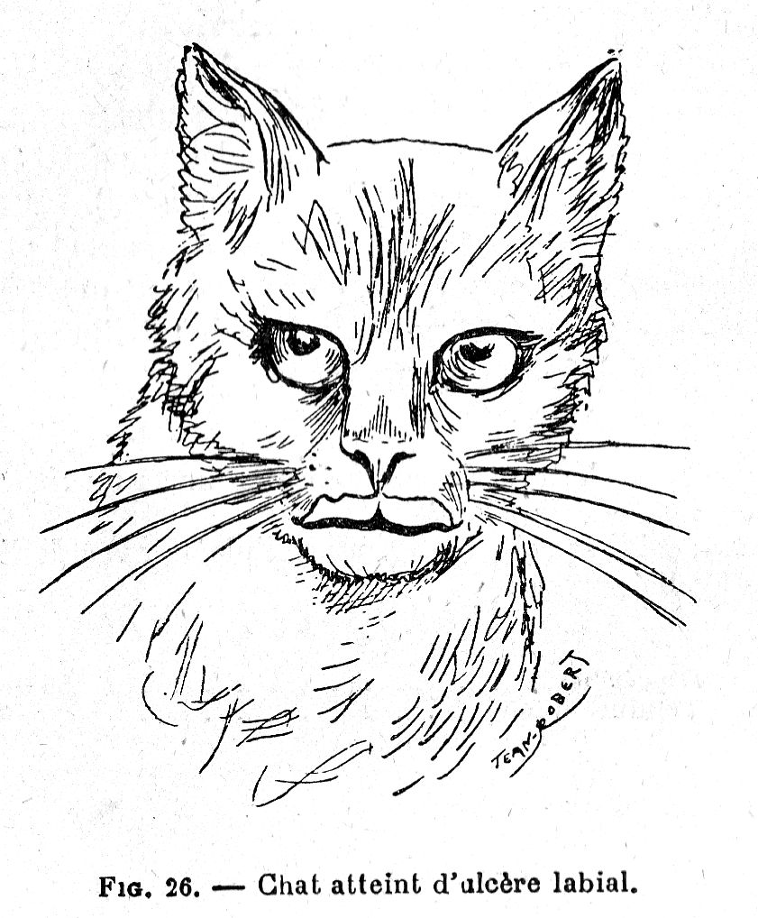
It almost always begins on the free edge of the upper lip with a small, concave, regular, rather dry greyish wound, soon accompanied by induration of neighbouring tissues; as it gradually spreads, the ulcer causes a semi-circular loss of tissue, up to 1.5 to 2 centimetres wide by 0.5 cm high and showing teeth and gum tissue; when the disease is located in the midline the nose may be affected over time. The impact on the ganglia is not very marked.
Sometimes the labial ulcer stops, recedes and heals, leaving the affected lip with a barely visible indentation; sometimes, tenacious and invasive, it slowly but constantly eats into the tissues, seeming to cause quite severe pains which lead to emaciation of animals, sometimes even death.
This condition appears to be due to irritation caused during licking by the tongue; moreover, this is how the animal can inoculate this ulcer on other parts of the body, particularly on the parts where the skin is thin (belly and inner thighs). The disease, contagious to other subjects (Cadiot, Gilbert and Roger), the necrosis bacillus often seems to be recognized as an active agent. It is not transmissible to humans. The prognosis is still serious.
Isolate the patient and support him with nutritive food, especially with meat. Morning, noon and night, brush the ulcer with hydrogen peroxide or with:
Glycerine - 50 gr.
Tincture of iodine - 5 gr.
or with:
Methylene blue - 3 gr.
Borax - 5 gr
Boiled water - 120 gr.
Cautery with a silver nitrate pencil or 1 in 50 silver nitrate solution can also work well, if done gently.
DENTAL FISTULAS
According to Professor Labat, dental fistulas may be considered the primary cause of irritation of the dental pulp established by crushing a hard body, usually a bone, which the animal wants to chew.
The pulp become inflamed, causing, by continuity of tissue, alveolitis and, if infection occurs, pus production; by necrosis of the alveolar cavity this pus passes through the jawbone and then forms an abscess under the skin. This results in a narrow fistulous wound, through which abundant pus drains, and which ends up on the root of a tooth, most often a molar. The gums are often swollen at this stage and the infected tooth is sensitive to impact.
The only intervention is to extract the diseased tooth under anaesthetic, taking care to shake it in small strokes to avoid fracturing the jaw; then disinfect the wound by dabbing it with tincture of pure iodine and keep the animals exclusively on milk until completely healed.
PERFORATION OF THE PALATINE VAULT [CLEFT PALATE]
A congenital malformation, often resulting from inbreeding, resulting in the presence of an opening of varying sizes in the midline of the palate. The perforation can be wide (the palatal arch being almost non-existent) or narrow, anterior, posterior or total; the anterior perforation is by far the less serious.
This deformity causes great discomfort in swallowing, because food can pass through the nasal cavities and be come out of the nose. An operation is the only intervention possible; very delicate, it can only be attempted on narrow fistulas and the results are always uncertain.
Anesthetize the animal and secure it on its back with its jaws open. Incise the palatal mucosa lengthwise on each side of the fissure to peel the two bands thus obtained from the underlying bone, preferably with fine scissors. Sharpen the inner edge of each flap and, using silk, make a split stitch suture with a very fine needle.
Feed the animal only with milk until healing is complete.
HARE LIP
A congenital malformation which may accompany cleft palate, consisting of a crack involving one or both lips; the harelip is single in the first case, double in the other. This deformity does not in any way prevent the animal from eating. An operation very often gives excellent results.
Anesthetize the patient with chloroform and secure it on its back. Sharpen the lips of the slit with a scalpel and dissect the mucosa joining the lip to the gum; join the two edges of the slit with a few silk stitches, choosing fairly large threads. Cover the wound with collodion.
To avoid scratching and tearing of the threads, it is prudent, during the first days, to secure the front legs or to provide the patient with a collar made of a piece of cardboard with an opening just large enough to put the cat’s head through at the centre.
OESOPHAGEAL SPASM
Spasm causing a contraction of the muscles of the oesophagus that prevents the swallowing of food: it is often due to injury of the oesophagus by a foreign body (fishbone or bone splinter).
First confirm the diagnosis by catheterization, which at the same time provides information on the position of the injury. If you notice the presence of a foreign body, immediately resort to oesophagotomy; otherwise, use faradization:
Run the two electrodes of a Gaiffe device quite briskly for a few minutes along each of the oesophageal gutters.
Improvement is often instantaneous. If there are no results, give one or two teaspoons of belladonna tincture syrup or Codex potassium bromide syrup.
[Translator’s note: Faradization - therapeutic application of faradic, or induced, electrical current.]
FOREIGN BODIES
Foreign bodies can, when swallowed, obstruct the oesophagus. They can also pass through this passage without stopping there and, especially when sharp, produce symptoms of gastroenteritis which cease only when the cause is removed.
FOREIGN BODIES OF THE MOUTH
We have been consulted several times for adult cats that were not eating, which salivated profusely and whose jaw seemed paralyzed: these symptoms were always due to a foreign body making swallowing difficult or impossible.
In three cases the foreign bodies were nothing other than needles implanted in the palate and came from the mistress’s work basket. All were threaded and it is probably this peculiarity which explains their presence in the patient’s mouth: playing with needle and thread, the thread had been swallowed, dragging the needle which was then impaled in the palate. The patients were dejected and lacked any appetite; the immobilized lower jaw produced abundant saliva with a foul odour.
First make the diagnosis by examining the mouth. Then hold the jaws apart with two strong leather ties and grasp the needle with forceps.
In another case, the foreign body consisted of a ring of sheep's trachea which encircled the base of the tongue and prevented any swallowing. As a result of the swelling of the tongue, the removal of the cartilage was laborious and delicate. Prophylactic treatment in this case consists of monitoring animals fed with pieces of lamb or mutton lung.
OBSTRUCTION OF THE OESOPHAGUS
Obstruction of the oesophagus is most often caused by the swallowing of hard bodies, of varying size (bone fragments, fish bones, needles, etc.). It manifests as anxiety, difficulty or inability to swallow, profuse salivation, nausea and sometimes coughing; a clearly circumscribed and painful swelling may be found in the cervical region.
When they are small and sharp (fish bones), foreign bodies sometimes come to a stop at the entrance to the oesophagus, causing particular disorders: the animal spins, tries to vomit, has accompanying dyspnoea accompanied by shortness of breath, it paws at its mouth as if wanting to tear off the body that bothers him. In some cases, foreign bodies can compress the base of the tongue, producing a very marked swelling and becoming embedded there, as in the case of a cat whose tongue had passed between two rings of a poultry trachea and which could no longer feed.
When the foreign body has stopped at the entrance of the oesophagus, its extraction can be attempted using forceps and spreading the jaws; it is usually enough to compress the base of the tongue with a spoon and push back the soft palate to see and extract the object. When it is already fully engaged in the oesophageal passage, induce vomiting by subcutaneous injection of:
Apomorphine - two to five millig.
Distilled water - 5 c.c.
If the foreign body is not ejected, administer a teaspoon of olive oil morning and evening for four days and then try to push it backwards into the stomach using an oiled catheter.
Use oesophagotomy as a last resort when possible:
Oesophagostomy. - Anesthetize the animal with chloroform and fit it with a muzzle. Secure it on the right side, head held forward, neck straight. Shave the hair at the protrusion formed by the foreign body and disinfect the skin. Incise the skin first, then the subcutaneous muscle layers over a length of 2 or 3 centimetres; part the lips of the wound, isolate the oesophagus, avoiding injuring the surrounding large vessels. Puncture it with a straight scalpel and carefully debride it on the probe; extract the foreign body using forceps. Suture the oesophagus through a few separate stitches with catgut; disinfect the wound with hydrogen peroxide and suture the skin with silk. Cover the wound with a collodized bandage.
Give only milk for the first week; add egg yolks the second week; from the third week you can start giving minced meat.
A fistula can form, most often lasting six weeks or two months.
FOREIGN BODIES OF THE STOMACH
When the swallowed foreign body has entered the stomach and is causing trouble, a gastrotomy should be performed.
Gastrotomy. - Secure the anesthetized animal in full supine position; shave and disinfect the skin of the hypogastric region. Make an incision of about 2 inches in the skin and muscles, starting at the xiphoid appendage and extending backwards on the white line. Grasp the peritoneum with forceps, puncture it with a straight scalpel and debride it on a grooved probe up to the corners of the muscular lips: the stomach presents itself by its great curvature. Draw it outside the lips of the wound, grasp it between the jaws of two Péan forceps placed 1 cm apart, incise its tunics in the gap between the two forceps and extract the foreign body. Carefully disinfect the stomach wound with warm (9‰ [ppm]) salt water; make two levels of catgut sutures, one on the mucosa, the other on the serosa. Push the stomach back into the abdomen, sew separate catgut stitches on the peritoneum, silk or Florence horsehair stitches on the muscles first, then on the skin. Cover the wound with a layer of sublimate collodion and make a dressing held in place with a bandage.
Leave the patient on milk for the first week, giving very little at a time for the first two or three days; add a few egg yolks during the following week and only start solid food (chopped meat, liver or spleen) around the third week and going very gradually.
STOMACH ULCERS
Stomach ulcers can be caused by the presence of foreign bodies, the ingestion of corrosive substances, or certain illnesses, such as tuberculosis. The patient shows symptoms of gastritis; it refuses the foods that are offered or immediately vomits any food ingested; at the slightest movement nausea occurs, sometimes accompanied by the blood-streaked vomit; foetid diarrhoea often occurs, the faeces having a blackish tint or being streaked with blood.
Completely stop giving meat and feed only milk, vegetable broths and eggs.
Vegetable broth 1:
Carrots - 80 gr.
Potatoes 60 gr.
Turnips - 60 gr.
Lentils - 30 gr.
Dried peas - 30 gr.
Dried beans - 30 gr.
Sea salt - 5 gr.
Water - 2 litres
Boil gently for two hours and strain; if necessary, make up to half a litre by adding boiled water.
Vegetable broth 2:
Carrots - 60 gr.
Potatoes 60 gr.
Turnips - 60 gr.
Parsnips - 60 gr.
Leeks - 60 gr.
Sea salt - 5 gr.
Water - 2 litres
Boil gently for three hours and strain; make up to half a litre by adding boiled water.
These broths should always be prepared in the morning for the whole day.
Calm vomiting by giving one to three teaspoons of opium syrup in a little milk each day. Fight intestinal bleeding by subcutaneous injections of:
Bonjean’s Ergotine - Three gr.
Sterilized glycerine - 16 gr
Distilled water - 16 gr
Inject 0.25 cc to 0.5 cc.
and calm diarrhoea by giving:
Bismuth sub-nitrate - 5 gr.
Quince syrup - 50 gr
Mint water - 100 gr.
One to three teaspoons each day.
or by injecting under the skin:
Ordenine Sulphate - Ten Centigrams
Boiled water - 5 tsp.
For one injection.
Take intestinal antiseptics at the same time:
Benzo-naphthol – 0.01 gr
Baking soda – 0.50 gr
Per dose. 20 doses. One packet morning and evening in a little milk.
GASTROENTERITIS
Inflammation of the lining of the stomach and intestines caused by the ingestion of indigestible or too abundant food, irritants or foreign bodies; it can also be due to food poisoning caused by spoiled food or complications of an infectious disease.
The patient becomes somewhat dejected and restless, gets up frequently, complains; it no longer eats, it tries to drink often, vomits; the breath becomes foul, the mouth dry; constipation is noted first, then diarrhoea, and then exaggerated arching of the loins is observed; the belly is hard, retracted. In some cases there is ulcerative stomatitis with perforation of the cheeks or partial or total gangrene of the tongue. Certain severe forms may be complicated by nervous, cerebral or spinal cord symptoms and individuals may succumb to a coma or seizure.
Combat vomiting by eliminating all solid food and feeding only boiled milk with the addition of one third of Vais water (Source Saint-Jean) or baking soda:
Baking soda - 1 gr
Per dose. 10 doses. One dose per day.
Give, before each meal, a teaspoonful of:
Soda citrate - 1 gr
Distilled water - 50 gr
then, when the vomiting has subsided, give some vegetable (See p. 151) or cereal broth:
Cereal and vegetable broth:
Dried peas - 20 gr
Dried white beans - 20 gr
Lentils - 20 gr
Pearl barley - 20 gr
Cracked corn - 20 gr
Sea salt - 5 gr
Water - 2 litres
Let it boil for three hours and strain, then add the boiled water to make up to half a litre.
Cereal decoction:
Wheat - 20 gr
Barley - 20 gr
Oats - 20 gr
Rye - 20 gr
Corn - 20 gr
Water - 2 litres
Boil for three hours, strain through several layers of gauze and make up to half a litre by adding boiled water if necessary.
These broths, like the vegetable broths, should be prepared every day.
Then give a teaspoonful of fine flour dissolved in a little water to is added 3 drops of tincture of iodine; as soon as the stomach can take it, present the patient with small fragments of fresh sheep lung or liver, lightly grilled.
Monitor regular bowel movements. If there is constipation, give a small enema using a rubber enema bulb:
Glycerin - V drops
Boiled water - 5 cc
For an enema
or give:
Castor oil - 5-10 grams
Combat diarrhoea by adding a pinch of bismuth sub-nitrate to the milk in the morning and evening, or by giving morning, noon and evening, or a teaspoon of:
Bismuth sub-nitrate - 5 grams
Sydenham's Laudanum - Twenty-five drops
Julep [rosewater] gum - 150 grams
or a lacteol tablet in a little milk. If the diarrhoea, which is abundant, does not change, inject under the skin at once:
Ordenine Sulphate - Ten Centigrams
Boiled water - 5 cc
Support the patient's strength by injecting into the subcutaneous connective tissue, every day or every other day, 10 ccs of caffeinated physiological serum made up as follows:
Sea salt - 0.75 gr
Caffeine - 0.10 gr
Soda benzoate - 0.10 gr
Boiled water - 100 cc
Give three or four teaspoons of an infusion of tea or coffee during the day. Combat adynamia [lack of strength due to illness] with daily injections of:
Camphor - 5 gr.
Sterilized olive oil - 50 gr
Inject 1 cc each time under the skin.
or:
Camphor - 1 gr.
Ether - 10 gr
Sterilized olive oil - 10 gr
Inject 1 cc each time under the skin.
If the abdominal pains are very strong, combat them by giving every three hours
Chloroform - Four gr
Glycerine - 60 gr
A teaspoonful in a little water.
Pay attention to the diet and only gradually return to a normal diet.
ENTERITIS
Inflammation of different parts of the intestine, most often parasitic in origin (intestinal coccidiosis and lambliosis [giardiasis]) or microbial, resulting in more or less abundant liquid excreta, sometimes streaked with blood (dysentery); the belly is hard, tense, painful; the patient utters dull complaints, the stools are foetid, and fever and depression are often noted.
When the animal presents a very acute crisis with frequent completely liquid, foetid stools, administer:
Ordenine Sulphate - Ten Centigrams
either in a meatball if appetite is present, or by injection under the skin (in which case dissolve it in 5 ccs of boiled water). When the stools have become more or less normal, give a lacto-bacillin or lactéol tablet morning and evening, in a little sweetened milk. Keep the patient warm; give milk diluted in half with rice water as long as the diarrhoea persists and continue with rice pates (rice pudding).
Rice water. - Put two tablespoons of rice in half a litre of cold water; when the grains are well swollen add half a litre of boiling water and boil for twenty minutes. Pass through a fine cloth and collect in a container sterilised with boiling water and well stoppered. Rice water should not be stored for more than twenty-four hours.
Or:
Bismuth benzoate - 5 gr
Ratanhia powder - 4 gr
For 15 doses. One morning and evening in a meatball.
If diarrhoea persists, take:
Laudanum - Three to five drops
Medications that can also give very good results include raw egg white (one teaspoon every three hours), charcoal powder (half a teaspoon in a little water), 1% saccharin solution (one teaspoonful in the morning).
Combat weakness with subcutaneous injections of physiological saline (10 ccs every other day).
Then avoid indigestible, coarse foods and overly large meals,
INFECTIOUS DIARRHEA OF YOUNG CATS
Always due to umbilical infection, it is accompanied by inflammation, often suppurative, of the umbilical cord; it is extremely serious, the kittens may succumb in a few hours.
The only precaution to be taken, when possible, is to disinfect the cord at birth by touching it with tincture of iodine. If diarrhoea appears give morning, noon and evening a tablet of lactéol in a little sweetened milk.
CONSTIPATION
Common in the elderly, it is often due to lack of exercise or to disorders of the digestive tract; it can also accompany febrile illnesses.
The stools, hard and scarce, are compressed and their expulsion requires prolonged and painful efforts. Over time, they may form a hard body in the large intestine and rectum, noticeable on palpation and rectal exploration (coprostasis); defecation then becomes almost impossible. There is sensitivity in the stomach, the animal no longer eats, its gait becomes more and more difficult and it starts vomiting. When the coprostasis is dependent on a mechanical obstacle blocking the evacuation of excreta, such as the backflow of the intestine by a callus below the spine, the kidneys, ureters and bladder can also become compressed by the intestine and distended by the pressure of the urine which can no longer flow; in this case, symptoms of uraemia quickly appear caused by the retention of toxic products in the body.
For a few days, give morning, noon and evening, a teaspoon of a mixture of equal parts castor oil and salad oil, or in a meatball:
Calomel 0.10 gr
Scammony - 0.10 gr [Convolvulus scammonia, a purgative]
Per dose; 10 doses. A dose morning and evening.
Using an enema bulb, administer a copious daily enema of marshmallow water or lukewarm flaxseed water to which 5 grams of glycerine is added. Boil 30 grams of marshmallow root or flax seeds in one litre of water for a quarter of an hour.
Apply hot compresses under the stomach. If necessary, try to evacuate the rectum with your finger or using a blunt curette.
Remove the meat and substitute a refreshing diet: clear mash, vegetable broth, prune juice, milk.
LIVER CIRRHOSIS
Due to the encroachment of sclerous tissue in the, this can result from either small whitish pea-sized nodules, caused by a parasite, or from chronic inflammation of the bile ducts accompanied by diffuse hypertrophy of surrounding connective tissue, caused by chronic intoxication or heart damage.
Patients generally lose weight and gradually weaken, there is oedema of the limbs, ascites and sometimes jaundice; when the liver is enlarged, it can be detected on palpation, on the right side, behind the last rib and towards its upper region.
Give the patient milk diluted in half with Vals water, and administer:
Calomel – 0.02 gr.
Powdered sugar – 0.50 gr.
Per dose. 25 doses. A dose morning and evening in a little milk.
JAUNDICE
Characteristic symptom is yellow colouration of the mucous membranes and sometimes of the skin due to the passage, in the blood, of bile pigments: it is most often a complication of an infectious disease, gastroenteritis or distemper.
The affliction begins most of the time with lack of appetite; then vomiting occurs, accompanied by intense thirst, the urine is dark, the stools discoloured; constipation develops. The patients are depressed, drowsy, lose weight quickly and eventually succumb in a coma.
Keep the patient warm and only give him, as food, boiled milk cut in half of Vals water, if necessary by dessert spoonfuls or by mouth. Each day, give:
Calomel – 0.05 gr
Powdered sugar – 0.50 gr
Per dose; 15 doses. A dose in the morning in a spoonful of milk.
and inject under the skin:
Physiological serum - 10-20 grams
CHRONIC CHOLECYSTITIS
Inflammation of the gallbladder which may be produced by partial obstruction of one of the ducts, usually the common bile duct, and over time results in enlargement and sclerosis of the liver.
There is an increase in the volume of the abdomen, progressive slump and weight loss, very pronounced jaundice of the mucous membranes and skin. Death is the almost inevitable ending.
If this condition is suspected, immediately put the animal on a completely milk diet and administer urotropine:
Urotropin - Five grams
Distilled water - 120 gr
One to two spoonfuls per day.
LIVER PARASITES
Embryos of Ollulanus tricuspis can be found in the liver, and in the gallbladder and bile ducts, fluke (Distoma truncatum and D. lanceolatum) and Amphistoma truncatum. Weight loss, weakness, and symptoms of anaemia and cachexia are often noted, but the true cause is usually not recognized until autopsy.
DISEASES OF THE PANCREAS
Invasion of the pancreas with sclerotic tissue or pancreatic cirrhosis may accompany hepatic cirrhosis; it results in a fluctuating swelling in the hypochondrium and the right epigastrium and in progressive cachexia leading to the death of the patient over time; it is most often due to the invasion of the pancreatic ducts by a fluke.
Inflammation of the pancreas or pancreatitis can accompany fatty degeneration of the liver and kidneys and is almost always due to microbial or parasitic infection. In addition to the above symptoms one may encounter fine droplets of fat in the faeces and glycosuria.
Diseases of the pancreas are seldom recognized during the animal’s life. Put the animal on a milk diet and give intestinal antiseptics:
Benzo-naphthol – 0.01 gr
Bicarbonate of soda – 0.50 gr
Per does; 20 doses. A dose morning and evening in a little milk.
STEATONECROSIS
Fatty necrosis resulting in patches of a whitish, dry, very dull appearance, comparable to candle stains, varying from the size of a pinhead to that of a 0.50 franc coin which may appear on the omentum, mesentery and peritoneum. These are due to the digestion of fat by pancreatic juice leaking into the peritoneum.
This condition, which is encountered in very obese subjects, is most often accompanied by changes in the pancreas and often in the liver. There may be some abdominal pain, lack of appetite, depression, fever, sometimes a little lethargy.
Any treatment is illusory, even when the condition can be suspected, which is rarely.
ACUTE PERITONITIS
Most often due to tumours, trauma or tuberculosis of the peritoneum, inflammation of this serosa can also be a complication of metritis.
It causes depression with loss of appetite, vomiting, abnormal tenderness of the stomach; walking is slow, painful, the loins become stiff; one may also notice a more or less abundant effusion in the abdominal cavity, sero-fibrinous, haemorrhagic or purulent, causing a deformation of the belly and being able, by its volume, to produce dyspnoea.
Apply warm compresses under the stomach, frequently renewed. Give morning and evening, in a little milk:
Foxglove leaf powder - 0.05 gr
Calomel - 0.05 gr
Per dose; 8 doses. Stop after eight days and then resume.
Puncture the abdomen (paracentesis) if the effusion is too abundant.
Paracentesis. - Cut and shave the hairs on the lower part of the side, a little behind the umbilicus and in the vicinity of the white line; disinfect the area. Quickly implant a small trocar or hollow needle while limiting penetration with the index finger; remove the mandrel and slowly drain the liquid. Remove the cannula or needle, cover the wound with collodion and apply a cotton ball.
If the liquid is clear, inject 2 - 5 cubic centimetres under the skin (auto-serotherapy).
Support the patient by injections of camphor oil at 1 part in 10 (2 - 5 cc per day).
ASCITE
Ascites or dropsy of the peritoneum is most commonly caused by a chronic condition of the heart, lungs or liver. The belly is distended, drooping, fluctuates on palpation, and the limbs are often swollen; sometimes the enlarged liver is easily perceptible. Paracentesis usually results in sero-fibrinous fluid; much more rarely one can have a milky, alkaline liquid, resembling chyle (chylous ascites).
Rule out tuberculosisby injecting tuberculin.
Boost the energy of the heart with daily injections of 1 in 10 camphor oil (2 - 5 cc). Administer diuretics for five days: digitalis wine composed (X to XX drops per day), or squill wine (1/2 to 2 teaspoons per day), tincture of digitalis (V to X drops per day). Stop ten days and start again in the same way. Also take:
Calomel - 0 gr. 05
Powdered sugar - 0 - 50
Per dose; 10 doses. A dose in the morning in milk
When there is difficulty in breathing, perform paracentesis, far enough back to avoid injury to the liver.
INDIGESTION
A sudden and generally transient disorder of the digestive function, appearing for a variable time after a meal, caused either by the ingestion of too much food, poor quality food, or by paralysis of the digestive viscera. The condition is usually mild due to the ease with which the cat can vomit.
Symptoms are uneasiness, agitation, some restlessness, then symptoms of colic appear: the patient stamps its feet, lies down, gets up, makes complaints.
Administer by subcutaneous injection:
Apomorphine – Two - five milligr
Boiled water - 5 cc
to induce vomiting, or give:
Syrup of ipecac - 30 gr.
A teaspoonful every quarter of an hour until effective.
Keep the animal warm and, for a few days, give only milk; then monitor its diet.
If the colic is too severe, calm it down by giving:
Chloroform - Two gr
Glycerin - 30 gr
A teaspoonful every three hours in a little sweetened water.
POISONING
Common in cats due to its voracity, it is often caused by the ingestion of mice poisoned by arsenic or phosphorus, which form the basis of products used to destroy rodents, or meatballs containing strychnine. In all poisonings, you must first induce vomiting by injecting:
Apomorphine - Five milligr.
Boiled water - 5 cc
under the skin, then give skimmed milk as the exclusive food for a few days. If the poison is unknown, give a teaspoon of Jeannel’s antidote every quarter of an hour, consisting of a mixture of equal parts of the following two solutions:
Solution n ° 1
Iron sulphate - 60 gr
Distilled water - 250 gr
Solution n ° 2
Calcined magnesia - 15 gr
Distilled water - 250 gr
If the poison is known, use the specific antidote.
1. Arsenic. - The animal presents with profuse salivation, colic, very great weakness and finally paralysis.
Every quarter of an hour, give a teaspoon of albuminous water (3 beaten egg whites per half litre of water) or Jeannel's mixture.
2. Phosphorus. - The mucous membrane of the mouth becomes swollen, the thirst is intense; vomiting occurs and produces material that may phosphoresce in the dark and present an onion-like odour; jaundice can occur; after some time, tremors and great weakness of the hindquarters appear.
Administer a teaspoonful of:
Iron sulphate - 0 gr. 50
Distilled water - 100 gr
Remove milk from the diet and do not use oils as a purgative.
3. Strychnine. - As the ingested dose is generally very strong, symptoms occur suddenly. There is anxiety, discomfort, then extreme sensitivity, general rigidity, stiffness of the ears and limbs; the animal falls and seizes.
If we can intervene early enough, inject 0.5 - 1 cc of the following solution under the skin:
Atropine Sulphate – Five milligr.
Morphine hydrochloride - Ten centigr.
Distilled water - 20 cc
or:
Potassium iodide – 0.50 gr
Boiled water - 1 cc
For an injection.
Give a teaspoon of chloral syrup every hour.
4. Mercury. - May be caused by using calomel as a purgative and applications of mercury ointment. There is profuse salivation first, with foul breath; then soon the teeth loosen, ulcerations appear on the inside of the lips and cheeks, and a rash may even occur; there is stupor, tremors and, finally, paralysis.
Every two hours, give a teaspoonful of an emulsion of 3 egg whites beaten to stiff peaks in 200 grams of milk.
5. Carbolic acid. - The cat can be poisoned by very low doses of carbolic acid, so this antiseptic should be absolutely forbidden at home. The appetite decreases, then disappears completely; death is preceded by convulsions lasting several hours and affecting the muscles of the extremities, trunk and head, during which the flexors and extensors come into play alternately.
Administer a teaspoonful of albuminous water every quarter of an hour. Inject under the skin, morning and evening:
Camphor oil at 1 p. 10 - 0.5 cc
Put the animal on a milk diet, which will continue for several days after recovery, and gradually return to the usual diet.
6. Benzo-naphthol. - Cats are particularly sensitive to benzo-naphthol. At a dose of 10 centigrammes per kilogramme, naphthol fatally kills animals; doses of 5 centigrammes per kilogramme cause very frequent sneezing, salivation and tearing, so that 1 centigramme per kilogramme should hardly be exceeded as the therapeutic dose; moreover, since the saliva does not contain naphthol, the hypersecretion appears to be due to reflex movement and not to the elimination of the poison by the glands.
Twenty to thirty minutes after ingestion of benzo-naphthol, very profuse salivation is observed; then there is intense tearing and frequent sneezing. The urine, turns from light yellow to brownish-yellow, then brown, but there is no haemoglobinuria. The excrement is soft, blackish, without bad odour. Breathing becomes painful towards the last moments and death, which is relatively slow, occurs in seven to twelve hours.
Contrary to popular belief, naphthol, which must be regarded as a poison of the nervous system, is not an anthelmintic in cats because it kills cats before killing the parasites.
HELMINTHIASIS (WORMS)
We find, in the stomach or the intestine of the cat, a certain number of worms capable of causing in the long run seizures, retraction of the abdomen, progressive weight loss, irregular appetite, itching around the anus causing the animal to scratch, alternating diarrhoea and constipation, sometimes vomiting.
In the stomach we may find:
The robust gnathostome (Gnathostoma robustum), a small cylindroid worm, averaging 15 to 25 millimetres long, enclosed in small tumours that communicate with the stomach cavity through a narrow opening.
The three-pointed ollulan (Ollulanus tricuspis), a nematode of about 1 millimetre, seated in the stomach lining which appears red, soft and bruised; this worm produces embryos, some of which will encyst in the pleura, liver, diaphragm and lung, forming small tumours, while others, being expelled with the faeces, are ingested by small rodents and encyst themselves in their muscles. This is how cats can become infected by eating mice.
Spirura gastrophila and Spirocerca sanguinolenta have also been found on several occasions.
In the small intestine we can see: Whiskered roundworm (Ascaris myxtax), especially frequent in young cats, a roundworm with a curved head and whitish body, 4 to 10 centimetres long and which, by curling up, can sometimes obstruct the lumen of the intestine; it can also go up into the stomach and cause intestinal perforations.
The oxyuris compar, a cylindroid worm 8 to 65 millimetres long [pinworm].
The trigonocephalic hookworm, also called uncinar, dochmia (ankylostoma trigonocephala), 10 to 20 millimetres long, has a whitish or reddish body, cylindrical and slender; by feeding on blood, it determines pernicious anaemia in cats.
The fairly common thick-necked tapeworm (Taenia crassicolis) is a flatworm, 35 to 60 centimetres long, with a large head continuing without constriction by and equally wide neck, the last rings measuring 8 to 10 millimetres long by 5 to 6 wide; it comes from the fasciolar cysticercus (cysticercus fasciolaris) which inhabits the livers of rats, mice, voles, water rats, etc. It can cause very serious epidemics as was observed in 1874 in the Black Forest.
The elliptical tapeworm (Taenia elliptica), rarer than the previous one, is 10 to 30 centimetres long and 3 millimetres wide.
The echinococcal tapeworm (Taenia echinococcus), very difficult to identify because it sinks into the mucosa, is only 3 to 4 millimetres long and usually has only four rings. The fairly frequent presence of this worm in cats, reported by Barbagallo, indicates that cats should be prevented from entering slaughterhouses since they can serve as agents of transmission of hydatid cysts to humans.
The botriocephalic tapeworm (Botriocephalus felis) is a flat worm, with a broad head, flattened and lanceolate, 15 to 22 centimetres long, whose eggs, expelled with faeces, will hatch in water producing embryos which, absorbed by fish, can then infect cats; this worm does not usually cause illness in cats.
Treatment varies depending on whether roundworms or tapeworms have been found; but, in all cases, it is essential, before administering an anthelmintic, to put the animal on a diet for at least twenty-four hours and to purge it the day before.
ROUNDWORMS. - Administer over four or five days:
Santonine - 2-5 centigr.
Sugar - 5 gr.
Per dose; 5 doses. A dose each day in a little milk.
(Santonin can also be given in biscuit or tablet form; each tablet contains one centigramme of santonin.)
or give every morning, for three days, in jelly or sweetened milk:
Artemisia powder - 1-2 centigr.
or :
Chenopodium essence - 15-20 centigr.
Take an elastic pill filled with the essence (it usually contains 30 centigrammes, pierce it and run the necessary amount against the animal's palate or tongue.
Give only milk and, three hours after the last dose, give:
Castor oil - 10-20 grams.
For pinworms, complete the treatment by giving a cold enema morning and evening with 20% vinegar water.
Combat hookworms by first giving:
Calomel - 10 centigr
Scammony - 10 centigr
and, twenty-four hours later:
Thymol - 0.25 gr
Per dose. 4 doses. Give each dose an hour apart in a hot infusion.
Two hours after the last dose, purge with:
Calomel - 0.05 gr
Sugar - 0.50 gr
in a little milk.
TAPEWORMS, - Administer:
Ethereal extract of male fern - 0.50 gr
Chloroform - One gr.
Castor oil - 10-20 grams
Four times, an hour apart.
or :
Ethereal extract of male fern - 20-50 centigr.
Mint syrup - 5-10 grams
In two instalments, two hours apart.
Purge four hours after the last dose with castor oil.
If the same patient presents with both roundworms and flatworms, we can give:
Ethereal extract of male fern - 0.50 gr
Chloroform - One gr
Simple syrup - 10 gr
Mint water - 40 gr
Four times, an hour apart.
Then purge as above.
RESPIRATORY SYSTEM
CORYZA
Coryza or inflammation of the nasal mucosa is most often caused by cold or humidity. It results in frequent sneezing, an abnormal tenderness of the mucous membrane causing the patient to rub or scratch his nose, the production of discharge, first serous, then mucous, finally mucopurulent; breathing may be hampered by the narrowness of the nasal cavities and the arrangement of the turbinates which make it difficult for phlegm to flow and can even cause obstruction of these cavities. Although this condition is generally mild, however, when neglected it can develop into a chronic state.
Clean the nostrils morning, noon and evening with lukewarm boric water:
Boric acid - 40 gr.
Boiled water - 1 litre
and coat them each time with mentholated petroleum jelly:
Menthol - 1 gr
White Vaseline - 60 gr
or guaiacolea:
Guaiacol - 2 gr.
White Vaseline - 60 gr
If there is marked respiratory discomfort, thin the secretions by giving steam inhalations:
Pour a teaspoon of one of the following solutions into a container placed in the patient’s room and containing 250 grams of boiling water:
Eucalyptus tincture - 30 gr
Benzoin tincture - 30 gr
Alcohol at 60 proof - 30 gr
or:
Eucalyptol - 3 gr
Menthol - gr
Guaiacol - 10 gr
Cinnamon tincture - 15 gr
Benzoin tincture - 90 gr
Place the patient's head over the container and cover it with a towel.
or either by giving morning and evening a teaspoonful of syrup of ipecac, or by injecting under the skin 1 cc of
Apomorphine - Five centigr
Distilled water - 10 gr
ANGINA [THROAT INFECTION]
A usually acute disease of the upper respiratory tract characterized by inflammation of the mucous membranes of the back of the mouth (pharynx and larynx) and the lacrimal mucosa; it sometimes complicates coryza and is generally recognized as caused by distemper [cat flu], changes in temperature (cold and especially humidity), irritation of the mucous membrane by vapours or fumes; finally, angina can complicate various infectious diseases.
There is a nagging cough, first dry, then phlegmy, easily brought on by the arrival of cold air; there is often abnormal tenderness in the laryngeal area (laryngitis). Soon, a foamy discharge appears and swallowing becomes more difficult (pharyngitis). In a few cases the appetite wanes and disappears while there is low fever, depression and lethargy. The condition, when left untreated, can progress to a chronic state: the cough becomes strong, hacking, discharge is less profuse and often even intermittent.
Keep the patient warm at all times and give him lukewarm drinks. Wrap the neck either with a hot compress that is renewed every two hours and held in place by a flannel tie, or with a cotton dressing attached in the same way. Give benzoin and eucalyptus inhalations (See p. 170) and give:
Tolu syrup - 60 grams
Half a teaspoonful morning and evening.
If the condition becomes chronic administer:
Potassium iodide syrup - 120 grams
A teaspoonful morning and evening.
Preferably feed with hot milk as long as swallowing remains difficult.
DIPHTHERIA
Also called Malignant Angina, it is characterized by the presence of false membranes in the back of the throat (larynx and pharynx); always of microbial origin and often caused by Staphylococcus albus, it does not appear to be transmissible to humans.
The animals suddenly show very poor appetite and great prostration; they lie down, complaining whenever you want to move them; their hair becomes dull and prickly, the gait is shaky, the pupils dilated; a little discharge appears in the nostrils, the breath becomes foul and some vomiting may occur. The inflamed soft palate is red with whitish membranes in the pharynx and larynx.
Keep the patient in a heated room and give only hot milk. Give benzoin and eucalyptus inhalations (See p, 170) and touch the back of the throat with:
Methylene blue - 1 gr
Glycerine - 50 gr
Coat the nostrils with mentholated petroleum jelly at 1 p. 60.
ACUTE BRONCHITIS
Caused by inflammation of the large bronchi, it is often due to a chill, but can also be due to infection. The patient becomes sad, dejected and seeks cold water; it presents with a low fever with accelerated respiration and circulation, then develops a strong, dry cough, a mucous or mucopurulent discharge; auscultation denotes a harsh respiratory murmur. In capillary bronchitis, where the inflammation extends to the bronchioles, Respiratory discomfort and fever are more pronounced, the cough is painful, profuse discharge and noise from the lips is observed, due to the fact that breathing, instead of taking place through the nose, is done through the mouth.
Leave the patient in a warm room and cover them as much as possible. Have them take, morning and evening, a teaspoonful of:
Soda benzoate - 1 gr
Diacode syrup - 40 gr [diacode syrup = weak opium syrup]
Tolu syrup - 80 gr
or:
Terpine - 4 gr.
Mineral kermes - 5 gr
Gum arabic - 20 gr
Simple syrup - 50 gr
Distilled water - 100 gr
If, from the start, the problem appears to be very pronounced give an emetic:
Ipecac syrup - 30 gr
A teaspoonful every quarter of an hour until vomiting occurs.
When the cough is frequent give a teaspoonful, morning and evening, of:
Diacode syrup - 20 gr [weak opium syrup]
Tolu syrup - 100 gr
Cherry laurel water - 2 gr
or:
Potassium iodide - 3 gr
Mineral kermes - 3 gr
Codeine syrup - 20 gr
Simple syrup - 100 gr
Combat weakness with alcoholic coffee or tea infusions (one teaspoonful of brandy or rum per cup).
CHRONIC BRONCHITIS
This sometimes follows acute bronchitis, it is often tuberculous in nature. It results in a phlegmy cough, occurring in fits and a mucopurulent discharge; auscultation of the lung indicates the presence of mucous and wheezing rattling.
Rule out tuberculosis by injecting tuberculin. (See p. 237.). Give abundant food (milk, raw meat) very freely. Modify bronchial secretions by administering:
Terpine - 1.10 gr
Per dose; 10 doses. A dose morning and evening in honey, jelly or in a ball of minced meat.
or:
Potassium iodide syrup - 100 grams
A teaspoonful morning and evening.
If there is a strong coughing fit, give:
Strontium bromide - 0.50 gr
Diacode syrup - 40 gr [weak opium syrup]
Tolu syrup - 80 gr
A teaspoonful morning and evening.
Give cod liver oil (two teaspoons per day) and Fowler's liqueur (I to II drops morning and night in a little milk).
BRONCHO-PNEUMONIA
Often infectious in nature and follows cat flu, it can also be caused by the accidental arrival of foreign bodies (food, drugs) in the bronchi and lungs; it can also be caused by cold and humidity and can complicate capillary bronchitis.
It begins with depression; the animal is weak, its appetite decreases, its thirst becomes unquenchable; there is a dry cough, infrequent, accelerated circulation and respiration with a noticeable rise in temperature; the nose is hot. After a short time breathing becomes more difficult, there is a viscous, bloody discharge; percussion detects dullness, auscultation the presence of mucous rales and sometimes a tubal murmur. Complications of jaundice or enteritis can occur, and death almost always occurs from asphyxia or uraemia.
Protect patients from cold and humidity; treat them as soon as they show the first symptoms of coryza or angina.
As soon as the disease is diagnosed, make a fixation abscess by injecting, after cutting the hair, 0.25 cc of turpentine under the skin, at the level of the sternum. Apply iodine tincture to the sides of the breasts and against the direction of the fur or wrap the chest in wet compresses that are repeated every three hours. Inject every day under the skin 1 cc of camphor oil at 1 p. 10 or the following solution:
Caffeine - 1 gr
Soda salicylate - 1 gr
Distilled water - 5 cc
Dissolve, then add:
Camphorated alcohol - 5 cc
Fight the infection by injecting 0.5 to 1 cc of collargol or electrargol under the skin, or 5 ccs Leclainche and Vallée polyvalent serum or polyvalent antistreptococcal serum.
Morning and evening, give a teaspoonful of:
Terpine - 4 gr.
Kermes - 5 gr
Gum arabic - 20 gr
Simple syrup - 50 gr
Distilled water - 100 gr
or:
Soda benzoate -1 gr.
Diacode syrup - 60 gr [weak opium syrup]
Tolu syrup - 60 gr
Support the sick with raw meat, milk, slightly alcoholic tea and coffee infusions; if the appetite has completely disappeared, give the following as food enemas:
Milk - 100 gr.
Peptone - 20 gr
Egg yolk - 1
Laudanum - Five drops
After first emptying the rectum with a warm water enema.
PNEUMONIA
Very rare in cats, straightforward pneumonia is always brought on by a chill. We note depression with fever, loss of appetite, unquenchable thirst, accelerated breathing, painful, sticky cough; then mucous discharge appears, streaked with blood (rusty discharge); percussion finds dullness on one or both sides, auscultation finds crackling breaths with, after a few days, the production of a tubal murmur. Pneumonia can be complicated by pleurisy (pleuro-pneumonia) and sometimes endocarditis.
Indicated treatment is the same as for bronchopneumonia.
ASTHMA
Ordinary asthma is a neurosis characterized by dyspnoea, mainly manifested by great difficulty in breathing; nasal asthma, due to abnormal irritability of the nasal mucosa, is caused by the retention of phlegm in the turbinates and is characterized by repeated sneezing with bouts of dyspnoea consisting mainly of a series of deep, convulsive breaths.
Calm attacks of ordinary asthma by making the patient breathe a few drops of pyridine poured into a saucer or better onto the corner of a handkerchief, or by giving a teaspoon of the following every two hours:
Ether - 2 gr
Diacode syrup - 20 gr [weak opium syrup]
Orange blossom syrup - 20 gr
Simple syrup - 120 cc
until the seizures disappear. Ten days a month, give a teaspoonful of potassium iodide syrup every morning before a meal. For the next ten days, give I to II drops of Fowler's liqueur in a little milk on an empty stomach.
Fight nasal asthma by administering:
Ipecac syrup - 30 gr
A teaspoonful every quarter of an hour, until effective.
or by injecting under the skin:
Apomorphine - One - two milligr
Distilled water - 1 cc
to induce vomiting. Then facilitate the expulsion of phlegm by causing sneezing by light inhalations of ammonia.
Pour a few drops on a cloth and bring it carefully to the nostrils.
PLEURISY
Inflammation of the pleura is sometimes caused by prolonged cooling, but most often by various infectious states (distemper, tuberculosis, etc.); it can be unilateral or bilateral.
The animal, depressed or weakened, presents with a very marked rise in temperature with chills, intense thirst, decrease or loss of appetite, more or less marked respiratory discomfort, small, dry, painful coughs, abnormal sensitivity of the ribs, dullness in the lower regions of the chest demarcated by a horizontal line and shifting when the patient is standing on his feet, rapid heartbeat, small pulse.
Confirm the presence of the exudate by an aseptic puncture made with a hollow needle and Rule out tuberculosis by an injection of tuberculin. Rub iodine tincture against the direction of the fur on each side of the chest, and at the same time perform a fixation abscess:
Inject 0.25 cc of turpentine under the skin.
Take each day, in a little honey or jelly:
Calomel - 0.05 gr
Per dose; 6 doses.
and stop as soon as diarrhoea appears. Support the heart by injecting morning and evening 1 cc of:
Camphor - 2 gr.
Ether - 18 gr
If the dyspnoea is intense perform thoracentesis,
Thoracentesis. - After shaving and disinfecting the area, puncture aseptically between two ribs, at the 6th or 7th intercostal space, limiting the penetration of the needle with the index finger. Slowly drain the liquid, withdraw the needle and cover the wound with a collodion bandage.
If the exudate is clear, inject 1 or 2 ccs under the skin (auto-serotherapy). Promote the absorption of the liquid by daily injections of pilocarpine:
Pilocarpine azotate [nitrate] - Two centigr.
Boiled distilled water - 25 gr.
Inject 1 cc under the skin each time.
LUNG PARASITES
Some parasites, by entering the general bloodstream, can enter the lung tissue and lodge there. Thus, verminous [wormy] bronchitis or verminous pseudo-tuberculosis has been reported, resulting in the formation in the lung of granulations of variable volume, up to the size of a pea, caused by the encystment of embryos of Allulanis tricuspis, a nematode that can be found, at the same time, in the adult state in the stomach mucosa.
Frequent attacks of coughing are noted, often followed by vomiting; the patient becomes depressed, thin, his hair becomes prickly; after some time diarrhoea occurs and the patient goes into a decline.
Combat diarrhoea with subcutaneous injections of:
Ordenine Sulphate - Ten Centigr.
Distilled water - 5 cc
Combat coughing with creosoted cod liver oil (A dessert spoonful morning and evening).
Healing is always very uncertain.
Feline verminous pneumonia has also been described, characterized by foci of purulent miliary [millet-like] pneumonia containing large numbers of lungworm eggs or embryos.
Same medication as for ordinary bronchopneumonia.
Finally, a cysticercus (Cysticercus Bailleti) has been found in the pleural cavity of the cat, a small worm 16 to 100 millimetres in length, wider at its anterior part where it measures 1 to 2 millimetres more than at its posterior part.
Although in some cases microscopic examination of the phlegm may detect the presence of eggs, the exact cause of the conditions is usually not diagnosed until autopsy and death is the usual outcome.
CIRCULATORY SYSTEM
As a result of its sedentary life, the cat rarely presents with cardiac afflictions (pericarditis, myocarditis, endocarditis); more often than not, the cardiac problems are complications of infectious diseases, whose prognosis they worsen.
ANAEMIA
Anaemia, characterized by a somewhat considerable decrease in blood cells, is often due to irrational feeding, insufficient food, or food consisting of foods lacking in vitamins; it can also be a consequence of abundant or repeated haemorrhages, to acute or chronic diseases or even to parasitic affections.
It results in the pallor of all mucous membranes and progressive weakening of the animals; appetite decreases, becomes irregular, and often the pulse is shallow.
First, look for the cause of the anaemia (tuberculosis, cancer, nephritis) and fight it, if possible, with appropriate treatment. If the appetite is normal, give a meat diet. If it is decreased, take:
Cola tincture - 1 gr
Gentian tincture - 5 gr
Cinchona tincture - 10 gr
Simple syrup - 100 gr
A teaspoonful morning and evening.
or:
Cinchona fluid extract - 10 gr
Kola - 10 gr
Coca - 10 gr
Rhubarb tincture - 15 gr
X drops, in the morning on an empty stomach in a little milk, for three weeks or a month.
Give a teaspoonful or dessert spoonful, depending on the patient’s size, of cod liver oil daily.
When the patient is very depressed, inject under the skin 5 ccs of physiological serum:
Sea salt - 1.50 gr
Filtered boiled water - 200 gr
and support the heart with subcutaneous injections of camphor oil:
Camphor - 1 gr
Sterilized olive oil - 10 gr
Inject 1 cc every day.
PERNICIOUS ANEMIA
Caused, as in dogs, by hookworm or Uncinaire trigonocephalic which attaches itself to the intestinal mucosa to feed on blood, but much rarer in cats, it also results in depression, decline, a dull prickly coat, pale mucous membranes; the nose leather soiled by light discharge, often bloody; finally, intense blackish diarrhoea appears, constantly staining the tail; lack of appetite, vomiting, progressive weakness occurs, and death occurs from cachexia. The diagnosis can be made with certainty by microscopic examination of the faeces which shows the presence of the parasite or its eggs.
Administer to the animal on an empty stomach:
Ethereal extract of male fern - one gr
Chloroform - one gr.
Castor oil - 20 gr
In two batches, fifteen minutes apart. Renew the medication three times at four days intervals.
BLOOD PARASITES
Surra is a fairly rare condition caused by a specific trypanosome entering the bloodstream. It first manifests as oedema of the face and neck, then as eye disorders (oedema of the cornea and lens, small haemorrhages on the iris); increasingly progressive weight loss, becoming complicated, after a certain time, by muscular weakness, which forces the animal to a permanent recumbency. The spleen is also enlarged, and death is the almost inevitable conclusion.
Leishmaniasis, also very rare, is a generalized disease, still confined to the Mediterranean coast, caused by the invasion of the organism by a protozoan (Leishmania furunculosa); it abounds in the body of fleas which appear to be the vector of transmission from animals to humans. There is progressive anaemia with the appearance of small ulcers on the mucous membranes of the mouth and nose, conjunctiva and on the skin, mainly around the eyelids, lips and ears.
The only treatment to try in both cases are injections of Trypan bleu or Novarsenobenzol:
Trypan blue - Ten centigr.
Boiled water - 10 cc
Make three injections of 1 to 2 ccs under the skin, at two day intervals.
Novarsenobenzol – Two - five centigr.
Boiled distilled water - 2 cc
For intramuscular injection. Repeat after eight days.
PERICARDITIS
Inflammation, acute or chronic, of the external serosa of the heart, most often due to the localization of toxins during infectious diseases (pneumonia, illness of young age, tuberculosis, etc.).
The acute form is manifested by weakness, anxiety, rather marked respiratory discomfort with accelerated breathing, erratic appetite; then the cardiac shock subsides sharply and a large zone of dullness appears in the heart region, indicating the extent of the effusion, with tenderness at this level. In the chronic form, which may follow on from the previous one or be established straight away, the start, at least in the latter case, are very vague; we first note weight loss, inappetence, marked paleness of the mucous membranes, lethargy, irregular breathing and a weak pulse; then the cardiac shock disappears and the percussion indicates a fairly extensive zone of dullness; there is a very distinct venous pulse with dilation of the jugulars and oedema of the limbs; finally chronic diarrhoea appears, quickly leading to wasting and death.
Whenever possible, apply daily tincture of iodine to the dull area for four or five days. For a few days, give the following in a little milk:
Calomel - 3 - 5 centigr
Sugar - 50 centigr
For a package; n ° 5.
and:
Foxglove tincture - Five gr
Simple syrup - 95 gr
A teaspoonful in the morning and evening every other week.
In the intervening weeks inject:
Caffeine - 1 gr
Sodium salicylate - 1 gr
Distilled water - Q S p. 5 cc
After dissolution add
Camphorated alcohol - 5 cc
Morning and evening, 0.5 cc under the skin,
or:
Camphor - 2 gr.
Sterilized olive oil - 20 gr
Morning and evening 1 cc under the skin.
Support the patient with milk and broth as soon as the appetite disappears.
In the chronic form additionally resort to iodides:
Sodium iodide - 2 gr. 5
Simple syrup - 150 gr
Every other week, give a teaspoonful in the morning.
MYOCARDITIS
Acute or chronic inflammation of the heart muscle, always of toxi-infectious origin.
Often unrecognized at first, acute myocarditis begins with a strong pulse and rapid breathing; then the heartbeat quickly weakens and becomes intermittent; healing may occur, but more often than not the pulse subsides and the dyspnoea increases, resulting in death. Chronic myocarditis, which can establish itself immediately, often follows the acute form, producing shortness of breath, heart palpitations and finally intermittences; death usually occurs from wasting or cardiac syncope.
In the beginning, eliminate toxins by injections of physiological serum:
Sea salt - 0.75 gr
Boiled water - 100 cc
Inject 10-15 cc of solution under the skin every day.
then support the heart with caffeine injections:
Caffeine - 2.5 gr
Soda benzoate - 3 gr
Distilled water - 25 cc
One cc, morning and evening, under the skin,
and camphorated ether:
Camphor - 1 gr.
Ether - 9 gr
Combat dyspnoea with digitalis:
Foxglove tincture - Five gr.
Simple syrup - 95 gr
A teaspoonful, morning and evening, every third week.
In the chronic form, combat lesions by administering iodides:
Sodium iodide - 2 gr.
Simple syrup - 150 -
A teaspoonful in the morning, every other week.
ENDOCARDITIS
Acute or chronic inflammation of the inner serosa of the heart, almost always caused by infectious disease.
The acute form begins abruptly with depression, prostration, lack of appetite and low fever; the heartbeat is violent and tumultuous, the pulse irregular; there is also respiratory discomfort. In the chronic form there is weakness with decreased appetite, dejection, rapid dyspnoea, irregular and variable pulse; effusions often occur in the pleura, pericardium and peritoneum. In both cases, auscultation reveals a heart murmur with doubled heart sounds.
When the symptoms are still mild, give:
Potassium iodide - 2 gr.
Simple syrup - 150 gr
A teaspoonful in the morning every other week.
When the palpitations become very strong, administer, for a week:
Potassium bromide - 2 gr.
Simple syrup - 100 gr
A teaspoonful morning and evening.
As soon as dyspnoea and effusions appear, give:
Foxglove tincture - Five gr.
Simple syrup - 95 gr
A teaspoonful in the morning and evening every third week.
When the contractions of the heart are very weak, inject:
Caffeine - 2.5 gr
Soda benzoate - 3.5 gr
Distilled water - 25 cc
One cc as an injection, morning and evening.
When the appetite decreases, support the patient's strength with milk and broth.
LYMPHADENIA
Rather rare condition of the lymphoid organs, appearing to be infectious in nature, characterized by rather great enlargement of the lymph nodes in one or more regions.
The superficial ganglia appear enormous, hard, bumpy, sometimes joined together; weight loss occurs, then weakness; the mucous membranes weep. Complications of nephritis may occur and an enlarged spleen (splenomegaly) may be found.
Give morning and evening a teaspoonful of:
Cola tincture - 1 gr.
Gentian tincture - 5 gr
Cinchona tincture - 10 gr
Simple syrup - 100 gr
and give cod liver oil by teaspoonfuls. Administer every other week, twice a day, in a little milk I to IV drops of:
Fowler's liqueur - 10 gr
Double tartrate of iron and potash - 10 gr
URINARY SYSTEM
KIDNEY ABSCESS
Complication of a previous disease, almost always of infectious origin, it is detected only by an abnormal tenderness of the area accompanied by an intense fever: the urine examination shows the presence of globules of pus. Death is the rend result.
KIDNEY CONGESTION
Usually appearing during an infectious disease, it can be caused by trauma to the area, prolonged ingestion of irritants, or cold.
Congestion of the kidneys first causes difficulty in walking; the hind limbs are spread, the back is arched; at the same time, there is accelerated breathing, and the onset of mild colic; urination is difficult although the urine emitted is very abundant (polyuria} and there is tenderness in the lumbar region. In severe cases colic persists, there is progressive weakness, the pulse weakens, and the patient succumbs.
Keep the patient at rest and warm. Only allow it to take milk mixed with an equal volume of Vichy water. If the pain is severe, apply hot compresses to the lumbar region and give a teaspoonful of:
Camphor monobromide - 1 gr.
Alcohol at 60 proof - 30 gr
Glycerine - 30 gr
ACUTE NEPHRITIS
Acute inflammation of the kidney which most often has an infectious origin (distemper, bronchopneumonia, ascites, spoiled food, etc.), but which can also be the result of trauma to the region, congestion of the kidney or poisonings.
The disease begins with depression, lack of appetite, dull colic; then the movements of the hindquarters become difficult; urination is frequent, painful, scanty, and produces thick, mucilaginous urine, sometimes red-coloured (haematuria); breathing quickens, the stomach tightens, the patient weakens and loses weight; constipation and oedema occur in declining patients. When the secretion of urine completely stops, toxic build-up in the body (uraemia) is detected by the appearance of vomiting and nervous disorders (convulsions, muscular weakness).
Keep the animal warm and put it on a milk diet; apply warm compresses to the kidney area and administer a purgative in the morning on an empty stomach:
Castor oil - 10-20 grams
Inject every day under the skin 10 ccs of physiological serum:
Sea salt – 1.50 gr
Filtered boiled water - 200 gr
Give lightly alcoholic tea or coffee infusions.
CHRONIC NEPHRITIS
Frequent ailment, developing slowly and insidiously, so that it is often not unrecognized; it is generally the consequence of a faulty diet (abuse of meat, spoiled food) leading to repeated gastroenteritis which affects the functioning of the kidneys, which, sometimes atrophied, are most often enlarged. Chronic nephritis can also be due to heart and lung conditions, or tuberculosis.
At first the symptoms are very vague; there is a more or less marked albuminuria with frequent but scanty emissions (pollakiuria [frequent daytime urination]) of urine, an accelerated, tense pulse, lassitude, a fairly great sensitivity to cold, small nosebleeds and, often, some itching ; later, the albumin level increases in the urine which also contains urine casts, the urine output becomes more abundant (polyuria), arterial hypertension is observed with enlarged heart; haematuria, retinal haemorrhages, oedema, dyspnoea, vomiting and diarrhoea then occur. Although eating well, the animal loses weight, quickly gets out of breath, has stiff loins when it moves and may even present with real lameness from the pain it feels. Death is almost always the conclusion.
Keep the patient warm and on a milk diet; give the following during the day in a little milk:
Bicarbonate of soda - 2 grams
Raise the energy of the heart by giving, during the day,
3 to 5 teaspoons of tea or coffee; add in one of them:
Tincture of strophanthus - II to IV drops
KIDNEY TUMORS
The kidneys can be the site of various tumors, carcinomas, sarcomas, etc.
Tumours can sometimes be detected on abdominal palpation, but in most cases they are not diagnosed until autopsy; they may be localized to the kidneys or arise from the generalization of tumours of other organs. In July 1901, Professor Petit presented, a very interesting observation from this point of view to the Central Society of Veterinary Medicine. A female cat, previously operated on for a mammary tumour, died shortly after the operation. At autopsy, all organs were found to be healthy except the kidneys. The left, of normal size, hidden under a thick layer of fat, contained only a few neoplastic nodules the size of a pea which protruded slightly from the capsule; on the contrary, the right kidney was affected in its entirety, had tripled in size, and appeared whitish with a lumpy surface; it was not surrounded by fat. In this case, it was a spindle cell sarcoma.
At the autopsy of a 3-year-old cat destroyed because it was visibly thinning and its belly had developed an exaggerated bulge, mainly in its upper part, Auger and Riquet found kidneys transformed into yellowish-grey masses, almost completely covering the sub-lumbar arch, the left kidney weighed 175 grams, the right 190 grams: these tumours were cancerous in origin.
One of us had the opportunity to autopsy a cat whose kidneys had been invaded by tumours similar to those seen in other organs; the animal had succumbed to wasting.
It is obvious that, under these conditions, one cannot institute any treatment, neither medical nor surgical.
CYSTITIS
Inflammation of the bladder lining may be due to neighbouring inflammations (nephritis, peritonitis, metritis), ingestion of irritants, the presence of stones, urinary retention, trauma, etc.
In acute cystitis we observe depression with inappetence, difficult or even painful urination, urine containing mucus, albumin, white blood cells and epithelial cells from the bladder, abnormal sensitivity of this organ, fever, constipation, and decreased or loss of appetite. Chronic cystitis results in the release of purulent urine and marked pain at the end of urination.
Remove meat from the diet and replace it with milk or vegetable broth (See p. 151) diluted to half by barley tea. Give daily in milk:
Bicarbonate of soda - 0.5 - 1 gr
to promote diuresis. Calm the pain of urination with:
Camphor monobromide - 10-15 centigr.
Per dose; 10 doses. One dose per day in milk.
Disinfect the bladder by giving, always in milk:
Urotropin - 10 to 20 centigr.
Salol - 10 to 20 centigr.
Soda benzoate - 5 gr
Per dose; 20 doses. A dose morning and evening, twenty minutes before the meal.
Combat constipation with lukewarm daily enemas:
Salad oil - 10 gr.
Flax seed water - 100 gr
For an enema.
or:
Glycerin - 8-12 gr.
Lukewarm boiled water - 100 gr
For an enema.
If the urine becomes purulent, catheterize the bladder using a rubber catheter (which has been boiled beforehand) coated with boric vaseline, to evacuate it and then inject the following into the bladder, to wash it :
Salicylic acid - 2 gr
Soda borate - 2 gr
Boiled water - 250 cc.
In lukewarm solution.
URINARY RETENTION
This condition is common in cities among castrated adult cats; lack of exercise and holding onto urine due to cleanliness predispose them to it, with bladder compression eventually leading to paralysis of the bladder; but various ailments (compression or narrowing of the urethra, enlarged prostate, lesions of the nerve centres), can also produce it; finally, the retention may be due to a mechanical obstacle formed either by an accidental ligature of the penis by matted hairs from the coat, or most often by a deposit obstructing the urethral canal at its end, or also by prolapse of the bladder.
The patient presents a pained expression and makes plaintive meows; he has difficulty lying down, preferring to keep the quadrupedal position to avoid contraction of the abdominal muscles; often, the body is bent in an arch to induce the relaxation of these muscles. Urination is difficult, often even impossible despite frequent efforts; the animal is anxious, walks with legs apart and shows symptoms of colic; often the appetite disappears. On palpation, the bladder appears painful when pressure is applied, distended, sometimes so hard that it could be mistaken for a tumour. The opening of the foreskin may be plugged by mucous matter containing an aggregate of hair.
First check with a catheter if the urethra is free. If there is sediment, massage the penis lightly, which often results in expulsion; then arouse bladder contractions with a few massages and by giving morning and evening, in a little milk:
Nux vomica tincture - V to X drops
If it is impossible to extract the urethral plug by massage, debride the urethra using a fluted probe or even, using a fine trocar or a hollow needle, puncture the bladder in the prepubic region after shaving and disinfecting the skin.
Perform a urethrotomy if there are stones.
Urethrotomy. - Insert a sterilized probe into the urethra to locate the blocked point. Shave and disinfect the skin at this level and make, using a convex scalpel, a 2-3 cm incision; divide the skin and underlying tissue, cut the urethral wall, and extract the stone with blunt forceps. Do not suture. Apply boricated glycerine; a fistula often forms which persists for several weeks.
Then disinfect the bladder by administering:
Urotropin - 0.20 gr
Per dose; 10 doses. One per day in a little milk.
URINARY INCONTINENCE
Involuntary evacuation of urine, which usually flows drop by drop, due to various causes: meningitis, epilepsy, lesions of the bladder, urethra, vagina, etc.
The only rational treatment is to treat the cause; when it remains unknown, give the following each day, in a spoonful of sweetened milk:
Nux vomica tincture - V to X drops
REPRODUCTIVE ORGANS
CONGESTION OF MAMMARY GLANDS
Normal at the end of gestation, it is often seen when all of the young are removed from the mother immediately after giving birth; the cat retains all the appearances of health, but one or more mammary glands are hot and swollen; pressure does not cause pain, but rather makes milk flow.
Put the animal exclusively on a milk diet. Purge it with:
Buckthorn syrup - 10 - 15 grams
and make three or four astringent lotions each day with Eau Blanche:
Liquid lead acetate - 5 gr.
Fresh water - 500 cc.
or an application of:
Spanish White[chalk] - 15 gr.
Vinegar - 10 gr
Water - 10 gr
or better, apply wet dressings with lukewarm boric water in the morning, at noon and in the evening. Remove the dressing as soon as it cools and apply the following to the entire area:
Aristol - 4 gr.
Vaseline - 20 gr
If the pain is too severe apply:
Cocaine hydrochloride - Five gr.
Vaseline - 50 gr.
Lanolin - 50 gr
When engorgement persists after heat and sensitivity have subsided, apply daily applications of iodide ointment made with Codex iodized potassium.
Keep the mammary region in a state of absolute cleanliness.
MASTITIS
Inflammation of the mammary glands, often a complication of congestion of these organs, but usually due to small wounds allowing infection of the mammary.
This becomes hot, painful, swollen and produces greyish, purulent milk on pressure; there is fever and loss of appetite. When pus forms, we find a more or less marked fluctuation at the boundary of the organ. The condition can progress to a chronic state: the glandular tissue turns into sclerous tissue and the enlarged mammary gland becomes hard and fibrous.
If the cat is nursing, remove her young, give her milk and purge her with:
Buckthorn syrup - 10-15 grams
Apply hot boiled water compresses to the mammary glands and hold them in place with a band around the chest and belly; renew them as soon as they are cold.
Monitor the formation of abscesses and puncture them as soon as the exudate is clear; clean the cavity with hydrogen peroxide and dab it with pure tincture of iodine or with:
Methylene blue - 1.25 gr
Borax - 2 gr
Boiled water - 100 gr
Protect the area with a bandage.
Although the treatment of chronic mastitis is almost pointless, one can try applications of iodine-iodide ointment:
Iodine - 1 gr
Potassium iodide - 5 gr
Water - 5 gr
Soft lard - 40 gr
MAMMARY TUMOURS
Mammary tumours are quite common, especially in the elderly. One finds benign tumours, generally constituted by fibro-adenomas and malignant tumours or cancers, due most often to epitheliomas; the former evolve slowly, remain painless and do not cause fever; the latter, on the contrary, ulcerate, lead to loss of tissue, spread to the sub-lumbar nodes and sometimes to the abdominal wall, surrounding themselves with an area of infiltration and eventually causing fatal wasting.
The only treatment is broad ablation, which should only be done when the tumour is causing significant discomfort or is accompanied by an area of infiltration.
Perform general anaesthesia. Shave and disinfect the area to be operated on and make two incisions on each side of the tumour, delimiting an elliptical flap encompassing the tumour and the infiltrated area. Dissect out the skin and the tumour, pinch off and tie the blood vessels with catgut. Explore nearby nodes and remove them if they appear affected. Suture the skin with silk or Florence horsehair and cover with a wadded bandage held in place by a body bandage, renewed after 48 hours.
VULVO-VAGINITIS
Inflammation of the vagina (vaginitis), which is often accompanied by inflammation of the vulva (vulvitis), is usually due to a difficult parturition; but it can be due to mating or the presence of tumours in the vagina.
It is manifested, in the acute state, by marked swelling of the lips of the vulva, an initially bloody discharge, then mucopurulent discharge, red colour of the vaginal mucosa; in the chronic state, the swelling of the lips is not very pronounced, but the vulva exudes a mucopurulent liquid that clumps the hairs of the lower commissure.
Remove any tumours with small curved scissors.
Combat acute vaginitis with warm injections, given morning and evening, with a solution of boric acid (30 grams per litre of boiled water) or potassium permanganate (0.50 gr per litre of boiled water). Watch for abscesses in the walls of the vagina or the thickness of the labia and puncture them. Give the following each day, in milk or mash:
Baking soda - 2 gr.
Per dose; 15 doses.
When the condition becomes chronic, give injections of:
Crystallized alum - 10 gr
Boiled water - 500 gr
Or :
Zinc sulphate - 0.5 gr
Boiled water - 500 gr
METRITIS
Metritis, or inflammation of the uterus, can be acute or chronic; it is most commonly caused by abortion, retention of a dead foetus, non-delivery and starts with infection of the vagina.
Acute metritis is characterized by a mild discharge, bloody or foetid, with depression, intense fever, tenderness of the stomach and loss of appetite. In chronic metritis, a fairly abundant purulent fluid flows and the patient loses weight quickly. In pyelometritis, the pus, which can no longer flow, dilates the uterus and thins the walls; the belly increases in size, its palpation is not very painful, and may give the sensation of fluctuation or the distended uterine horns can be felt; percussion gives a straightforward dullness; the appetite disappears, slight constipation occurs, the patient becomes thinner and eventually succumbs to wasting.
The affection is always very serious.
From the start, apply hot compresses under the stomach. Wash the uterus daily using a syringe fitted with a catheter, containing a litre of hot boiled water and fitted with a previously boiled rubber probe; end with an injection of potassium permanganate:
Potassium permanganate - 0.25 gr
Lukewarm boiled water - 250 gr
For an injection.
Build up the patient’s strength by giving teaspoonfuls of tea or coffee with a little alcohol added (one teaspoonful of cognac or rum per cup); if the depression gets worse, inject under the skin 1 tsp of:
Camphor - 1 gr
Sterilized olive oil - 9 gr
If pyometritis occurs, try catheterization followed by injection with 1 p. 2,000 potassium permanganate solution. 2,000; puncture the uterus if necessary.
Surgical treatment of metritis consisting of total hysterectomy is not advisable as it almost always has to be performed on debilitated and weakened subjects and usually results in their death.
Hysterectomy. - Leave the patient on an empty stomach for 24 hours and operate on the anesthetized animal. Shave and disinfect the area; cover it with a windowed compress.
Incise in the midline and behind the skin, muscle layers by pinching or ligating the blood vessels; grasp the peritoneum at the level of the posterior commissure of the wound, puncture it, slide a grooved probe underneath, and debride it over the entire length of the wound; disengage the uterine horns and spread them out on an aseptic towel. Ligate on the one hand the body of the uterus, on the other hand each of the ovarian pedicles; first sever the broad ligaments from front to back by pinching the blood vessels, then the body of the uterus in front of its ligature. Ligate the vessels, disinfect the uterine stump with a hot salt water solution of 8 parts per thousand, then suture it by invagination. Make three layers of suture with separate points, with catgut for the peritoneal plane and the muscular layer, with silk for the skin. Cover the wound with salol [Phenyl salicylate] or boric acid and apply a wadded dressing, held in place with a body bandage, renewed twenty-four or forty-eight hours later.
DIFFICULT BIRTH
When, during parturition, the uterus remains completely inert, its contractions can be induced by injecting under the skin, at three times, during the day, 3 ccs of pituitrine (pituitary gland extract); the action begins to be felt 10 to 20 minutes after the injection and lasts for an hour and a half.
When the cat makes futile efforts, use a speculum to perform a vaginal exploration which can make it possible to recognize the twist of one of the horns: in this case, the bottom of the vagina appears deviated, completely closed and without any folds.
As long as the kitten is still in the uterus, any direct intervention is dangerous; forceps should never be introduced into the uterus and use of this instrument should be limited to cases where the foetus is already in the vagina. If the expulsion is only slowed down and pain persists, apply light pressure with your finger on the floor of the vagina at the level of the pubic symphysis to provoke the expulsive effort. When the foetuses cannot enter the vagina, perform a caesarean operation.
Hysterotomy or caesarean operation. - Perform general anaesthesia and disinfect the area to be operated on. Incise the right flank, equidistant from the last rib and the external angle of the ilium, as for laparotomy. Extract the uterine horn from the abdomen, cut longitudinally through the serosa and muscularis of the horn, avoiding large vessels, puncture the mucosa and then divide it from inside out on a fluted probe. Extract the foetuses, then the placentas, using long-jaw haemostats and clean the surgical site with an irrigation of hot salted boiled water at 8 parts per thousand. Suture the horn with catgut and, if necessary, perform the same operation on the other horn. Then suture the peritoneum and the muscular layers with catgut and the integument with silk or Florence horsehair. Cover the wound with boric acid or salol, [Phenyl salicylate] and apply a cotton dressing, held in place by a body bandage, which will be renewed after three to four days.
ECTOPIC PREGNANCY
Although this has rarely been observed, examples of ectopic pregnancy have nevertheless been cited; in a case where the cat had succumbed to metritis caused by the presence, in the uterus, of a dead foetus, another foetus was found in the abdominal cavity, behind the spleen, enveloped in the omentum and mummified. Although it is sometimes difficult to determine its exact location, palpation can sometimes recognize the existence of the foetus.
The only possible treatment is laparotomy under general anaesthesia.
Laparotomy. - Remove the animal from a full diet for twenty-four hours. Fix it on the side after anaesthesia, shave and disinfect the area, cover it with a windowed compress. Incise, at the level of the flank, the skin and muscular layers; tie or plug the blood vessels; puncture the peritoneum at one of the corners of the wound and debride it either with scissors or with a scalpel by sliding a grooved probe underneath. Go in search of the foetus and extract it quickly. Suture the peritoneum with catgut, the muscle layer with fine silk or catgut, the skin with Florence horsehair or silk. Cover the wound with saline collodion or boric acid and make a cotton dressing that is held in place with a body bandage. Renew the dressing after 48 hours, covering the wound with salol [Phenyl salicylate] or boric acid.
CASTRATION
An operation of convenience, performed almost exclusively on males, very rarely in females (ovariotomy) and which can be done as soon as the testes have descended.
Immobilize the animal and have the tail lifted to the side by a helper. Incise the scrotum in the midline and bring one of the testes to the level of the wound, its major axis corresponding to the incision; then incise the other tunicae, enucleate the testis, raise the envelopes as high as possible and remove the gland by twisting using two haemostats. Then bring the second testicle to the level of the scrotal incision and operate as for the first.
Bleeding, which is exceptional in young subjects, is a much greater concern in adults; in large subjects it is prudent to tie the cord.
CRYPTORCHIDY
Genital malformation resulting in the testis remaining in the abdomen or inguinal path on one (single cryptorchidism) or both sides (double cryptorchidism).
Anaesthetize the animal on an empty stomach, shave and disinfect the area then cover it with a windowed compress; incise the flank (skin and muscle layers) over a length of 3 centimetres and puncture the peritoneum; search for the testicle with your finger, bring it outside and excise it. Suture the peritoneum and the muscle wound with catgut, the skin with Florence horsehair or silk. Repeat the operation on the other side if necessary and dress as in a laparotomy.
FUNICULITIS
Inflammation of the testicular cord following castration resulting in more or less abundant suppuration and the appearance, under the scrotum, of a small, hard, slightly painful fistulous tumour at the end of the cord.
Dab the wound with tincture of iodine every day. If no improvement occurs, resort to removal of the tumour.
After anaesthesia and disinfection of the area of operation, incise the scrotum at the level of the fistula, dissect the cord high enough with scissors and resect the entire diseased part. Brush the wound with tincture of iodine and cover with a collodion bandage. Renew the dressing after forty-eight hours.
NERVOUS SYSTEM
CONGESTION OF THE BRAIN [STROKE]
Often caused by head trauma, sometimes by prolonged exposure to the scorching sun or very severe cold, stroke can also be a complication of distemper.
The animal at first appears overexcited and sometimes tries to bite; he may present with vomiting and convulsions, and there is always increased breathing and circulation and a narrowing of the pupil; then drowsiness, dazedness occurs; the gait becomes unsteady, uncertain, the patient falling to the ground every moment. The temperature remains roughly normal.
Keep the animal in a cool room and at absolute rest. Place cold compresses on its head, renewed every half hour. Administer an energetic purgative:
Buckthorn syrup - 5-10 grams
and every three hours, a cold enema.
Calm the phenomena of excitement by giving: morning and evening, a teaspoonful of:
Strontium bromide - 2 gr.
Bitter orange peel syrup - 100 gr
Support the heart by giving, during the day, 4-5 teaspoons of tea or coffee with the addition of a little brandy or rum (one teaspoon of alcohol per cup of liquid).
CEREBRAL HAEMORRHAGE
Cerebral haemorrhage can occur during a stroke, after damage to the brain vessels, or immediately after trauma. It manifests as twisting of the head on the neck, rolling eyes, slowed breathing, weak pulse, and sometimes uncoordinated movement. When the haemorrhage occurs in the medulla, paralysis varying with the compressed point can occur. The temperature remains normal, intelligence and appetite are not affected.
The treatment, which is very hazardous, consists of promoting the resorption of the clot by taking iodide:
Potassium iodide - 1 gr.
Sodium arsenate - One centigr.
Bitter orange peel syrup - 150 gr.
A teaspoonful morning and evening, five days a week.
EPILEPSY
Chronic disease characterized by convulsive attacks with loss of consciousness and awareness, which may be basic and of unknown cause or symptomatic and caused by intoxications, the presence of parasites in the ear (auricular acariasis), in the intestine (helminthiasis) or in the meninges (cysticerci).
The attacks occur suddenly and usually last four to five minutes, more rarely lasting for fifteen to twenty minutes; the patient is gripped by tremors, staggers and falls stiffly, shaken by convulsions; the eyes stare and bulge wildly, roll in the orbit, the pupil dilates; teeth rattle, sometimes injuring the tongue; foamy and sometimes bloody saliva flows from the mouth; limbs are stiff, tense; urine and faeces are often expelled involuntarily; the pulse is shallow, slow, irregular. Then the convulsive movements subside, the patient gets up and returns to normal.
Treat auricluar acariasis and helminthiasis as soon as they are recognized. If the cause is undetermined, give:
Chemically pure boric potassium tartrate - 10 gr.
Boiled water - 150 gr
A teaspoonful morning and evening.
ECLAMPSIA
Acute disease which can appear in cats in the days preceding or following giving birth and is also seen in young subjects: in the first case it is recognizes as caused by an infection of uterine and renal origin, in the second it is an auto-intoxication due to gastrointestinal disorders or distemper.
The animal, at first anxious, goes in all directions uttering hoarse and eerie meows, bumping into any object it encounters; its breathing and circulation get faster; soon it falls, the body shaking with convulsions, the limbs stiff, jaws pressed clenched, frothy drool staining the lips, urination and defecation are suspended and its awareness is somewhat disturbed. The convulsions persist for several hours, sometimes a whole day, but are interrupted by remissions; then they gradually diminish and disappear, giving way to a state of drowsiness which fades more and more.
Completely isolate the animal and administer sedatives:
Chloral or pure chloroform - Five gr.
Simple syrup - 100 gr
A teaspoonful, four times a day for cats. Halve the dose in youngsters.
Eliminate solid food and give only milk.
HYSTERIA
Nervous disorder without appreciable injuries, especially common in females, but may also be seen in males; it appears in youth, during sexual development, a few weeks before the first signs of heat. Heredity, certain conditions of the uterus and ovaries, appear to play an important role in the aetiology of this condition.
There are nervous crises more or less close together, appearing almost without reason; the animal, anxious, hides or rushes at what it encounters, bites, claws, rolls on the ground with clenched teeth, foamy mouth, convulsively clenched limbs; the cat can take all the attitudes of coitus and partial paralysis and muscle contractures can occur. The attack is usually short-lived and followed by a period of depression of varying length after which everything is fine. Signs of degeneration can be found in hysterical cats: deformations, tics, numb areas and spasmogenic areas, pruritus, attenuation of the pharyngeal reflex, etc.
The condition becomes more serious as the patients become older.
The most effective treatment is the castration of males and mating and pregnancy in the females. In young animals, when symptoms are still mild, give:
Potassium bromide - 5 gr.
Bitter almond peel syrup - 100 gr
A teaspoonful morning and evening, or else:
Camphor monobromide - 1 gr.
Alcohol - 30 gr
Glycerine - 30 gr
A teaspoonful morning and evening. Give every other week.
PARALYSIS
Loss of muscle contractility and sensitivity caused by lesions of the brain, spinal cord or nerves.
Paralysis due to cerebral lesions (haemorrhages, softening) always occurs on the side opposite the lesion and is accompanied by sensory and motor disorders, sometimes localized to the head, but which can extend to a lateral half of the body (hemiplegia); lesions of the cerebellum cause incoordination of movements resulting in a staggering gait; those of the spinal cord produce paralysis of the hind limbs (paraplegia), unless they are located in the region of the neck where they cause paralysis of all four limbs (diplegia); when there is just a limited lesion it may lead to paralysis of a single limb (monoplegia) only, or even just the paralysis of a group of muscles. Paralysis resulting from nerve damage is usually confined to a single nerve and most often involves only a very small group of muscles.
First use rubs of eau de Cologne or camphor alcohol. Then give iodides:
Potassium iodide - 2 gr.
Bitter orange peel syrup - 100 gr
A teaspoonful in the morning and evening every other week,
and strychnine:
Nux vomica tincture - 30 grams
X drops morning and evening in a little sweetened milk.
If there is no improvement, use galvanic electricity to act on the brain and medulla, faradic to act on the nerves and muscles; in both cases the current should be of low intensity and not cause pain. Make two applications per day of one to two minutes with galvanic electricity and one application of three to four minutes with faradic electricity, the interruptions must be slow when acting on the muscles, frequent when acting on the nerves. This action must be completed by massages with camphorated alcohol.
INFECTIOUS BULBAR PARALYSIS [PSEUDORABIES]
Fast-moving paralysis, always occurring as a result of infection, usually caused by a filterable virus, and most often from ingestion of sick mice.
In a few hours the animals become listless, remain squatting, appear tired; they make continual complaints, the lower part of the neck and the limbs are soon soiled with copious saliva flowing from the mouth. Intense pruritus occurs, usually located on one side and in certain parts of the head (nose, jaw, forehead, temples) leading to hair removal by scratching, or even abnormal tenderness at these points. There is a decrease in the pupillary reflex with unequal dilation of the pupils, as well as tendon and skin reflexes with, at the same time, a general decrease in awareness; Often there is a contraction of the muscles of the head, neck and lower jaw. There is never excitement, fury or a tendency to bite as there is in rabies.
The prognosis is very grave, with patients usually dying within twenty-four to thirty-six hours, and there is seldom time to intervene effectively.
[Note: Aujeszky's disease, suid herpesvirus 1 (aka Mad Itch)]
CHOREA
A very slowly progressive disease, which can last for months and even years, characterized by the involuntary contraction of certain muscle groups, which usually occurs as a toxico-infectious complication of distemper.
Initially convulsions appear, light at first, which increase more or less rapidly, extending to the whole body or to a single region, continuous, persisting during sleep, most often sudden, rapid, rhythmic, rarely disorderly; at first, the pain results in complaints. Walking is awkward, awareness is preserved, muscles generally limp; sometimes there is incontinence of urine and faeces due to the relaxation of the sphincters.
The prognosis is always very serious because, while the convulsions may subside over time, patients sometimes lose weight, stop eating and die.
Feed a meat diet and give a dessert spoonful of cod liver oil every morning. Keep the patient warm and away from moisture. Reduce convulsions with:
Potassium bromide - 2 gr.
Simple syrup - 100 gr
A teaspoonful morning and evening.
and fight lesions of the nervous centres with:
Potassium iodide - 2 gr.
Soda arsenate - One centigr.
Bitter orange peel syrup - 150 gr
A teaspoonful in the morning, every other week.
MENINGO-ENCEPHALITIS
Inflammation of the envelopes of the brain caused either by trauma or by an infectious complication, mainly during distemper, or by suppuration of the eye, ear or sinuses spreading to the meninges.
In the acute form, restlessness, complaints and excessive sensitivity are noted first; the eye becomes staring, the pupil is narrowed, the patient no longer eats, even vomits and does before long has convulsions or epileptiform symptoms; then there is drowsiness and depression; the patient staggers along, its awareness dulls, paralysis appears. The fever is usually very high, and the patient most often succumbs in a coma.
In the chronic form there are signs of dizziness, limb weakness, convulsions, disturbances of awareness, signs of depression and paralysis; it usually ends with death in a coma or due to brain haemorrhage.
Isolate the patient in a dark room and feed him exclusively with milk. Place frequently renewed cold compresses on the head (every half hour). Administer a purgative.
Calomel - 0.10 gr
Scammonée - 0.10 gr
Per dose; 3 doses. Give a dose in a spoonful of milk.
or:
Compound jalap tincture - 10 grams
Senna syrup - 10 gr
A teaspoonful every hour until it takes effect,
and constantly maintain the freedom of the bowels with enemas. Monitor urine output and catheterize the bladder as needed. Calm nervous crises with:
Potassium bromide - 5 gr.
Bitter orange peel syrup - 100 gr
A teaspoonful morning and evening.
and support the heart with slightly alcoholic coffee or tea given by a teaspoon.
MENINGO-MYELITIS
Inflammation of the envelopes of the medulla can also be caused by trauma, poisoning and infection.
The acute form begins with excessive sensitivity in the spine, symptoms of excitement, contractures, sometimes retention of urine and faeces; then phenomena of depression appear, localised paralysis, diplegia when the lesion is located in the cervical region of the cord, paraplegia when it is located lower down, incontinence of urine and excrement through paralysis of the sphincters.
Most of the time the chronic form appears immediately after the acute form, resulting in limb weakness, painful walking, partial paralysis of the hindquarters; paraplegia becomes complete and the affected muscle groups gradually wither; there is urine retention at first, followed by incontinence of urine and diarrhoea.
Appetite and temperature remain normal.
Apply cold compresses to the spine and administer a purgative; empty the bladder, if necessary, by catheterizing (See p. 191), and the rectum by giving hot enemas. Administer potassium bromide syrup and, in the chronic form, promote absorption of exudates with iodide:
Potassium iodide - 2 gr.
Simple syrup - 100 gr
Every other week give a teaspoonful in the morning and evening.
Fight paralysis by giving, morning and evening:
Nux vomica tincture - 30 grams
X drops morning and evening in a little milk.
or by using electricity (See p. 207).
LOCOMOTORY SYSTEM
FOREIGN BODY OF THE FOOT
Thorns, splinters from floors, the pointed end of a needle or a pin sometimes get stuck in the ends of the legs; they can become encysted and go unnoticed, but they can also cause inflammatory lesions and tenderness with lameness.
Extract the foreign body with blunt forceps or pressure forceps, debriding the area if necessary; give lukewarm baths (at 30 or 40 degrees) in water with a quarter of bleach at 8 degrees chlorometric, or hot baths in 1% salt water or boric water at 4%. If there is a wound, brush it with iodine tincture or 2.5% methylene blue solution.
CONTUSIONS
These are traumatic lesions that do not cause open wounds, accompanied by simple bruises, or wounds with more or less abundant blood effusions, or even crushing of tissues with or without fracture depending on the severity; they can be accompanied by oedematous swelling in the region, myositis [aching muscles] with difficulty in walking which may suggest paralysis, and finally real paralysis when the nerve trunks or the medulla have been damaged. When there were blows to the skull, nervous disorders due to cerebral anaemia and constituting concussion can be seen: the injured individual falls to the ground, limbs stiff, jaws clenched; breathing is difficult, noisy, loss of awareness, sphincters relaxed; sometimes the symptoms, less severe and transient, result in dazedness, unstable balance and slow breathing.
Apply white water compresses to light bruises:
Liquid lead acetate - 5 gr.
Fresh water - 500 gr
or an astringent solution:
Crystallized alum - 15 gr.
Boiled water - 500 gr
held in place by a dressing if necessary. Aseptically puncture large blood blisters, evacuate the blood or serum and immediately inject diluted tincture of iodine:
Tincture of iodine - 10 gr.
Potassium iodide - 5 gr
Boiled water - 100 gr
Combat concussion by pouring cold water on the head and by injecting under the skin 1 cc of:
Sterilized olive oil - 9 gr.
Camphor - 1 gr
or:
Caffeine - 1 gr
Soda benzoate - 1 gr
Boiled distilled water - 20 cc
One injection per day.
Raise the patient's energy by making it drink tea or coffee containing a little alcohol.
SPRAIN
Constituted by the incomplete displacement of the articular surfaces and the partial tearing of the ligaments, a sprain is almost always caused by external violence; it results in rapid swelling of the injured joint which becomes hot, oedematous, tender, and in severe lameness.
Give very hot baths; when the pain begins to subside, massage the joint very lightly after smearing it with oil or petroleum jelly, or apply tincture of iodine.
DISLOCATION
Permanent displacement of the articular surfaces accompanied by ligamentous tears, the dislocation results in a deformation of the joint which presents as abnormal mobility and very great sensitivity; it is almost always due to trauma.
Proceed with reduction after diagnosing the situation of the displaced extremities; if the swelling is too severe and prevents examination, apply warm compresses held in place with a band and renewed every two hours.
Attempt, by continuous traction performed in the opposite direction and varying the manoeuvres, to bring the articular extremities back to their normal position.
Apply a tar or potash silicate dressing to prevent recurrence and keep the animal at rest as much as possible. Remove the bandage after about three weeks and then massage the area for ten to fifteen days.
WOUNDS
Traumatic lesions accompanied by broken tissue, which may remain limited to the skin or mucous membranes (superficial wounds), or involve thick layers of tissue (deep wounds), or which may end in a splanchic or joint cavity (penetrating wounds), the wounds may present edges that are barely bruised and lightly soiled (simple wounds), or soiled and torn (complicated wounds); we can also see haemorrhage by opening an artery or vein, the severity of which varies with the diameter of the vessel.
Several varieties are recognised, the main ones being: cuts where the lips of the wound, very clean, are evenly parted and which can be accompanied by profuse bleeding; stings, narrow and deep wounds, except when produced by a curved body; contused wounds with irregular edges, fractures, often involving underlying tissues as well as the skin, almost always infected and in which the bleeding is usually very slight; bites consisting of multiple punctures cutting and tearing tissue. Wounds caused by firearms, envenomed wounds (viper bites), and poisoned wounds are very rare in cats.
For small wounds and stings, cut the hair, disinfect with 1% salt water or 1/3 bleach, dab with tincture of iodine or with:
Methylene blue - 3 gr.
Borax - 5 gr
Boiled water - 120 gr
and cover with salolated collodion:
Salol - 1 gr [Phenyl salicylate]
Ether - 1 gr
Elastic collodion - 10 gr
When the cut is deep, sew a silk or Florence horsehair suture with separate stitches or use one or two staples, and bandage in the same way.
If there has been profuse bleeding, inject physiological serum under the skin:
Sea salt - 0.4 gr.
Boiled water - 50 gr
As an injection. Renew it as needed after forty-eight hours.
Cover wounds on the limbs with padded bandages.
Penetrating injuries to the thorax and abdomen require careful disinfection with or without suturing and a dressing securely held in place by a body bandage; the vessels involved are ligated and the herniated organs washed with lukewarm boiled water and put back in place; muscles and skin are sutured separately.
Contused wounds must be cleaned perfectly, rid of foreign bodies or crushed shreds; the depths of the wound and peeled tissues are debrided and drained, washed with lukewarm salt water at 8 part per thousand and dressed with Leclainche and Vallée's polyvalent serum; or disinfected with a solution of sodium perborate:
Soda perborate - 50 gr.
Boiled water - 1.000 gr
or hydrogen peroxide and sprinkled with perborate powder or boric acid powder. If the area allows, cover with a bandage wrap renewed every two to three days.
JOINT WOUNDS
Almost always on the limbs, joint wounds are almost always quite serious because they are complicated by lesions of the synovial tendons or joints. They first exhibit the characteristics of ordinary wounds, then the pus becomes yellowish and viscous, following a discharge from the synovium; when the inflammation spreads to the joint (traumatic arthritis) the area swells and becomes tender; one or more fistulas may form, leaving the synovium to drain: the support stops, a fever occurs, the appetite disappears. Traumatic arthritis most often ends with ankylosis of the joint.
Treat joint wounds aseptically: cut the hair, dab with methylene blue or iodine tincture, protect the areas of the limbs with a closed bandage. Debride too narrow fistulas and administer antiseptic injections: bleach at 1 p. 4, salt water at 1%, potassium permanganate at 1 p. 2,000; dab them with tincture of iodine or make dressings with sodium perborate or, after washing in salted water at 8 parts per thousand, dress them with Leclainche and Vallée polyvalent serum.
ARTHRITIS
Arthritis, or inflammation of the joint, is often produced by bruising and can accompany sprains, dislocations and fractures of the bone ends. Simple arthritis is accompanied by swelling of the joint area which becomes hot, tender, painful; the patient’s movements are hampered and can be completely stopped and, when it sits on the limbs, very severe lameness occurs, sometimes with complete loss of support; it ends either with a cure or with chronic arthritis, in which the joint remains a little sensitive and regains part of its movements, but where periostitis forms, or with purulent arthritis which presents all the symptoms characteristic of traumatic arthritis.
Combat uncomplicated arthritis with lotions or hot baths; for chronic arthritis, use repeated applications of tincture of iodine. When purulent arthritis occurs, proceed as for traumatic arthritis: debridement of fistulas, antiseptic washings, dressings with perborate, tincture of iodine or Leclainche and Vallée polyvalent serum.
FRACTURES
Interruptions in bone continuity which may be incomplete and affect only the superficial parts, or complete and then affect its entire thickness; in this case they are simple when the bone is only broken in one place, multiple when it has several fracture lines, comminuted when the bone presents several small fragments or splinters, complicated when there is at the same time a wound communicating the fractured bone with the outside.
While extreme youth and extreme old age predispose indivduals to fractures as well as do certain general diseases such as rickets, the immediate cause still lies in external violence. Fractures can be detected by tenderness, deformation and a perceptible crackling sound when trying to move the fractured bone; fractures of the limbs are usually accompanied by abnormal mobility of the region, shortening of the radius and loss of support; in those of the vertebral column we may see paraplegia with paralysis of the bladder and rectum.
Although simple fractures can heal on their own, it is always best to treat them to avoid a faulty weld and bulky callus.
Reduce the fracture as quickly as possible by doing traction in the opposite direction to align the ends back in place. For limb fractures, hold them in position with two cardboard splints going down to the bottom, resting on a thick layer of cotton and fixed with a bandage wound from bottom to top soaked in a mixture of equal parts of modelling plaster and water, or a solution of silicate of potash or of dextrin:
Dextrin - 100 gr.
Brandy - 60 gr
Hot water - 40 gr
For fractures on the body, remove the splints and apply a dressing made with several layers of string tape soaked in plaster, silicate, dextrin or coated with pitch melted in a hot water bath.
The dressing should remain in place for about a month, except when severe pain occurs and appetite disappears, it then becomes necessary to remove it in order to find out what complications have arisen.
When the fracture is complicated, remove the splinters if there are any, leave the wound bare by cutting a window in the occlusive dressing, and treat it as a regular wound.
EYES
Adult cats are not often prone to eye disease if kept in good hygienic conditions; but, despite all possible care, kittens are rather prone to conjunctivitis, ophthalmia and other inflammatory diseases.
As the eye is a very delicate organ, the treatment of its diseases should be immediately entrusted to a veterinarian. In all eye afflictions, the patient should be kept in a dimly lit room, for semi-darkness is essential to lessen the pain and to hasten recovery.
To instil eye drops, the eyelids are held open and the liquid dripped into the centre of the eyeball so that the eye is completely filled. The best method is to use a dropper; you can also saturate a piece of cotton wool with eye drops and then squeeze it slowly over the eye.
Before applying any medication, always lotion the eyes with lukewarm 4% boric water, being careful never to use the same piece of cotton twice. After the treatment, apply a little olive oil or petroleum jelly to the eyelids to prevent cracks which are always painful.
CONTUSIONS
These most often result in swelling and closing of the eyelids, tearing, redness and tenderness of the conjunctiva (conjunctivitis); in more severe cases erosion may occur accompanied by opacity of the cornea (keratitis).
Apply lotions three times a day to the diseased eye using a warm solution of sea salt at 7 parts per thousand.
CONTUSIONS
They most often result in swelling and closing of the eyelids, lacrimation, redness and tenderness of the conjunctiva (conjunctivitis); in more severe cases erosions may occur accompanied by opacity of the cornea (keratitis).
Make lotions three times a day with a warm solution of sea salt 7 parts per thousand on the diseased eye.
Combat conjunctivitis by instilling morning and evening a few warm drops of:
Zinc sulphate - 0.5 gr
Distilled water - 50 gr
If keratitis has occurred, instil each day, using a dropper, a few drops of:
Collargol - 1 gr.
Distilled water - 30 gr
or if there is great tenderness, instil:
Atropine Sulphate - Ten Centigr
Cocaine hydrochloride - Ten centigr
Distilled water - 10 gr.
Dab the corneal spot very lightly with a silver nitrate or copper sulphate pencil.
TRAUMAS
Often caused by scratches, these can affect the eyelids, cornea and even the iris and lens by perforation of the eye; a retinal tear with subsequent haemorrhage and choroidal tear caused by a violently thrown stone has also been reported.
Eyelid and conjunctiva wounds are usually mild and require the same care as ordinary wounds. If conjunctivitis occurs and even, with the cornea being affected, keratitis, combat them with hot lotions or lukewarm instillations of eye drops.
When, as a result of the injury, a corneal ulcer occurs, wash, morning and evening, with:
Bi-iodide of mercury - 10 centigr.
Potassium iodide - 0.25 gr
Boiled water - 500 gr
and dab it lightly each day with a silver nitrate or copper sulphate pencil. If the ulcer becomes invasive, make daily applications with:
Aristol - 1 gr.
White Vaseline - 30 gr
When the eye is punctured and suppurating, wash twice a day with the lukewarm mercury bi-iodide solution and, if the suppuration does not dry up quickly, remove the eyeball.
Extirpation of the eyeball. - Anaesthetize the animal. Incise the conjunctiva circularly at some distance from the cornea and detach it to a depth of about 1 centimetre; introduce a strabismus hook under each of the tendons of the four rectus muscles and cut them near the sclera. Grasp the eyeball outward, insert the curved scissors through the outer corner of the eye, cut the posterior rectus muscle and the optic nerve near the eyeball; cut the two oblique muscles after dislocating the globe.
Dab the cavity with gauze and fill it with a moderately compressed gauze pad; close the eyelids with two stitches. After forty-eight hours remove the stitches and renew the dressing; give lukewarm irrigations every day with:
Mercury bi-iodide - 10 centigr
Potassium iodide - 0.25 gr
Boiled water - 500 gr
and dab the eye socket with methylene blue solution until completely healed.
ENTROPION
More or less pronounced inwards turning of the eyelids, which may occur on one or both eyelids and on one or both eyes; it is often the result of scabies or eczema of the eyelids. Treat the causal condition first. If no results are obtained, resort to surgery.
Trim the hairs and disinfect the area. Using curved scissors, excise, near the free edge of the eyelid and on the upturned part, a flap of skin of width proportionate to the degree of the affliction.
Although healing can occur spontaneously with simple antiseptic treatments, it is best to bring the close the wound with a few stitches.
ECTROPION
The turning outward of the eyelid, most often located on the lower eyelid, usually caused by loss of substance from the eyelid or swelling of the conjunctiva. Combat conjunctivitis with hot boric lotions. Perform surgery when the condition is due to a skin scar:
Using curved scissors, remove a flap of the conjunctiva near the free edge of the eyelid and parallel to it. Disinfect the eye, morning and evening, with a lukewarm 4% boric water solution.
BLEPHARITIS
Inflammation of the eyelids, often caused by scabies or eczema, most commonly seen in elderly or poorly cared for animals.
It can be detected by depilation of the eyelids, which are slightly swollen and oedematous; the eyes are bleary, tearful, the eyelashes sometimes agglutinated by a viscous material; the glands at the ciliary border may form abscesses (styes) and the meibomian glands may become inflamed, producing a small tumour (chalazion).
Give, morning and evening, hot lotions with:
Borax - 4 gr
Boiled water - 200 gr
and gently peel off the scabs; in the evening, apply:
Calomel - 20 centigr.
White Vaseline - 10 gr
If a stye occurs, puncture it and then treat as a simple sore. Extract the chalazion through an incision after local anaesthesia.
FUSED EYELIDS
Fusion of the eyelids by their ciliary edge, known as ankyloblepharon, can be complete or incomplete, and is most often congenital. The only possible operation is surgery.
Lift the eyelids, make a light buttonhole on the line of fusion, introduce a grooved probe underneath and debride along the groove.
On the following days, wash daily with lukewarm boric water.
The adhesion of the eyelid to the eyeball (symblepharon), generally very limited, may occur on the sclera and in no way interferes with vision, or on the cornea; it is the result of eye injury or entropion. The affected eye is usually irritated, red, and weeping. The only treatment is to break the adhesions.
Use general anaesthesia or local anaesthesia of the eye. Cut the adhesions with a thin knife and completely free the eyelid.
TUMOURS OF THE EYE
Known as dermoid of the cornea, the production of a cutaneous island sometimes covered with hairs, most often located in the external angle of the opening of the eyelids, but can also appear on any point of the transparent cornea; this type of tumour is almost always very vascularized. The only treatment is general anaesthesia and excision under a running water.
We had the opportunity to observe a tumour of the eyeball presenting the same characteristics as that described by G. Petit and Breton in the Bulletin of the Central Society of Veterinary Medicine of 1902, p. 38. The neoplasm protruded the size of a mandarin in front of the orbit, fleshy, bloody, presented superficial ulcerations filled with secretions and making the animal hideous; it nevertheless retained the appearances of health, for, although thin, it had a good appetite and was unworried by the tumour which was, moreover, without sensation. Generalization had not yet occurred because the subglossian nodes were normal; moreover, the eyelids did not form part of the tumour, which precluded the idea of conjunctival cancer; histological sections enabled Professor G. Petit to diagnose globocellular sarcoma without pigmentation of connective cells. Again, excision was the only possible treatment.
CATARACT
Opacity of the lens, often due to bruises or sores of the eye, but which can also be due to old age, appearing around the age of ten. The lens is opalescent or dotted with white or greyish spots, of various sizes, taking up part or all of the organ.
Only very delicate surgery, which consists in moving or extracting the lens, can give some results when it is deemed appropriate to intervene.
LUXATION OF THE CRYSTALLINE LENS
The dislocation of the lens into the anterior or posterior chamber can be congenital and due to a disorder in the development of the eye or acquired and caused by trauma (scratching). Dislocation of the lens is diagnosed by examining the Purkinje Sanson images which detects only the image formed by the anterior aspect of the cornea, to the exclusion of the other two images. There is no cure.
PURULENT OPHTHALMIA
An illness of young cats whose mothers have vaginitis, this inflammation of the eye is very contagious and usually develops from the first to the twentieth day; it is rarely recognized until the little ones open their eyes, that is, not before the ninth day.
Even when still closed, the eyelids are thick and allow a yellowish-white purulent material to filter out; if they are forcibly parted, the pus drains, the conjunctiva appears an intense red, often herniating between the edges of the eyelids and obscuring the cornea. Later the latter becomes cloudy, ulcerated and abscessed; there may be a perforation of the eye, with or without an iris herniation, which can empty. It is not uncommon for the patients to die of exhaustion.
Treatment is only effective when the cornea is still unharmed.
A few days before, during, and after the birth, and throughout the suckling period of the young, give the mother daily warm antiseptic injections into the vagina:
Potassium permanganate - 0.25 gr.
Boiled water - 500 gr
Per injection.
Immediately after birth, wipe the eyes of newborns with absorbent cotton and instil into each one a drop of lemon juice or silver nitrate eye drops:
Silver azotate [nitrate] - 1 gr.
Distilled water - 50 gr
On the following days, practice daily eye wash with:
Potassium permanganate - 0.10 gr
Boiled water - 500 gr
As soon as suppuration becomes established, cauterize the conjunctiva of each eyelid, morning and evening, with a brush dipped in a solution of silver nitrate at 1 p. 50; immediately afterwards and in the same way, stroke with another brush soaked in a saturated solution of sea salt:
Sea salt - 10 gr.
Boiled water - 40 gr
to neutralize the excess nitrate. In the meantime, wash the eye twice during the day with:
Thebaic extract - Five centigr [paramorphine, codeine methyl enol ether]
Boiled water - 500 cc.
For a wash.
Gradually decrease the concentration of silver nitrate solution as soon as suppuration decreases.
EARS
CONTUSIONS
Bruises of the pinna, usually caused by scratching or biting, most often consist of a detachment of the skin with serous or blood effusion between it and the cartilage.
Puncture the tumour, drain the liquid and then inject diluted tincture of iodine using a glass syringe:
Tincture of iodine - 20 gr.
Potassium iodide - 5 gr
Boiled water - 20 gr
EXTERNAL OTITIS
Caused by inflammation of the external auditory canal, it is often a localization of eczema or scabies and also a manifestation of arthritis; it can also be due to an exaggerated accumulation of earwax.
In acute otitis, the ear is warm, red, tender, and shows seropurulent discharge; the patient tries to scratch himself and perforation of the eardrum with complications on the side of the brain may occur. Chronic otitis or auricular catarrh, usually tenacious, results in thickening of the base of the ear canal with oozing and sometimes ulceration of the mucous membrane; it is due either to an eczematous localization or to symbiotic mange (atrial autocariasis).
Check, by microscopic examination, that the condition is not of parasitic origin.
Clean the ear every day with lukewarm boric water and soap it as needed with glycerine soap; if the pain is severe add laudanum:
Laudanum - Five gr.
Boric acid - 15 gr
Boiled water - 500 gr
Then instil a few drops of iodized glycerine in the ear:
Tincture of iodine - 10 gr
Glycerine - 20 gr
or carbolic:
Crystallized carbolic acid - 1 gr
Glycerine - 10 gr
Reduce the meat ration whenever the condition appears to be due to eczema and give each day, in milk:
Bicarbonate of soda - 0.25 gr
Per dose. 20 doses. One dose per day.
Every third week, cut the milk with one third of La Bourboule water.
GENERAL DISEASES
DISTEMPER
[Note: known as "malady of young age" this often referred to both cat flu and infectious enteritis]
A contagious affection of young subjects, inoculable, appearing due to a filterable virus which, in 1779, wreaked great havoc in France, Italy, Denmark and during the winters of 1782, 1783 and 1784 decimated, among the farmers the entire feline population in the area around Chartres; in 1835, it raged in Brie in the form of a very deadly plague.
It manifests itself in more or less pronounced general symptoms, soon followed by localizations on the skin, the respiratory system, the digestive system which can be complicated by eye events or nervous phenomena.
The eruptive form begins with the appearance, in areas where the skin is very thin, of small ecchymotic spots [small haemotomas], quickly transforming into vesico-pustules by lifting of the epidermis, first exuding clear serous liquid, soon after, very watery pus. Localized or generalized, this rash can develop in successive outbreaks, sometimes accompanied by an exanthema [widespread rash] spreading to certain regions which first results in the agglutination of the hairs, then in the formation of adherent crusts taking on a greyish tint. There is a little dullness and depression, accompanied by a slight rise in temperature.
In the respiratory form, the patient, dull, eats less well, sneezes frequently, rubs its nose; it presents with a marked fever and, after three or four days, there is a mucopurulent discharge, greyish, adhering to the nostrils; the breathing is wheezy, accelerated, the eyes rheumy, the coat dull and prickly. Although these symptoms can recede and disappear in fifteen to twenty days, they can also worsen and the patient then develops all the signs of bronchopneumonia: extreme weakness, appearance of a labial murmur, rusty-coloured discharge, weak and painful cough, short and rapid breathing.
The gastrointestinal localization is detected first by dullness, inappetence, a sharp thirst, a marked febrile state, dryness and fetid odour of the mouth, constipation with tucked up abdomen soon followed by vomiting and profuse, foetid, bloody diarrhoea; the gums take on a leaden colour and become covered with ulcers; sometimes jaundice occurs.
A frequent complication is caused by eye symptoms: swelling of the eyelids, oozing tears, infiltration of the conjunctiva which soon becomes purulent; then diffuse keratitis, ulceration of the cornea and sometimes perforation of the eye.
The nervous symptoms, which always indicate a very serious complication, are those of cerebral congestion, meningoencephalitis and meningo-myelitis; rather rare, they result in a state of intense depression followed by dazedness, the animals have a tottering gait and fall down at their first steps, breathing is short and slowed down, heartbeat is slowed, temperature is lowered and coma follows. Sometimes, though, meningitis is noted with periods of excitement, very variable in duration, when the animals become agitated, irritable, and which are followed by coma. Sudden or progressive chorea may occur, epileptic seizures, or spinal cord symptoms with progressive or sudden paralysis.
The course of the affliction is very variable. Sometimes the animals succumb within a few days to an often undetected super-acute form, resulting only in cerebral congestion without other localizations. In the acute form, symptoms can be mild or take a serious form from the outset; the thoracic form may result in chronic bronchitis and cerebrospinal complications may lead to deafness, local paralysis or paraplegia.
There is not yet a specific vaccine against distemper and neither that of Lignières nor that of Phisalix has any prophylactic action; however, we can annihilate the action of certain secondary microbes such as staphylococci and streptococci by subcutaneous injection of certain serums (antistreptococcal serum from the Pasteur Institute, serum from Leclainche and Vallée, serum from Dassonville and Wissocq modified) or from a selected yeast (Péry’s yeast).
Place the patient in a warm room. When the appetite decreases or disappears, have them take, with a spoon if necessary, milk and coffee with a slight alcohol content.
If pustules form, wash them with 4% boric water and sprinkle them with:
Amidon powder - 30 gr.
Zinc oxide - 10 gr
Boric acid powder - 10 gr
Combat coryza with antiseptic inhalations:
Eucalyptus tincture - 20 gr
Benzoin tincture - 20 gr
60 proof alcohol - 20 gr
or:
Menthol -10 gr
Eucalyptol - 10 gr
Essence of turpentine - 5 gr
60 proof alcohol - 100 gr
A teaspoonful in a bowl of boiling water.
For coughs, rub iodine tincture on each side of the chest, followed by cotton wraps and give:
Terpine - 0.10 gr
Per dose; 10 doses. A dose morning and evening in honey.
or:
Codeine - Twenty centigr.
Potassium iodide - 3 gr
Kermes - 3 gr
Simple syrup - 120 gr
A teaspoonful morning, noon and evening, or even:
Diacode syrup - 50 gr [weak opium syrup]
Tolu syrup - 100 gr
Aconite tincture - Two gr
A teaspoonful morning and evening.
If necessary, create a fixation abscess by injecting 0.25 cc essence of turpentine under the skin.
Calm vomiting by taking:
Opium syrup - Sixty gr.
A teaspoonful morning, noon and evening.
Combat diarrhoea by giving:
Bismuth sub-nitrate - 2 gr.
Ratanhia syrup - 300 gr
Mint water - 100 gr
A teaspoonful morning, noon and evening,
or better:
Lactic acid - 4 gr
Bismuth sub-nitrate - 4 gr
Quince syrup - 50 gr
Distilled water - 150 gr
A teaspoonful morning, noon and evening, half an hour after meals.
or:
Lactic acid - 4 gr.
Quince syrup - 40 gr
Distilled water - 160 gr
A teaspoonful every two hours, half an hour after the meal.
or, inject, into the subcutaneous connective tissue:
Ordenine Sulphate - Ten Centigr.
Boiled water - 5 cc
Per injection.
If jaundice occurs give:
Calomel - 0.05 gr
Powdered sugar - 0.5 gr
Per dose; 10 doses. A dose per day in a spoonful of milk.
and, as food, milk diluted to half with Vals water (source Saint-Jean).
Wash the eyes several times during the day with a warm 2% sodium borate solution; if corneal ulcers occur, instil a few drops of:
Collargol - 1 gr.
Distilled water - 30 gr
Against convulsive incidents, use:
Potassium bromide - 4 gr
Sodium bromide - 2 gr
Strontium bromide - 2 gr
Simple syrup - 50 gr
Distilled water - 100 gr
A teaspoonful morning and evening.
Combatt paralysis with:
Nux vomica - 30 grams
X drops morning and evening in a little sweetened milk.
When the weakness becomes very severe, use camphor injections:
Camphor - 1 gr
Ether - 1 gr
Sterilized olive oil - 10 gr
A cc morning and evening under the skin.
Aid convalescence by administering cod liver oil (one dessert spoonful each day).
RABIES
Contagious disease communicable to humans, caused by a filterable virus which is found in a pure state in the cerebrospinal system, and characterized by brain and spinal cord disorders.
Due to its flexibility and agility which allow it to escape the attacks of a dog, and due also to its wild nature, when a cat feels sick it runs off and hides to die in a gutter or behind a fireplace, the disease is seen much less frequently in cats than in dogs; it has only one cause, the inoculation, almost always by bites, of virulent saliva, and it can appear in its two modalities, furious rabies and paralytic rabies.
The furious form is characterized at the beginning by sadness, restlessness, over-excitement, the animal is agitated, comes and goes, makes unusual movements without apparent cause; it first ingests substances that are foreign to his diet, then his appetite decreases and disappears. Its voice becomes plaintive, its eyes take on a threatening expression, it jumps in a confused manner; it tears everything it finds with its claws: rugs, curtains, cushions, blankets; salivates profusely, arches its back, bites anything that is presented to it; if driven out of hiding it becomes excessively ferocious, pouncing on animals and even people, aiming at the hands, neck and face, making narrow, deep bites. The patient loses weight very quickly, starts staggering, falls into a coma, paralysis occurs, partial at first, then spreading very quickly. Death usually occurs two to four days after the onset of symptoms.
In the paralytic form there is first lameness with no apparent cause, quickly followed by paralysis of the affected limb, which then spreads to the opposite limb and soon becomes general. Sometimes it begins with the masseters muscles (sullen rabies): the mouth remains open, the lower jaw hangs down allowing the flow of saliva which can no longer be swallowed due to the paralysis of the jaws. This paralysis progresses very quickly, and death occurs within two or three days.
The incubation of rabies, which is highly variable, usually lasts from fifteen to sixty days; according to Nocard, in more than half of cases rabies appears within a month after the bite; in about four-fifths incubation does not exceed two months and it is rare for it to appear after four months.
Article 12 of the decree of October 6, 1904 on contagious diseases, reproduced in article 3 of the Ministerial circular of June 29, 1916, prescribes that "when cats have bitten people and there is reason to suspect rabies, these animals, if they can be caught without slaughtering them, are placed under observation under veterinary supervision until a diagnosis can be made. Cats bitten or rolled by a rabid animal or having been in contact with it are immediately slaughtered, by order of the mayor, in accordance with paragraph 2 of article 38 of the Rural Code."
Article 38 of the law of June 21, 1898 of the Rural Code indeed says that: "Rabies, when it is observed in animals, whatever species they are, entails their slaughter which cannot be deferred under any pretext. Cats suspected of having rabies should be slaughtered immediately. The owner of the suspect animal is obliged, even in the absence of an order from the administration officials, to ensure that this requirement is fulfilled."
In no case should rabies be treated, even from a prophylactic point of view, since a cat that has simply come into contact with a rabid animal must be slaughtered immediately.
TUBERCULOSIS
Virulent disease, transmissible to humans, caused by the introduction into the body of Koch's bacillus; according to Professor G. Petit, it is found in 2% of animals, which are most often infected by ingesting soiled refuse of any kind, or tuberculous meat or milk. It is most often of human origin and very clear cases of contagion from human to cat and, conversely, from cat to human have been cited.
Symptoms vary with the nature of the lesions. In pulmonary tuberculosis there is first noticeable weight loss accompanied by weakness; a dry, nagging cough is heard; breathing is short and jerky, temperature hovers around normal; weight loss becomes more pronounced, the cough, which discharges phlegm from the nose, becomes more frequent, a slight muco-purulent discharge occurs; auscultation often identifies lesions of bronchitis, pneumonia and even pleurisy; after alternating constipation and diarrhoea, the latter becomes continuous and bloody; the nasal discharge becomes profuse, foetid and purulent.
Abdominal tuberculosis always causes progressive wasting with weakness and marked weight loss; enteritis occurs with chronic diarrhoea and the abdomen distends either as a result of passive (ascites) or inflammatory (peritonitis) effusion, or as a result of enlarged organs.
Lymph node tuberculosis can occur without visceral lesions, mainly located on the mesenteric, bronchial, mediastinal and peripheral groups. Often ulcerative sores appear under the throat or on the face (lips, nose, forehead, eyelids, ears), hollowing and widening more and more, which can even destroy the upper wall of the nasal cavities and let out a pus rich in bacilli. Chronic arthritis can also occur, sometimes extending to all the joints of the limbs, most often localized to the hind limbs (hock and stifle).
According to J. Verge any cat with progressive debilitating emaciation, coughing, showing rapid breathing with dyspnoea and continued shortness of breath is already suspected of tuberculosis; if there is a spontaneous exacerbation of temperature with the presence of ascites, pleurisy or pericarditis, formation of ulcerative or fistulous wounds resistant to scarring on the nose or face, chronic hypertrophic arthritis arranged symmetrically in the limbs , tuberculosis is probable; but only the finding of Koch's bacillus in the sputum, nasal discharge, stools, or pus, can allow a definite diagnosis to be made.
Pulmonary tuberculosis is always unconfined, the bacilli are ejected with the nasal mucus or during coughing; moreover, as Calmette and Guérin have shown, some of the bacilli introduced into the blood are eliminated by the bile and are evacuated by the intestine even when there is no diarrhoea; the urine, in some cases, may also permanently contain bacilli.
The lesions are extremely variable. The lungs may have very fine granulations or foci of caseous pneumonia that are disseminated or confluent; the bronchial nodes are usually enlarged, sometimes even without pulmonary lesions: often the mediastinal and mesenteric nodes are altered. The liver, frequently affected, shows lesions of congestion, hypertrophy, sclerosis or degeneration; it may be found infiltrated with miliary [millet-like] tubercles or else containing large caseous tubercles. The kidneys are often invaded, the spleen a little less. In generalized tuberculosis we find nodular peritonitis, serous pleurisy and pericarditis. Enteritis lesions can also be found, and several cases of tuberculous retinal detachment have been observed. Finally, tubercles have been found in the testes and in the uterus.
According to recent statistics, out of 933 autopsied cats (250 males and 683 females) 110 were found to have tuberculosis, including 24 males and 86 females, which shows that, contrary to what exists in dogs, females are more often tuberculous than males: 4 tuberculous animals were less than 1 year old, 18 were 1 to 2 years old, 22 were 2 to 4 years old, 38 were 4 to 6 years old, 28 were over 6 years old; the most marked lesions were found especially in subjects aged 4 to 6 years. There were no differences in the cat breeds: they were almost always the ordinary domestic cat, except forsome Angora cats. The coat type did not create a particular predisposition.
The main lesions were found either in the rib cage or in the abdominal cavity.
Rib cage. - 25 times on the bronchial ganglion and lung; 11 times on the lung alone (usually with miliary tuberculosis); 19 cats presented with miliary pulmonary tuberculosis with caseo-calcareous lesions of the mesenteric ganglia; 2 cats with caseous pneumonia and 1 cat with pulmonary caverns also had lesions to the mesenteric ganglia; 1 cat, showing tuberculosis of the lung and mesenteric lymph ganglia, had tuberculous peritonitis at the same time; 2 cats had both lung and spleen tuberculosis; 4 cats showed lesions on the lung, spleen and mesenteric ganglia; a cat with tuberculosis of the lung, also had tuberculous lesions on the liver and on the mesenteric ganglia; one cat presented with tuberculosis of the spleen, kidney and mesenteric ganglia with pulmonary tuberculosis; another had lesions in the lung, pleura and liver; one cat showed tuberculosis of the lung, pleura, both kidneys and mesenteric ganglia; one cat with tuberculosis of the lung, at the same time had lesions on the mesenteric ganglia and the right adrenal capsule (lesions recognized by microscopic examination).
Abdomen. - 30 animals showed lesions of the mesenteric ganglia, without other alterations; In addition, there was one case of tuberculosis of the spleen and mesenteric ganglia, two cases of liver and mesenteric ganglia, and one case of tuberculosis of the mesenteric and inguinal ganglia.
Autopsy results cannot confirm whether the lesions are intestinal or respiratory in origin. Miliary tuberculosis of the lung, which does not exist in dogs, is common in cats and is reminiscent of the lesions of galloping consumption in humans. In addition, the diseased lymph nodes of the cat showed dry or caseo-purulent changes, while in the dog the calcareous form is the most frequent. Research is currently being done by Jost and Lydia Rabinowitsch to determine the type of tuberculous bacilli found in cats, in such close contact with humans.
Tuberculination. - The cat reacts badly to tuberculin; According to Professor Douville, while cats with tuberculosis may not show a reaction, others with simple organic lesions (cirrhosis or chronic nephritis) may react. In elderly cats, 0.3 cc to 0.5 cc of tuberculin in tenth is injected under the skin; the rise in temperature usually appears in a tuberculous animal five hours after injection, reaches 1 or 2 degrees and lasts four to five hours; the temperature must be maintained for a few hours at 40 degrees so that we can surely conclude that there is tuberculosis. The intra-dermo-reaction can be performed on the inner face of the fold of the flank or on one of the inguinal mammary glands in females, or on one of the lateral surfaces of the sheath in males; it is a bit delicate due to the thinness of the skin, gives even less certain results than subcutaneous injection, nor is it certain when it is negative.
The cat, an apartment animal, becomes very dangerous for those around it as soon as it presents the slightest tuberculous lesion; it is therefore necessary to keep away from the house any cat with chronic cough and diarrhoea, especially when, at the same time, it is losing weight and, all the more so, when it shows invasive wounds that are resistant to scarring on the neck and the face. If we want to treat it, it becomes absolutely essential to isolate it.
Feed the patient with meat.
Give morning and evening and alternately, a teaspoon of iodized cod liver oil:
Iodine - 0.50 gr
Cod liver oil - 500 gr
or medicinal creosote:
Medicinal creosote - 5 gr.
Cod liver oil - 500 gr
Combat coughing with terpine:
Terpine - 0.10 gr
Per dose; 20 doses. A dose morning and evening in a minced meatball or in honey.
If diarrhoea occurs give:
Benzo-napthol - four gr
Glycerine - four gr
Quince syrup - 60 gr
Water - 60 gr
A teaspoonful morning and evening.
or inject under the skin:
Ordenine Sulohate - Ten Centigr.
Boiled water - 5 cc
For an injection.
COCCO-BACILLARY PSEUDO-TUBERCULOSIS
Illness with nodular anatomical manifestations, caused by the cocco-bacillus of Vignal and Malassez; infection usually occurs by eating diseased rabbit liver.
There is a sudden lack of appetite with prostration, more or less irregular bowel movements, bloating, weight loss. On palpation, we find a bulky, lumpy liver; symptoms of gastroenteritis may appear without examination of the abdomen being able to provide precise information. Death is the usual ending.
Treatment for the liver changes is pointless.
Treat the cough by giving a teaspoonful of creosoted cod liver oil morning and evening:
Medicinal creosote - 5 gr.
Cod liver oil - 500 gr
Against diarrhoea administer:
Benzo-napthol - four gr
Glycerine - four gr
Quince syrup - 60 gr
Water - 60 gr
A teaspoonful morning and evening.
Increase the patient’s strength by daily injections of camphor oil:
Camphor - 2 gr.
Sterilized olive oil - 20 gr
Inject morning and evening 1 cc under the skin.
Support the patient with milk and a little alcoholic coffee.
TUMOURS
Tissue masses of new formation, tending to persist or o increase, found especially on elderly subjects, they can be due to shocks, repeated irritations or the involvement of infectious agents (yeasts, protozoa, etc.).
The appearance of tumours varies according to the tissue that gave rise to them. In connective tumours, fibroids are distinguishable, generally small, whitish and hard; sarcomas are rounded or bumpy, soft, grow rapidly, ulcerate nearby tissues and spread through the blood that carries their elements to form new tumours at a distance; angiomas are due to the multiplication of blood capillaries sometimes with formation of erectile tissue (cavernous angioma); chondromas are caused by the proliferation of cartilage tissue. Muscular tumours are represented by myomas, which grow at the expense of smooth fibres and have no tendency to generalize. Among the epithelial tumours are adenomas, formed by the exaggerated development of a gland, without tendency to generalization and which are found mainly in the mammary tissue; papillomas or warts are found on the skin and mucous membranes and are either arranged in cauliflower shapes or in multiple; epitheliomas or carcinomas which develop at the expense of epithelia of the skin, mucous membranes or glands, are predominantly malignant in nature and may be hard (scirrhus) or flabby (encephaloid tumours). Some tumours can combine to form mixed tumours (myxosarcomas, fibromyomas, fibroadenomas, naevoepitheliomas).
Tumours may be found in different parts of the organism, but some, by reason of their origin, affect only well-defined organs: myomas and fibromyomas have only been found on the uterus; adenomas and fibroadenomas in the vicinity of the mammary tissue; sarcomas and adamantine epitheliomas have been reported on the gums; angiomas are located in internal organs (liver) and can lead to sudden death by internal haemorrhage following rupture of the tumour wall; chondromas, which can calcify, are located in the cartilages, mainly in the limbs, which they manage to deform; sarcomas of the kidneys, gums and even the eyeball have been found; finally carcinomas, beginning in any organ, soon reach the others through the intermediary of the lymph vessels and their ganglia, showing up in the liver, intestine, kidneys, lungs, parotid, on the serous membranes, and very rarely on the oesophagus and tongue. While benign tumours (fibromas, papillomas, adenomas) show no tendency to generalize and do not recur after ablation, on the other hand malignant tumours (sarcomas and epitheliomas), also referred to as cancer, are always very serious due to their rapid development, their great tendency to spread throughout the body and to reappear; the same is also true of angiomas, which are rarely identified except at autopsy, due to the possibility of fatal bleeding.
In addition to local symptoms, with some malignant tumours rapid weight loss is observed accompanied by an increase in the volume of the belly; in this case, palpation often makes it possible to recognize lesions of an internal organ (liver, kidney, uterus, etc.). As a result of the generalization of cancer, it is common to find several organs affected at the same time and even lesions on certain serous membranes (pleura, pericardium, peritoneum, etc.).
When tumours do not cause discomfort there is no point in treating them; but when they increase rapidly, threatening to cause functional disturbances, they can be ligated at their base, if they are well demarcated, using a strongly tightened durable thread or an elastic tie, to cause mortification; when the tumour has fallen away, cauterize the wound with a hot iron. For malignant tumours it is necessary to proceed as quickly as possible with their wide ablation.
On the anaesthetized animal (local or general anaesthesia) shave and disinfect the surgical site. Delimit the tumour by two curved incisions involving the skin and the underlying connective tissue; dissect the skin on each side, then enucleate the tumour using scissors or, if heavy bleeding is feared, the crusher. Stop the bleeding by ligating the vessels or lightly cauterizing the surface, clean the wound with lukewarm boiled water, suture the skin lips leaving, if the extent and depth of the wound require it, a drainage window, and cover with a cotton dressing retained by a bandage.
In cases of myomas or fibromyomas of the uterus, hysterectomy can be performed if the lesion affects only the horns.
TRICHINOSIS
General illness caused by the invasion of Spiral Trichina (Trichina spiralis) into the body. Cats are hard to infect and it is almost always due to eating trichina infected rats, mice and field mice; very few muscular trichinae are found in cats.
The intestinal form, caused by the presence of adult trichinae in the intestinal mucus, results in inappetence accompanied by persistent diarrhoea: the back is arched, the stomach painful. The appetite then gradually returns and the muscular form occurs due to the encystment of larvae in the striated muscle fibres; there is discomfort in swallowing and chewing, hoarse voice, involuntary expulsion of diarrhoea, discomfort in locomotion, oedema.
We can only intervene in the intestinal form (which can only be recognized by microscopic examination of the faeces) by administering anti-helminthics.
Give in the morning, on an empty stomach:
Chloroform - One gr.
Ethereal extract of male fern - 0.50 gr.
Simple syrup - 10 gr
Mint water - 40 gr
Four times, an hour apart.
Four hours after the last dose administer:
Castor oil - 10-15 grams
If necessary, repeat the treatment after eight days.
SKIN DISEASES
I. - NON-PARASITIC CONDITIONS
PRURIGO
Characterized by intense itching, prurigo is often due to poisoning and is mainly found in castrated cats.
It results in the appearance of small conical papules, very itchy, especially in the evening or when the animal is exposed to heat, disseminated on the skin of the stomach, groins, back, limbs, tail, rarely on the head and face. The papules, miliary in appearance, release, as soon as they are scratched, a little blood and bloody serous fluid producing a small very hard crust, adhering to the hairs which eventually fall out; the skin is thus peeled off in places and most of the time we observe alternatives of aggravation during which the cat loses weight, and improvement during which it becomes fat. This very serious affection is exceedingly tenacious.
Calm the pruritus by washing the affected parts with:
Chloral hydrate - 2 gr
Glycerine - 20 gr
Boiled water - 180 gr
and sprinkle with:
Camphor spray - 5 gr
Zinc oxide - 15 gr
Amidon powder - 40 gr
PITYROID RASH [Pityriasis Rosea]
Skin rash following auto-intoxication, resulting in the appearance of small crusts and roughly circular scales, the diameter of which can vary between 0.5 mm and 2 millimetres, located on the lumbar region, the upper and outer parts of the thighs, on the back, chin, throat.
The skin remains normal under the scabs which are thin, flat, brown and may be penetrated by healthy, non-clumped hairs; they remain independent and never cluster together; on the other hand, the hair does not fall out and often this illness can only be recognized by running the hand against the grain of the fur on the animal’s body. The pruritus is little marked. At the same time there is either constipation or, more frequently, diarrhoea; food is vomited barely digested (lientheria) and there is abundant fermentation; the animal becomes bloated after each feeding and often intolerance of milk is observed.
The external treatment consists of simple care of cleanliness of the skin: brushing it while respecting the small adherent scabs. Internal treatment should aim to combat digestive disorders, which are generally very stubborn. If milk is not digested, give vegetable broth (See p. 151). Against intestinal fermentation, prescribe:
Calomel - 3-5 centigr.
Sugar - 50 centigr.
Per dose; 10 doses. One dose per day in a spoonful of water.
or:
Benzo-naphthol - Five centigr.
Per dose; 20 doses. One dose morning and evening in a spoonful of sugar water.
or a lacteol tablet, morning, noon and evening in a little sweetened water.
ECZEMA
Skin irritation, most often dependent on a diathetic state [predisposition] caused by irrational eating and lack of exercise, but which can be caused by repeated rubbing or scratching and dirty skin.
In the acute form, small red spots first appear which quickly turn into vesicles; once opened by scratching, they exude a seropurulent liquid, with a fetid odour, transforming, under the effect of desiccation, into yellowish crusts; itching is intense (wet eczema); sometimes the scabs thicken, adhere strongly to the suppurating skin (impetiginous eczema). Later, the inflammation subsides, the scabs dry out, and only a lot of peeling skin (scaly eczema) remains.
The chronic form, which is rarely seen except in the elderly, can become established straight away or follow the acute form. It results in depilation, thickening and wrinkling of the skin, rather marked pruritus leading to scratching and excoriations. Chronic eczema can localize to the inner surface of the ear and thus cause atrial catarrh.
Combat wet eczema by trimming the hair in affected areas and washing these patches daily with lukewarm boiled water; wipe them with a cotton ball and sprinkle them either with sublimated sulphur or with:
Starch - 45 gr
Zinc Oxide - 15 gr
Iris essence - X drops
or :
Talc - 30 gr
Zinc oxide - 30 gr.
Bismuth sub-nitrate - 10 gr
In the impetiginous form, loosen the scabs and wash the suppurating wounds with:
Sea salt - 4 gr.
Boiled water - 500 gr
then touch them with a hydrophilic cotton ball soaked in:
Tincture of iodine - 10 gr.
Glycerine - 30 gr
or apply:
Resorcinol - 1 gr.
Salicylic acid - 2 gr
Zinc oxide - 10 gr
Starch glyceride - 50 gr
In the scaly form, lotion, every morning, the diseased parts with:
Salicylic acid - 5 gr.
Boiled water - 100 gr
When the pruritus is very pronounced, in the morning, noon and evening, dab the regions affected with:
Chloral hydrate - 2 gr
Glycerine - 20 gr
Boiled water - 180 gr
and sprinkle each time with:
Powdered Camphor - 5 gr.
Zinc oxide - 15 gr
Starch powder - 40 gr
Reduce or even eliminate meat. Every other week give in a little milk:
Washed sulphur - 0.25 gr
Per dose; 15 doses. A dose in the morning.
and give at the same time:
Bicarbonate of soda - 0.50 gr.
Per dose; 15 doses. A dose each day in milk.
if there is a localization on the eyelids, wash them with a lukewarm salicylate solution:
Sodium salicylate - 5 gr.
Boiled water - 500 gr
and then apply:
Oxidized yellow mercury - 1 gr.
White Vaseline - 20 gr
or:
Cade oil - 40 centigr.
Lanolin - 10 gr
Vaseline - 10 gr.
If auricular catarrh occurs, soap the ear with lukewarm water and glycerine soap, dry it well and instil in the canal a few drops of one third iodized glycerine or carbolic glycerine;
Purified carbolic acid - 2 gr.
Glycerine - 4 gr
Rose water - 30 gr
In chronic forms, loosen crusts by washing with salt water at 8 parts per thousand and apply:
Cresyl - 50 gr
Vegetable tar - 50 gr
Green soap - 20 gr
Soap with lukewarm boiled water for eight days and repeat the application if necessary.
Modify the diet: give only milk, cut, every other week, with one third La Bourboule water or vegetable broth. Give bicarbonate of soda (0.50 to 1 gram per day) and Fowler's liquor:
Start with I drop in the morning, increase each day by I
drop to get to VI drops then reduce amount in the same way until back to I drop. Stop after eight days and resume in the same way.
Before starting treatment for eczema, it is prudent to make sure that the patient does not have fleas, and, if so, to get rid of them first.
OILY SEBORRHEA
Characterized by hypersecretion of the sebaceous glands, it begins in the upper parts of the body (neck, back, lower back, base of the tail). There is a thinning of the hair coinciding with the appearance, on the skin, of transverse folds; the integument, strewn with an infinity of black dots, is thickened without being hardened and remains painless and supple; the lightest pressure gives rise to twists of a semi-pasty, white, shiny material. There is no pruritus.
The treatment consists of lotioning the affected area, every other day, with:
Soda borate - 20 gr
Boiled water - 1 litre
or:
Potassium polysulphide - 20 gr
Water - 1 litre
ULCERS
In most cases, ulcerative sores on the skin are due to tuberculosis. Different in appearance, they secrete a greyish, bloody pus of varying abundance, sometimes - but not always – containing numerous bacilli. The diagnosis can usually be made accurately by inoculating the pus from these ulcers into a guinea pig.
The sacrifice of animals carrying bacillary ulcers is recommended. The treatment is as for tuberculosis.
TUMOURS
Less frequent in cats than in dogs, skin tumours are found in older cats; epithelial in nature, they lead to the onset of early stage chancre and spread through the lymphatic route, as secondary nuclei are often found in the lymph nodes.
They should not be confused with subcutaneous tumours (most often sarcomas) which are connective in nature and never cause metastatic foci, the cancer becoming generalized through the blood.
For benign tumours or those whose growth is slow, which does not interfere with any function, intervention may be avoided. For those requiring treatment, there is a choice between rubber band ligation, cauterization with caustics or hot iron, or excision by crusher.
For malignant tumours, the treatment of choice is early and complete ablation.
II. - PARASITIC CONDITIONS
ERYSIPELAS
The larva of the silky trombidion (Trombidium holocericeum), which can pass from dogs to cats and even to humans, is seen in late summer on garden animals and causes intense itching; these larvae are generally confined around the eyes and the nose, on the belly, the limbs, the sheath and can cause orange-yellow clusters simulating crusts at these points.
Wash the animal two or three days apart with:
Carbolic acid - 20 gr
Boiled water - 1 litre
The destruction of the parasites is generally obtained after a few washes.
LICE
Cat pediculosis, caused by the presence of the small-beaked Trichodecte (Trichodectes subrostratus) on the skin, is uncommon and rarely causes skin lesions; we note either the presence of the parasite, or that of its eggs or nits, attached to the base of the hairs; through their bites, lice can cause itching that leads to scratching.
Disinfect the crate or basket used by the cat and brush them with cresylated lime milk; remove and boil the covers used for its bedding.
Wash the animal with a decoction of the seeds of staphysaigre [Delphinium staphisagria] (50 grams per liter of water) or with:
Bleach at 8 chlorometric degrees - 200 gr.
Green soap - 20 gr
Lukewarm water - 1 liter
or lotion with camphorated alcohol, allowing the parasiticidal liquid to come into contact with the skin. Loosen nits by combing and brushing the animal.
FLEAS
Fleas (Pulex felis) are mainly found on young, puny subjects and on nursing mothers; they bite animals to feed on their blood, causing intense itching and making the animals scratch.
Disinfect the cat’s housing and brush it with cresylated lime milk; change the cushions and blankets. Wash the animal with:
Potassium pentasulfide - 30 gr.
Lukewarm water - 1 litre
or, spread pyrethrum, staphysaigre or wormwood powder into the fur. You can also use camphor alcohol as follows:
From a cotton sheet cut a square or rectangle large enough to completely wrap the animal, leaving only the head free, and place this on a same-sized square or rectangle of canvas. Spray camphorated alcohol abundantly all over the animal's body, place it in the centre of the piece of cotton fabric and bring the lateral sides of both the cotton and the canvas over the upper part of the cat’s body so as to completely enclose it with only the head remaining outside. Fleas will move to the head to escape the smell of camphor; remove them with a fine comb that is then dipped in boiling water. When you can no longer see fleas arriving, remove the cotton as it may still be holding some fleas and immediately burn it.
MANGE
Three kinds of mange are seen in cats: sarcoptic mange, caused by Sarcoptes notoedres, symbiotic mange caused by ecaudatus symbionts and follicular mange caused by Demodex folliculorum. Patients must always be isolated, and their housing disinfected.
Sarcoptic mange. - Also called notoedric scabies, the condition usually begins at the nape of the neck, spreads to the ears, forehead, extends to the entire head, but rarely extends beyond the anterior part of the neck.
From the start, we note pruritus of varying intensity; it forms vesicles giving rise to greyish, hard crusts, agglutinating the hairs which quickly fall out; at the same time, the skin thickens, becomes hard, wrinkled and can crack; as a result of rubbing or scratching, excoriations form, mainly on the nose and ears; eyelids look frayed, conjunctivitis occurs; the nostrils become blocked as a result of the deep thickening of surrounding tissue and breathing becomes difficult. The patient gradually weakens, becomes languid, dejected, takes on a repulsive appearance, and eventually succumbs to exhaustion. Very easy to cure when caught early, this form is easily passed on to humans.
Remove scabs by washing with lukewarm water; then apply to the affected area, overflowing it widely:
Cade oil - 10 gr.
White Vaseline - 50 gr
or:
Sublimated sulphur - 12 gr
Cade oil - 6 gr
Soft soap - 2 gr
Soft lard - 50 gr
Or:
Peruvian balm - 10 gr
Lanolin - 25 gr
Vaseline - 25 gr
Cleanse the head for eight days and repeat the application if necessary.
Symbiotic mange. - Also called, due to its location in the outer ear, parasitic otitis, auricular auto-cariasis, it causes intermittent nervous disorders resulting in signs of vertigo, convulsions, epileptiform attacks, and sometimes even vomiting.
There is intense itching causing the animal to scratch with the hind legs; often he shakes his head which he tilts to one side or the other; inside the ear there is chocolate brown earwax, sometimes scanty, other times abundant, containing many mites; there are no ulcers, scabs or discharge in the ear, but there are often excoriations at the base of the ear or in the occipital region.
Thoroughly disinfect the cat’s housing. Clean the ears with lukewarm soapy water to remove the earwax and give a lukewarm injection with:
Potassium pentasulphide - 2 gr
Boiled water - 200 gr
Dry the ear and every morning instil into the ear IV or V drops of:
Tincture of iodine - 4 gr
Glycerine - 40 gr
or:
Cresyl - 2 gr
Olive oil - 40 gr
or:
Peruvian balm - 20 gr
Glycerine - 20gr
Demodectic mange. - Usually localized to the head, it is benign and disappears quickly. It results in hair loss around the eyes, lips, cheek where the skin is covered with light dandruff (scaly form) or shows purulent and greyish pimples (pustular form) in which we meet Demodex. The pruritus is zero or little marked.
Clean the plates with ether or alcohol at 60 proof and make an application of:
Peruvian balm - 5 gr
Alcohol at 60 ° - 40 gr
repeated every two or three days.
RINGWORM
Due to fungi, ringworm is a skin conditions that spreads easily through brushes and combs. Ringworm, caused by Tricophyton tonsurans, and ringworm caused by Achorion Schoenleinii is found in cats. They can be transmitted to humans.
Tinea or tricophytic ringworm, quite rare, is characterized by the appearance, on all parts of the body, of small rounded patches, soon covered with first whitish, then yellowish or grey crusts, becoming brownish when they have been impregnated with blood as a result of scratching. The itching is most often trivial.
Favus ringworm more often affects the ends of the legs and the base of the claws, but can also start at the umbilicus and the sides of the chest; spreading first to the head, mainly the forehead, the base of the ears and the dorsal line of the nose, then the outer thighs and various parts of the body. The spots, rounded when they remain isolated, vary in size and present a slightly protruding outline, on the contrary their centres are depressed; crusts of variable thickness form, sulphur yellow when recent or brownish, slightly sticky, showing thin, smooth, ulcerated, often red, skin below. The pruritus is barely marked. The disease shows very slow progress and is transmitted not only to humans, but also to other animals; cats often contract it through mice.
Separate sick animals. Using tweezers, remove the scabs without making them bleed and burn them; if necessary, soften them beforehand by applying green soap or boric petroleum jelly. Then, every other day, apply cade oil or:
Peruvian balm - 10 gr
Alcohol at 60 proof - 40 gr
MISCELLANEOUS MEDICATIONS
RESTRAINT OF SUBJECTS
The cat being a most difficult and dangerous animal to hold onto, it is wise to take certain precautions. For small operations such as castration, the animal is gripped by an assistant who holds it on a table, with one hand by the skin of the neck, the other by the loins, pressing strongly so that the animal cannot scratch or bite; you can also put the head and the front legs in a heavy canvas bag and have the hind legs held in place by a helper.
For larger operations it is prudent to use anaesthesia.
When you want to give medication, put the cat in a thick bag so that the head alone remains out; then place two loops of fairly thick string in the mouth, behind the canines, and gently pull each loop in the opposite direction, to open the mouth.
ANAESTHESIA
When brief general anaesthesia is required, ethyl chloride can be used, which is harmless. For this, pour 2 or 3 ccs of anaesthetic on a cotton ball and place it at the bottom of a rubber mask or waterproof canvas: the whole is applied to the nose of the animal. Muscle relaxation occurs in about two minutes and lasts for two or three minutes; by then letting it breathe fresh air and renewing the dose several times, the anaesthesia can be prolonged for ten to twenty minutes.
The mixture of ether (9 parts) and chloroform (1 part) gives a longer lasting anaesthesia; to eliminate the excitation phase, initiate anaesthesia with ethyl chloride, then continue with the ether-chloroform mixture, starting with low doses and increasing gradually.
According to J. Lesage, a 1 p. 20 solution of chloral in water is toxic if ingested when using a dose of 0.15 gr per kilogram of animal; at a dose of 0.10 gr per kilogram it only produces drowsiness and not real sleep.
Anaesthesia is not as used as it should be in feline surgery. Many practitioners perform it only rarely, considering it, through ignorance, unnecessary, expensive and even dangerous, still preferring the traditional brutal methods of restraint.
However, it is the only way to protect against claws and teeth and allow the surgeon to operate conscientiously, with humanity and gentleness, in front of the owners; it is therefore essential for practicing feline medicine. At the practice of our colleague Chanvalon, from Lyon, and at one of our clinics, anaesthesia is an important part of all surgery and we have never seen a serious accident, especially since we use the device and the anaesthetic mixture indicated below.
1. Material. - The glass bell jar generally used is replaced by a wooden box, hermetically closed, 60 cm high, and 40 cm by 40 cm for the bottom; it is fitted on its front face with a door fitted with a strong window 50 cm by 30 cm; on the lateral sides, that is to say on those which are not opposite the door, there are two semi-circular cat flaps 10 cm high, above which there are two small glazed windows of 5 cm by 5 cm. All these openings, fully hermetical, are fitted with hinges and bolts. The useable capacity of the box is adjusted using a board that attaches to side rails, like the shelves of a cabinet. The bottom of the box is lined with a mat and the lower parts of the walls are padded.
The anaesthetic is introduced through a hole using a syringe or a vaporizer, and the subject is protected from its touch by a small interior board, fixed obliquely in front of the hole.
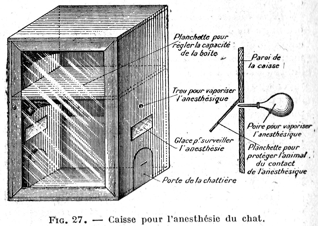
2. Preparation of the subject. - The preparatory injections recommended by some authors, apart from being generally difficult to perform, have given us deplorable results. Atropo-morphine or morphine, even in small doses, induces vomiting and post-operative excitement which may compromise the outcome of certain operations where suturing and dressings play a large role. They also seem completely unnecessary in carefully conducted ether anaesthesia.
The subject is placed in the crate, on an empty stomach as far as possible. If it is vicious, it is left in the basket it was brought in and placed inside as is after making enough space in the crate. Before closing the door, take care to give the patient the option of opening his basket, which he is sure to do with the first jet of ether.
3. Anaesthesia. - The mixture used is good quality ordinary ether with the addition of one tenth chloroform; it is introduced into the box as slowly as possible, in small quantities, to obtain anaesthesia after about twenty minutes. On can judge the cat’s reflexes by passing a hand or a wand through one of the cat doors; breathing should be monitored through the glass door.
When the operator judges that the anaesthesia is sufficient, two cases may arise:
A. The operation site is at the head. – Move the head out through one of the cat flaps up to the shoulder, after covering the opening with a towel to prevent the anaesthetic from being lost from the crate; put a cotton ball soaked in ether under the patient's nose. Monitor breathing through the glass door.
If the anaesthesia is insufficient, move the animal back into the crate by pulling it by the tail through the other cat flap, and complete the anaesthesia using the anaesthetic inside the crate, adding a little chloroform if necessary.
B. - The operating site is on the body. - Same manoeuvres, leaving its head inside the crate. If the anaesthesia prolonged, it is advisable to open one of the small windows so as not to rarefy the breathing air too much and to add a little anaesthetic.
In our opinion, this process has serious advantages over that of the bell jar:
1. The crate is less expensive and less fragile.
2. The anaesthetic is less bothersome to the operator and cannot produce frostbite which occurs frequently on skin that has been in contact with cotton soaked in ether.
3. The operator is protected from any accident in the event of incomplete anaesthesia.
4. Losses are kept to a minimum. With the bell jar, all the anaesthetic is removed along with the subject, and if there is insufficient anaesthesia, everything must be repeated from the beginning. This happens even more frequently because it is impossible to monitor the reflexes.
5. The cost of anaesthesia is kept to a minimum. The average capacity of the box being 5 litres, 90 ccs of ether and 10 ccs of chloroform at most are sufficient for a small operation; examination of natural orifices, castration, various punctures, fracture dressing, etc. For the longest operations, we have never used more than 200 ccs of anaesthetic.
All these considerations made us create and adopt this system which allows us to perform a number of anaesthetics without any accident.
DISINFECTION
Disinfection of the housing should be carried out whenever the animal has a parasitic skin disease or contagious disease. The housing must first be washed with a brush with very hot water containing 10 grams of soda ash (crystals) per litre, then three or four times weekly with cresylated hot water containing 30 grams of cresyl per litre of water. When the housing is made of wooden boxes brush them with cresylated lime milk:
Slaked lime - 50 gr.
Cresyl - 20 gr
Water - 500 gr
Boil the blankets and change the fabric of the pillows that the animals lie on.
ADMINISTRATION OF MEDICINES
Solid medicines (powders, granules) can be mixed with lukewarm milk, sweetened water, honey or a raw minced meatball.
Give fluids by spreading the corners of the lips or jaws and dropping them into the back of the mouth using a teaspoon or dropper.
When you want to give inhalations place the active ingredients (flowers, stems, roots, antiseptic solutions) in a container (bowl, salad bowl, saucepan) filled with boiling water and place the cat over this, its head covered with a towel.
BIBLIOGRAPHY
Grand Larousse Dictionary. – Cat article.
Grand Encyclopedia. - Cat, by E. Trouessart.
Conversational Dictionary. - Cat, by de Reiffemberg.
Nature. - Year 1900, book I, p. 150, - Cat without tail, by Dr Anthony.
Nature. - Year 1905, book II, p. 347. - The cat in Egyptian civilization, by Hippolyte Boussac.
Nature. - Year 1892, book I, p. 279. - Intelligence of the cat, by the Mis de Nadaillac.
The Intelligence of Animals, book III, by Romanes.
Animals and Proverbs, by Ch. Rozan.
Our Animals, by Dr H. Beauregard,
Natural History, book VI, by Buffon.
Prehistoric Man, by Sir John Lubbock.
On the Origin of Species, 3rd Edition, by Darwin.
On the Variation of Animals and Plants, 1879 edition, by Darwin.
Rabbits, Dogs and Cats, 1911 edition, by Diffloth.
The breeds of cats, veterinary doctorate thesis, by Ph. Jumaud.
The Cat, by A. Landrin.
Elements of Zoology, 1886 edition, by A. Raillet.
Treatise on Comparative Anatomy of Domestic Animals, 5th edition by Chauveau, Arloing and Lesbre.
Application of Anatomy to the Inspection of Butcher’s Meats by Lesbre and Panisset.
Treatise on Comparative Physiology of Animals, 3rd edition, by G. Colin.
Veterinary Ophthalmology by E. Nicolas.
Collection of Veterinary Medicine Reports from the Alfort School.
Collection of Bulletins of the Central Society of Veterinary Medicine.
The Book of the Cat, by Miss Simpson.
Cats for Pleasure and Profit, by Miss Simpson.
Les Chats journal, 1912-1914.
Our Friend the Cat, by P. Megnin.
Our Cats, by veterinarian Dr G. Hasse.