LIFE IN THE COUNTRYSIDE and "Farms and Chateaux" Combined
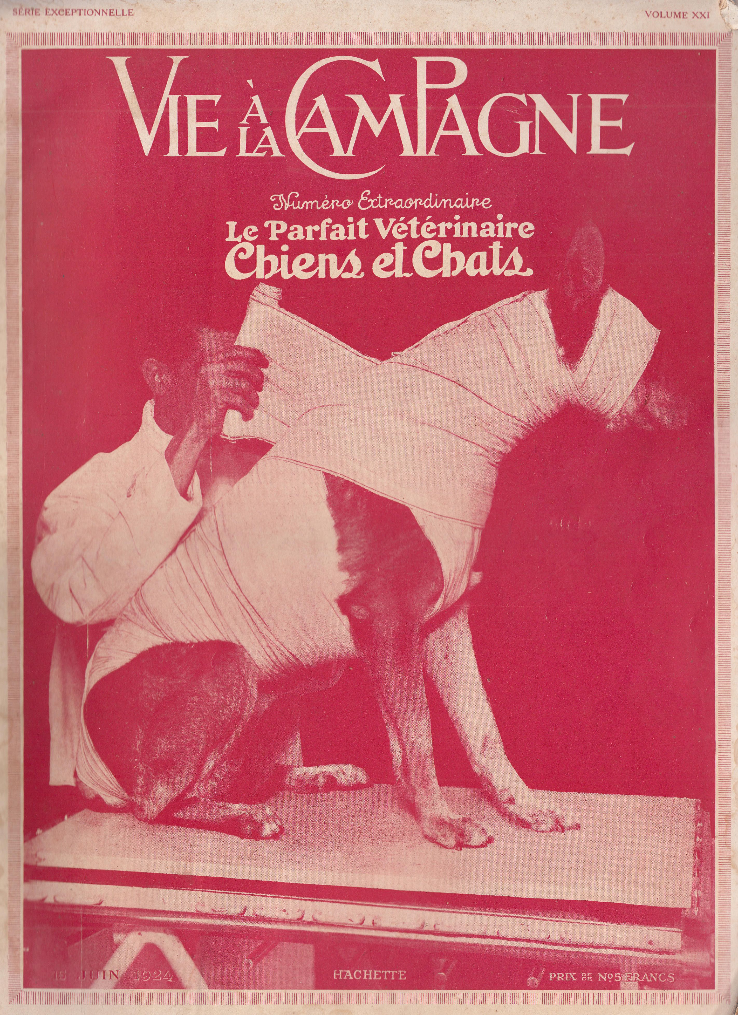
EXTRAORDINARY ISSUE - THE PERFECT DOG AND CAT VETERINARIAN.
Practical review above all, Published under the direction of Mr. Albert Maumené.
Subscription: 6 Nos: FRANCE: 25 Fr. Postal union: 32 Fr.
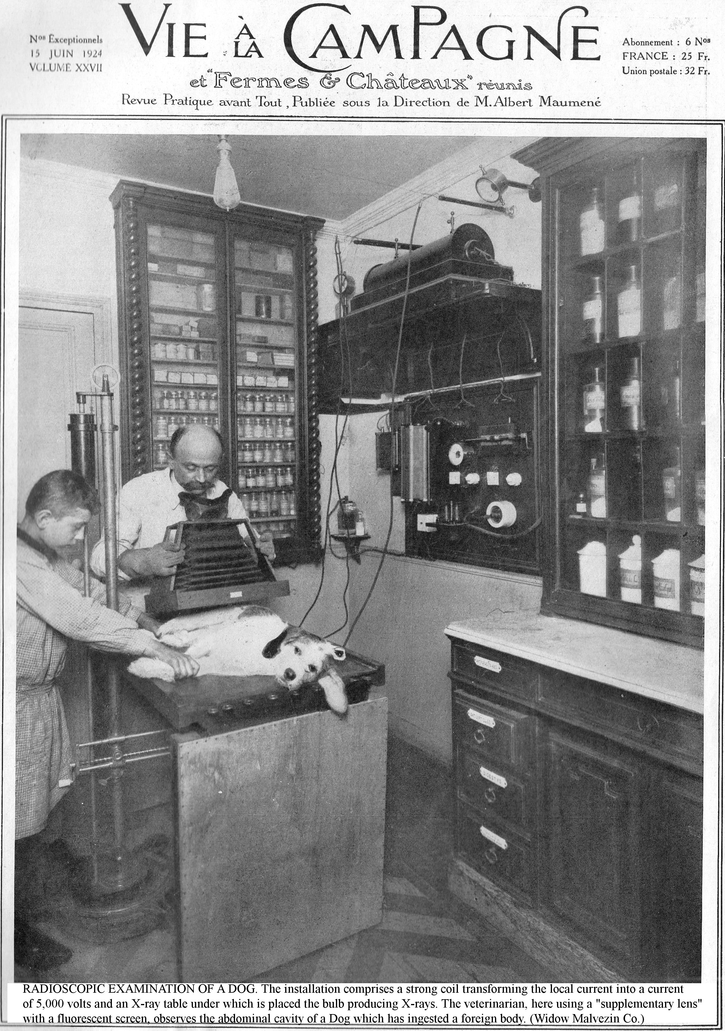
RADIOSCOPIC EXAMINATION OF A DOG. The installation comprises a strong coil transforming the local current into a current of 5,000 volts and an X-ray table under which is placed the bulb producing X-rays. The veterinarian, here using a "supplementary lens" with a fluorescent screen, observes the abdominal cavity of a Dog which has ingested a foreign body. (Widow Malvezin Co.)
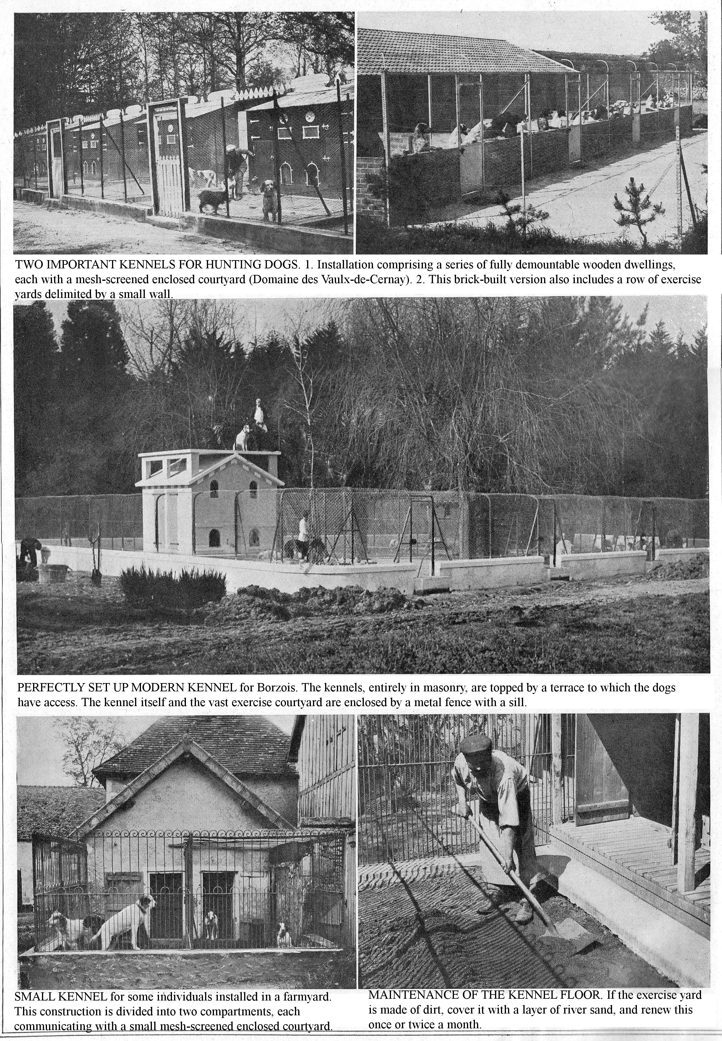
TWO IMPORTANT KENNELS FOR HUNTING DOGS. 1. Installation comprising a series of fully demountable wooden dwellings, each with a mesh-screened enclosed courtyard (Domaine des Vaulx-de-Cernay). 2. This brick-built version also includes a row of exercise yards delimited by a small wall.
PERFECTLY SET UP MODERN KENNEL for Borzois. The kennels, entirely in masonry, are topped by a terrace to which the dogs have access. The kennel itself and the vast exercise courtyard are enclosed by a metal fence with a sill.
SMALL KENNEL for some individuals installed in a farmyard. This construction is divided into two compartments, each communicating with a small mesh-screened enclosed courtyard.
MAINTENANCE OF THE KENNEL FLOOR. If the exercise yard is made of dirt, cover it with a layer of river sand, and renew this once or twice a month.
TO CARE FOR AND CURE DOGS AND CATS
This Volume Teaches you in the Text and Shows you Using Images:
WHY, this Manual of Hygiene, Medicine and Small Canine and Feline Surgery will make you the valued helper of the Veterinarian by teaching you to discern and detect specific benign or serious conditions.
HOW to ensure the hygiene of stock, housing and food in order to prevent epidemics and diseases of all kinds which could decimate the Kennel.
HOW, by knowing the symptoms and lesions, to treat your sick or injured animals, to give them first aid while waiting for the Veterinarian to arrive.
HOW to treat, care for and cure Dogs and Cats with intelligence and punctuality, by applying the Veterinarian’s prescriptions.
HOW to carry out small operations in the event of accidents of any kind, how to assist the Veterinarian, how to apply dressings, avoid cases of infection and hasten Healing.
THE PERFECT VETERINARIAN is the Vet himself, I wrote at the top of the presentation of "The Perfect Vet of the Courtyard." It is the same for everything concerning Ailments and Hygiene of Dogs and Cats. Do not try to be a substitute for the practitioner, but be the collaborator he needs by relating remarks of any kind that you may have noted, symptoms you have observed and that you need to look out for, the course of the illness, and, above all, strictly observe his prescriptions, follow his advice on hygiene measures, in the event of an epidemic, the treatment of sick individuals and prevention of spread to others.
While an ignorant person neglects to follow the given advice to the letter, because he often and without reason considers it useless, you, on the contrary, after reading this volume, will not omit anything, because you will know the importance of it. No veterinarian mindful of his task, rather than inflating his bills by the multiplication of visits and surgical interventions, does not prefer to be in the presence of an enlightened person, likely to assist him effectively, rather than some layman who is unable to comprehend the advisability of his prescriptions.
Certainly, I am aware that a few very rare and narrow-minded Vets, view this volume as a competitor; while, on the contrary, their more broad-minded and progressive colleagues view it differently. This is why valuable and quality practitioners have assisted in the preparation of this manual. They understand and realise that a work which exposes why and how to identify Canine and Feline Ailments in order to treat them better, like the preceding Volume and like those which will follow, are accessories of their profession.
Our collaborator Mr. Ed. Curot, veterinarian, in charge of preparing this volume, naturally tried to limit the descriptions of ailments to those most frequently observed in kennels. It would make your task more difficult if you were told about Ailments that are quite rarely seen or that specialists are still studying. However, he had to mention ailments that appear rather rarely: this is the case for rabies, where hardly any cases have been seen for over 2 years, as administrative measures have been observed.
The Ailments, Accidents, etc., are examined in their logical order for each case and, according to the nature of the Condition, the author describes it in a few words, follows the process, reviews in turn everything concerning the Etiology, Symptoms, Wounds, Diagnosis, Prognosis, Prophylaxis (preventive treatment), Curative Treatment, surgical intervention, when necessary, etc. Note that we have not repeated the conditions and accidents common to Cats and Dogs to avoid repetitions, but it has always been specified when such a condition is common to Dogs and Cats.
By pointing out the symptoms of Diseases, our goal is not to invite you to treat them yourself, but to show you how delicate, complex and difficult it is often to establish a correct Diagnosis, which would lead directly to effective treatment and prophylactic measures. By knowing and observing the Symptoms in a methodical way, you can often detect a serious condition which you had not otherwise paid attention to, and you can provide the Veterinarian with the essential elements for diagnosis.
In urgent cases, while waiting for the practitioner to arrive, the recommended treatments avoid the need to resort to using empirical measures (measures based on observation), so numerous in Canine and Feline Therapeutics, which are very often harmful and increase the severity of the prognosis. As well as noting the bad treatments, you will always find, if not the absolute remedy, at least the most appropriate treatment, with formulas of drugs for internal and external use. Obviously, drug doses are mostly given as a basic indication, because dosage often varies depending on Age, Race, Size and the individual subject and this is, essentially, the responsibility of the Veterinarian, to avoid serious accidents.
We explain at length the prophylactic measures in the field of hygiene and food that will help you reduce the frequency of internal diseases. Prophylaxis of contagious diseases (isolation, disinfection, vaccination, etc.), when judiciously applied, allow you to avoid epidemic outbreaks which all too often decimate breeding kennels and catteries.
In such a work, however, substituting scientific data for empirical data is often a rash attempt because, when the latter is badly applied due to lack of technical knowledge, they may be more dangerous than the former by assuming a pseudo-scientific character. Avoid this pitfall, the consequences of which (increased mortality, spread of contagious diseases) would be very serious, by calling your Veterinarian at start of an illness.
Our collaborator Mr. Ed. Curot, author of several important works in Veterinary Medicine, has tried to follow the guidelines and scheme of this Journal, by expanding on and substituting practical advice in place of considerations and empty and irrelevant sentences.
Images appropriate to the subject play an important role in this valuable practical work. We have used illustrations extensively in this Volume: photographs show you the characteristics of some ailments; other show characteristics, lesions and attitudes of sick subjects; many show you hygienic installations, expand on methods of treatment, administering drugs, surgical interventions, dressings, bandages, hygiene and grooming measures, etc., by presenting to you all the successive and logical phases, the whole process of carrying out the tasks which objectively informs you and engraves the subject forever in your memory. In this work, the photographs complete the various chapters and are so numerous that there are ten times as many as can be found in the most famous veterinary volumes.
Many of the original photographs were taken at the National Veterinary School of Alfort; others in the clinics and veterinary surgeries of J. Malvezin and J. Taskin.
Always at the forefront of progress, we wanted to encourage you to find new things that you looked for in vain elsewhere; we have, in fact, included in this Volume excellent radiology illustrations (fluoroscopy, radiography, radiotherapy). Radiology has a large role in the diagnosis and treatment of ailments in Dogs and Cats; so you should not be ignorant of what to expect from it. You will appreciate the synthetic radiography of Dr. André Lucy, radiologist at Hôtel-Dieu; another very remarkable one from U.S.A. doctor of veterinary medicine and radiologist, Jacques Tashin, as well as his special radiology facilities for small animals, and those of veterinarian J. Malvezin.
I am very grateful to these collaborators for supporting our efforts, for their interest and for the part they took in preparing this Volume, which I am convinced you will find a veritable Manual of Hygiene, Medicine and Small Canine and Feline Surgery, the Guide and the Advisor you have been waiting for.
Albert MAUMENE.
HYGIENE OF HOUSING AND SUBJECTS
A COMFORTABLE HOME WHOSE CLEANLINESS IS CONSTANTLY ENSURED AND PHYSICAL CARE IS THE BEST GUARANTEE OF A STATE OF GOOD HEALTH IN DOGS AND CATS.
Many ailments would be avoided if Dogs and Cats were provided with all the hygienic measures required by their breeding. Therefore we will first draw your attention to this point.
HOUSING HYGIENE.
Remember that lack of ventilation, damp and cold temperature in the kennels, the presence of drafts, the poor maintenance of litter, and the non-disinfection of premises, constitute the most frequent etiological causes of diseases; also, you cannot attach too much importance to the installation and perfect hygiene of your Kennel. It is through ignoring the importance of these issues that many farms, despite the animals’ lofty origin and a rational diet, are decimated by high mortality which often negates the breeders’ efforts.
HEALTHY HOUSING. The home of Dogs is, depending on the case, a Hutch or Kennel; all too often the Dog is housed in unsanitary conditions and suffers the adverse effects of cold and damp. Kennels are not recommended for all breeds. Miniature Dogs especially, when their births occur during winter, should be reared in apartments, up to the age of 5 or 6 months.
Here we consider the establishment of the Kennel from the point of view of hygiene; everything concerning technical questions (construction, fitting out of kennels, fixed or removable, enclosures, annexes, etc.) having already been dealt with (1). The Kennel needs air, light and space, a hygienic trinity, which directly affects the health of your subjects. When setting up your Kennel, do not sacrifice hygiene for luxury. Build it in a high place, surrounded by trees (pines, fir trees preferably) with loose and dry soil, exposed to the sun, sheltered from cold winds, with doors and windows facing east, south or west, depending on country and altitude. Give preference to a South-East orientation, which offers the maximum benefit, as your subjects, while enjoying the rising sun, are sheltered from freezing winds coming from the North.
(1) Vie à la Campagne: Chiens de service, Ëlevage et Dressage lucratifs (242 grav. Prix: 6 fr.). (Country life: Service Dogs: Lucrative Breeding and Training (242 engravings. Price: 6 fr.).
Ventilation, one of the most important points in Kennel hygiene, exerts a variable action on the inhabitants according to the degree of atmospheric purity. Depletion of the air, apart from having a depressive action on the body, promotes contagion in the event of any outbreak. The atmosphere is renewed by means of natural ventilation (openings, windows, fireplaces, etc.).
While ventilation is a physiological necessity, avoid over-ventilation which would lower the inside temperature beyond the degree compatible with the conservation of animal heat. Puppies are extremely sensitive to cold, their health means avoiding drafts skimming the ground; design the exposure and arrangement of openings to achieve this.
The dampness of the air in the kennels is one of the worst conditions for the health of young animals; it chills them more than a lower temperature in dry air, and predisposes them to rheumatism and rickets. So build your Kennel, or locate the hutches, on dry ground a little above ground level. Heat the Kennel during severe seasons, when you require great expenditure of strength, for pack dogs for example. The kennel of a medium-sized dog should be 1 metre in height and length, and 75 cm wide; do not exceed these dimensions, indeed, if a kennel is to be sufficiently ventilated, it should not be too spacious, as it becomes cold. For economy, use strong barrels, preferably those that contained petroleum oils because, due to their antiparasitic action, they repel the pests that commonly infest kennels.
For hygiene, establish the Kennel double-walled, empty wooden Kennel in double-walled wood with the space filled with sawdust. Cover everything with roofing felt; cement the ground; ventilate it through fairly high openings and fit it with benches. Arrange the beds around the perimeter of the area, except near the door; raise them 30 or 40 cm. above the ground. Avoid the Dogs getting their claws caught in the gaps by a grooved and jointed plank construction. Leave a border of 7 to 8 cm. to hold the straw and, on the opposite side, have a hinge to lift the sleeping benches for cleaning.
Complete this layout with a courtyard of concrete, brick or even asphalt which allows effective disinfection; draw a few markers that you foresee to be useful. Surround the courtyards with railings so that the Dog is not always tempted to jump up to see the horizon, a position that can distort the hindquarters.
Experience shows that, despite strict hygiene, gatherings of dogs are frequently centres of epizootic outbreaks, a consequence of numerous changes due to purchases, exchanges, trips to competitions, etc. For these various reasons, a removable Kennel is preferable to a fixed kennel, as it allows easy disinfection and immediate removal.
Plan for sick dogs under observation, sled dogs, bitches with puppies, and other special accommodations; arrange these as comfortably as possible, depending on their purpose. In a large Kennel the annexes will include, in addition to the kitchen and attic, an infirmary, a pharmacy, an operating room, and a bathroom with a cement bathtub, showers or water jets. Above all, plan the installation of an isolation room. Put any foreign subject or any animal returning from a competition or trip, in a special room for 15 days to monitor their health status, and apply the same measures if one of your own Dogs shows symptoms of illness: depression, inappetence, digestive or respiratory problems. Keep the isolation room as clean as possible, line the bunks with plenty of straw, constantly renewed; spread sawdust on the floor to allow dry cleaning; its odour repels the parasites that like to infest sick individuals. Disinfect the room frequently and urgently in cases of contagious diseases. By applying these basic hygiene principles, you can anticipate and quickly limit the contagious outbreaks that often decimate Kennels.
Do we need litter; which litter to choose? The role of litter is complex; it must absorb excreta, keep the premises dry, and protect the Dogs from damp and cold. The best litter for Dogs is fern; alternatively, use wheat or rye straw and add a few walnut leaves, the smell of which eliminates parasites. Oat straw is too short and quickly crumbles into dust. If a Dog has developed the bad habit of soiling the straw where he sleeps, dissuade him by putting some soiled litter on the floor of the kennel, and let him lie down on a bare bench. Change the litter at least once a week, more often for Puppies who dislike damp; to this end, lay down a thick layer to preserve them from the cold; put Puppies, at least for the first month, on good hay, and never use peat litter for them because it is very powdery and could block the noses of young animals and cause them to suffocate.
HYGIENE MEASURES. Purchases, exchanges, trips for competitions or for breeding, often require the movement of the subjects. Observe the following hygiene rules: avoid shipping the Dog after a large meal, and especially during hot or cold weather. Have the Dog travel in a special crate or strong basket; attach feeding and drinking troughs inside it; line the bottom with good litter, and double its food ration two days before the expedition in anticipation of post-travel fasting. Secure the top of the crate and provide ventilation with a small metal mesh frame. On arrival, report on the subject's health status, and if it is sick or has died, report this.
Traveling in railway kennels exposes Dogs to drafts and colds due to their lack of hygiene and comfort.
Kennel hygiene. The upkeep of the animals and the cleanliness of their kennels have a marked influence on their state of health. The hygiene of the Kennel includes the absolute cleanliness of the premises, the frequent renewal of the litter placed on the benches, and the grooming and bathing of the residents.
Disinfection, the best preventative of contagious diseases, is most often applied haphazardly. Insufficient disinfection explains the frequency, persistence and spread of contagious outbreaks. However severe it may be, isolation is never perfect and can only be an incomplete preventative measure. In fact it is not sufficient to avoid contagion by rigorous measures, it is also necessary to destroy the germs; disinfection fulfils this goal. However, it is only effective in hygienic premises; its effects are uncertain in the insanitary and badly arranged Kennels.
Do serious cleaning every day as soon as the Dogs have eaten their slop, put them in the exercise run or give them their freedom. While they are away, sweep the floor, rid the Kennel of excrement, then wash it with a brush with plenty of water. Finally sprinkle it with cresylated water to neutralize bad odours. Some breeders are in the habit of dusting the ground with sawdust or ashes to facilitate cleaning; do not use this process as it has the disadvantage of frequently dirtying the fur and sawdust gets into the food. Change the straw or bedding material frequently, especially if you are dealing with animals that have a bad habit of urinating in their kennels. Finally, dismantle and thoroughly clean the removable cabins every month in Winter and every two weeks in Summer. By doing so, you significantly reduce the number of parasites that infest Dogs. Bring your subjects in when the ground is perfectly dry and the water gutter is clean and covered with its grating.
To disinfect the Kennel, brush all the walls of the kennels [niches] with one of the following solutions which have the advantage of not attacking the paint or varnish: cresyl solution at 3% or 4% solution of sublimate at 1 part per thousand; wash the floor with a brush and a boiling solution of soda crystals at 10%, and swill it with cresylated lime milk comprising 10 parts slaked lime, 3 parts cresyl, 100 parts water. Preferably use removable wooden kennels because these are easy to clean thoroughly; rapid singeing or carbonyl disinfection immediately sanitizes them, something impossible with brick or cement boxes. Also disinfect the feeders, brush them, wash them with plenty of water, and leave them for at least twenty-four hours in bright sunlight, weather permitting. Sanitize the litter, stable manure, slurry and excrement with 10% milk of lime and add a quantity equal to that of the material to be disinfected.
ANIMAL HYGIENE.
Grooming, bathing or washing and exercising are valuable means at your fingertips to ensure the health of your animals. Do not neglect these.
PERFORM GROOMING. Grooming is a procedure that rids the skin of the grime and impurities deposited on the body's surface. In in common canine breeds it is all too often considered superfluous, but it meets an imperative need and contributes, to a large extent, to the maintenance of health. Lack of grooming allows the accumulation of epidermal waste, predisposing them to parasitic skin conditions. The skin of a healthy animal living in freedom, unlike one living in the kennel, is generally in a satisfactory state of cleanliness.
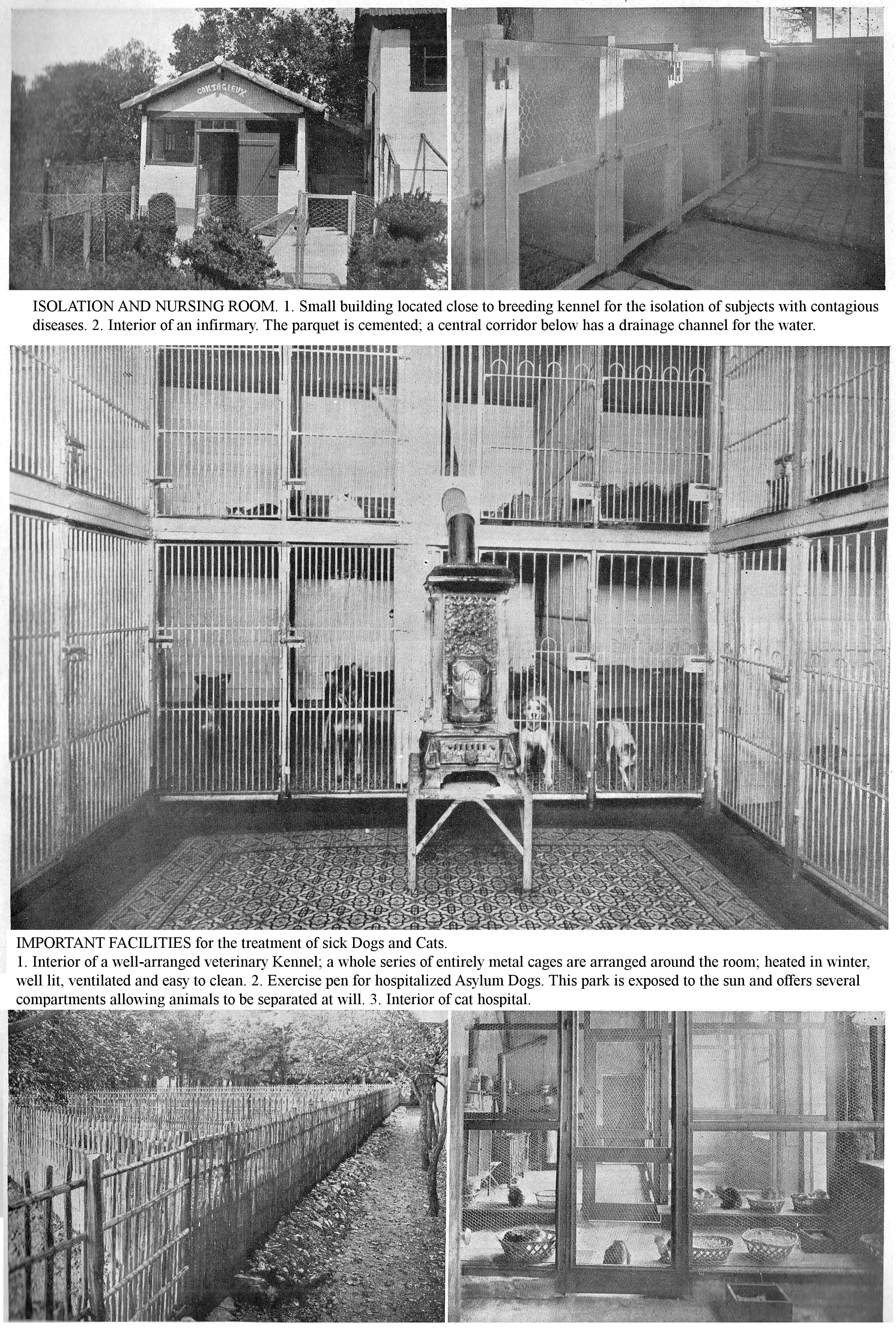
ISOLATION AND NURSING ROOM. 1. Small building located close to breeding kennel for the isolation of subjects with contagious diseases. 2. Interior of an infirmary. The parquet is cemented; a central corridor below has a drainage channel for the water.
IMPORTANT FACILITIES for the treatment of sick Dogs and Cats. 1. Interior of a well-arranged veterinary Kennel; a whole series of entirely metal cages are arranged around the room; heated in winter, well lit, ventilated and easy to clean. 2. Exercise pen for hospitalized Asylum Dogs. This park is exposed to the sun and offers several compartments allowing animals to be separated at will. 3. Interior of cat hospital.
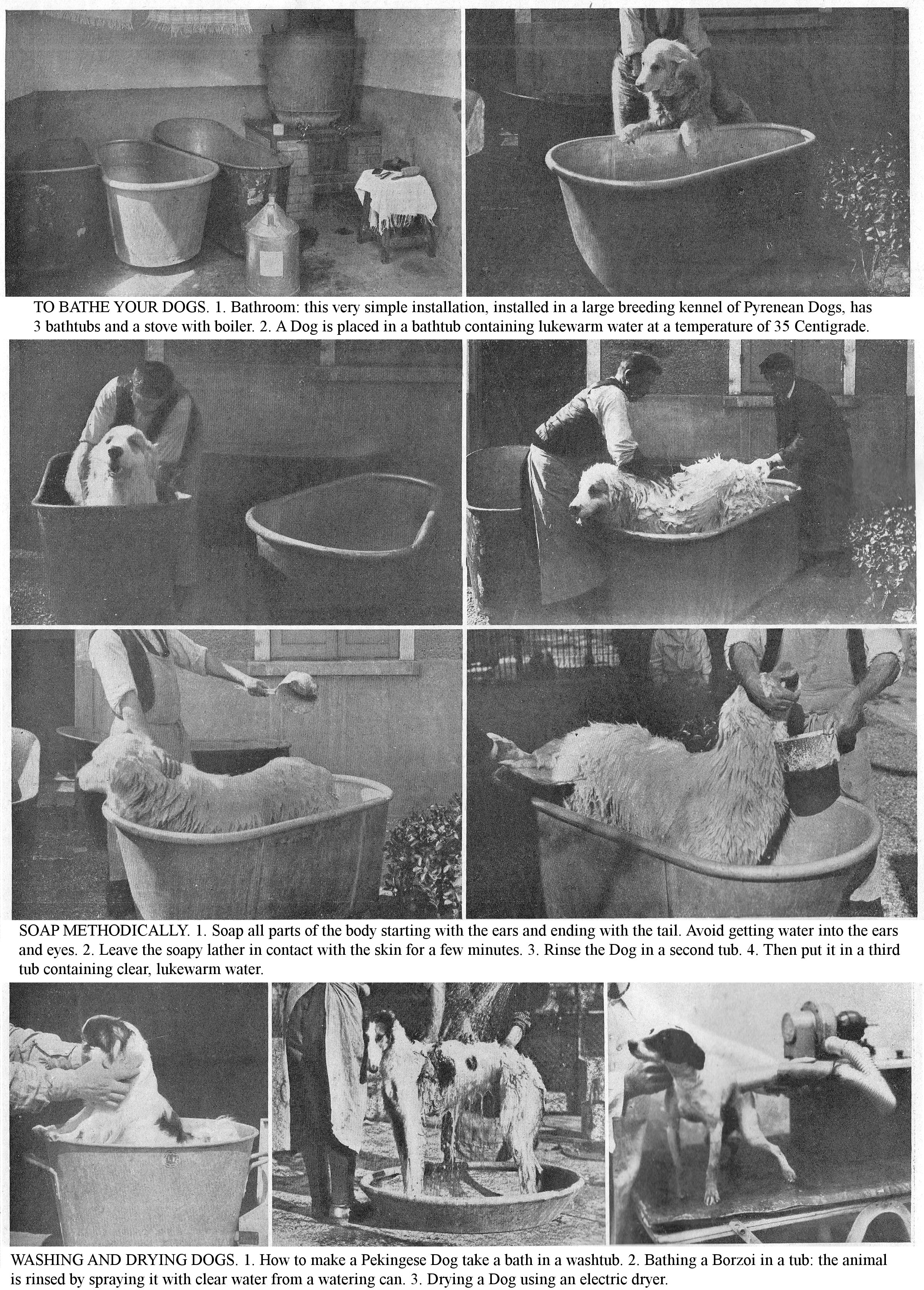
TO BATHE YOUR DOGS. 1. Bathroom: this very simple installation, installed in a large breeding kennel of Pyrenean Dogs, has 3 bathtubs and a stove with boiler. 2. A Dog is placed in a bathtub containing lukewarm water at a temperature of 35 Centigrade.
SOAP METHODICALLY. 1. Soap all parts of the body starting with the ears and ending with the tail. Avoid getting water into the ears and eyes. 2. Leave the soapy lather in contact with the skin for a few minutes. 3. Rinse the Dog in a second tub. 4. Then put it in a third tub containing clear, lukewarm water.
WASHING AND DRYING DOGS. 1. How to make a Pekingese Dog take a bath in a washtub. 2. Bathing a Borzoi in a tub: the animal is rinsed by spraying it with clear water from a watering can. 3. Drying a Dog using an electric dryer.
Give a regular daily grooming with a horsehair brush if the hair is long, or with a pig bristle brush if the hair is short. Follow this grooming with a general massage, performed with the free or gloved hand. Regular use of a brush, handled expertly, often makes washing unnecessary; washing removes the oily substance intended to lubricate the skin and hair and to maintain these in the necessary state of suppleness. Use an angled bristle brush for grooming short-haired dogs: terriers, etc.; strong whalebone, quack-grass or wire, half on rubber for long-haired dogs; silky bristles for luxury dogs. Awkwardly directed combs can irritate the skin and damage the coat; use combs with wide teeth and very blunt ends, in black rubber or aluminium. There are curry-combs for rough-haired dogs; dry them well after use. For hygiene reasons, and to prevent the transmission of skin diseases, disinfect these items often.
Grooming Dogs to appear in an exhibition has a few peculiarities: perfect the moulting of the winter hair by removing any dead hair, the fluff that eventually becomes felt, leaving only the young, shiny fur. Proceed with a somewhat stiff quack-grass brush, rubbing in all directions; then carefully use an unknotting comb, either metal or horn. This operation finished, continue with a fine comb, gently pulling out any dead hair. These preparations finished, give a bath, rub the Dog in all directions, and comb it before the fur is completely dry using a special curry comb or a curry-glove.
The hygienic care given to a Cat, due to its normal state of independence, is almost nil. The Cat, by using its tongue, grooms itself automatically; it successively and frequently, licks the various parts of its body for hours on end and its hair presents a shiny and silky appearance due to successive massages. However, perform a light brushing each morning, followed by a light hand massage.
Do not bathe Cats, due to this species’ aversion to water and the respiratory ailments to which they are exposed if they are wet.
BATHS – WASHES
Skin hygiene, apart from grooming, can be achieved by washing and bathing; these can be hygienic or medicinal. The therapeutic effect varies with the temperature of the water and the duration of immersion. Cool water (10 to 15 Centigrade), exerts a tonic action which resonates, by reflex, on other organs, by the reaction it causes on the skin.
Do not use baths before the age of 6 months; give lukewarm baths, around 35 centigrade, monthly in summer, and every two or three months in winter, in a warm room. Take care to stir up the subjects well during the bath, and above all rinse and dry them thoroughly at the end, avoiding any chills and drafts. Only give baths 2 or 3 hours after meals to avoid digestive upsets.
To soap it methodically, place the Dog in a tub containing hot water; soap all parts of the body starting with the ears and ending with the tail. Avoid getting water into the ears or eyes; leave the soapy lather on the skin for 7-8 minutes to give it time to kill fleas or other parasites. Then rinse the Dog in a tub, remove it and put it in a second tub of clear lukewarm water; wipe it down with tea towels, and let it run in a meadow or sandy place where it can lie down without getting dirty. Carry out this procedure carefully; never let a dog that has just been washed lie down in the kennel without first ensuring that it is completely dry. During the cold season, rub the Dog down; wrap it in a woollen blanket; or better, place it in front of a good fire to avoid any organic repercussions due to cooling.
Despite the hygienic effects of baths, do not overuse bath-therapy, especially among delicate breeds; remember that methodical and regular grooming avoids the need for frequent bathing.
Complete the grooming by washing the eyes, ears, nipples, and take special care of the digital region. Wash the eyes every day with a little lukewarm boric water; keep the inside of the ears very clean by washing with soapy water then dry them carefully; by this hygienic practice you will avoid auricular catarrh, a very stubborn affliction. Clean the nipples because, while nursing, Puppies can contract the parasitic diseases from the mother resulting in death or stunted growth: wash the nipples with a sponge soaked in a solution of salicylic acid at 1 part per 1000.
You will read with interest the following articles which have already appeared in Vie à la Campagne: How to choose a hunting dog, no. 230; What You Can Expect From The Gordon Setter, no. 231; The Breton Spaniel, a remarkable hunting dog, no. 237.
ENSURE EXERCISE. Exercise is necessary for the Dogs you want to keep healthy, fit and resilient. It prevents an accumulation of fat between the muscle fibres; it maintains the normal relationships that should exist between the different parts of the musculoskeletal system (bones, muscles, tendons, ligaments, joints); it ensures their regular play to a fair extent. In methodically measured sessions, exercise gradually acts most successfully by increasing respiratory capacity, promoting regular digestive function and it constitutes a preventive treatment for obesity and constipation which are frequently seen in apartment dogs which are kept almost completely inactive. Therefore, the duration of exercise must be in proportion to the Dog’s temperament, age and abilities. Give Service Dogs one hour in the morning on the leash and half an hour in the playground in the evening, releasing them two or three times. Take them for a daily walk in freedom to keep them in good condition; give Hunting Dogs a route of 5 to 6 km but take care that this exercise never take place straight after meals.
To stimulate the Dog, make it run behind a rider or a cyclist, preferably in the morning, especially in summer; the walk and the exercise enclosure are insufficient for service dogs. If you have more than one Dog, take them out together so that they train each other. To get an Apartment Dog to take healthy exercise, give it a rubber ball; he will fetch it, developing a particular eagerness in this game. When returning from a walk, wipe the Dogs dry if they get wet; wash their paws if they are muddy. Then shut the subjects in a kennel lined with dry straw or a cushion, as appropriate.
If the Dogs are kept in a too narrow place, take them for a walk after their meal so that they can frolic and void their wastes; once in a while, in the morning as much as possible, to a meadow, take them along a grassy road, so that they can eat the soft grass, which is the best purgative for them.
BASICS OF RATIONAL DOG FEEDING
UNDER WHAT CONDITIONS TO DISTRIBUTE FOOD, ACCORDING TO THE AGE AND CONDITION OF ANIMALS, TO AVOID NUTRITIONAL DISORDERS AND DISEASES OF THE DIGESTIVE SYSTEM.
The Dog’s diet must be both economical and hygienic. The cost of the daily diet is an important factor which directly depends on part of the envisaged profit; currently, a dog costs more than 400 francs in food per year.
Even more, or in the same way as hygiene, food plays a very big role in breeding; an irrational diet, both in quantity and quality, can cause serious digestive disorders and a predisposition to nutritional diseases (rickets) and in general to all the infectious diseases that often decimate kennels. Morbidity and mortality factors are largely due to food hygiene.
The Dog is a predator, but domestication has made it an omnivore whose diet includes animal and plant material. The terms of this mixed diet, which is a physiological necessity, are imposed by the structure of the dog's digestive system and by the particular conditions in which the animal lives at various times of its existence.
Chewing is very cursory in the Dog; mixing with saliva is minimal and food enters the stomach without undergoing any noticeable changes. The dog's stomach is spacious and its entire surface is covered with a mucous membrane endowed which actively secretes gastric juice. Stomach digestion, quite slow and very powerful, is an important act. In consequence of this long digestion time, and its intermittent nature, hunger returns at long intervals so meals should therefore be infrequent.
The Carnivore’s digestive system has a much smaller capacity than that of herbivores. The intestine, in particular, is significantly shorter and narrower; it is remarkable for its brevity and small volume. Large stomach, short intestine, cursory chewing, superficial mixing with saliva and very powerful gastric digestion are the physiological data which you must take into account in canine dietetics. From the point of view of hygiene, the dog's diet plays a big role: carefully avoid either excess or stinginess.
Common food-related illnesses (dyspepsia, stomach dilation, gastritis, gastroenteritis, etc.) show the importance of diet.
Dyspepsia is most often caused by an irrational, poorly regulated, insufficient or too copious diet. Stomach dilation is seen mainly in naturally voracious adult dogs that are heavily fed and accustomed to only one meal per day. Gastroenteritis and Indigestion are common following repeated ingestion of coarse, irritating, spoiled or toxic foods. Overconsumption of meat causes a predisposition to inflammation, most commonly eczema and auricular catarrh.
Examination of the faeces constitutes the best criterion of the diet used: look for a moulded, homogeneous, dark-coloured stool, with a typical odour, all signs of a well-functioning digestive tract. Prevent fluid faeces, consisting of undigested residue, discoloured or with a red appearance, bloody traces and a foul odour, indicating digestive disorders and incorrect diet.
DAILY DIET. The amount of food to be given to a Dog is not usually determined by any specific rule. Some are overfed, fat, plump, lazy, and predisposed to skin conditions; others, who are sparingly fed, are thin, fussy, physiologically poor, and predisposed to disease or parasitic invasion. Fix the dog's daily diet at about one-fifteenth of its weight; set it to about one-twelfth during the growing period and for those who tire; reduce it to one-twentieth for adults, the elderly, and the sedentary. But take into account that the daily diet is subject to variations depending on the degree of activity of the digestive functions, the constitutional state or the temperament of the animals.
A good diet must, in order to satisfy the creature’s needs, contain the necessary nutritional principles quantitatively and qualitatively, include easily digestible materials palatable to the animals, and have suitable nutritive values. When substituting foodstuffs, generally done for economic purposes, take into account the requirements expressed above, and also be aware that any substitutions must be applied carefully and gradually in order to avoid accidents.
In general, a combination of carefully chosen and complementary products is, in terms of nutrition, the best way to avoid deficiencies and the physiological and economic double wastefulness that can result from a defective make-up of the diet. It should not be too watery, containing more than 75% water; preferably make it up by combining elements of animal and plant origin, as digestible as possible and containing a minimum of cellulose.
Vary the food often to stimulate appetite and digestive functions. Remember that a uniform diet causes food aversion and has a depressive effect on the stomach and intestinal function. In the diet, take into account the importance of ancillary factors, hormones, vitamins: substances still poorly determined but whose absence from the diet can lead to serious deficiency disorders: acute, subacute, and chronic diseases (scurvy, rickets, anaemia, various weaknesses). These are destroyed by the handling of the specialties recommended for dog food and by temperatures above 110 to 120 Centigrade. Also supplement prepared foods with natural and fresh foods, in particular with tubers, roots and vegetables.
Vary the number of meals, their importance to health being considerable, with the age of the animals and the nature of the food. After weaning, give 5 meals; from 2 years old give 2 meals a day: one in the morning, another in the evening, to avoid the ingestion of excessive amounts of food at one time. Never give a heavy meal immediately before or immediately after strenuous exercise. Whenever possible ensure the meals are regular. Give lukewarm food to young dogs, cold or lukewarm food to adults during the winter season.
COMPLEMENTARY FEEDING. A milk diet, however physiological it may be, is no longer sufficient to ensure the rapid and steady development of puppies; achieve nutritional balance by complementary feeding which is really the first stage of weaning. The delicate structure of their digestive organs, their smaller volume, their incomplete development, and the state of secretions, are all health factors that you must take into account during this period.
The use of complementary foods must, therefore, be introduced slowly and progressively; carefully avoid overfeeding which leads to serious digestive disorders. At first it is more of a food substitution than an addition. "Often and a little at a time" is the rule you must observe.
Base the choice of foods on their nutritional value and digestibility; the cereals and pâtés serve as a transition between the milk diet and meat diet and allow a gradual adaptation of the digestive tract to the new diet. In the case of numerous young and towards the 4th or 5th week, the period when breast milk decreases in quantity and quality, make up for this deficit by giving boiled milk at a temperature of 30 to 35 centigrade. At this time, the basis of the diet should be raw and grated mutton, a teaspoonful of oil with a few drops of cod liver oil; soups with rice and vegetables, milk, pepsinated or baby cereal, grated veal bones. Distribute this ration in 5 or 6 meals; with very regular intervals between them, because of the reduced digestive capacity of the subjects.
FEEDING AFTER WEANING. The future of Puppies is ensured by breastfeeding that is sufficient in abundance and in duration, by a weaning conducted without a difficult transition and by an intensive and easily digestible feeding, after this period. After weaning, give milk soup morning and evening and meat soup at noon. Add, in addition, a little raw or cooked meat, grated or cut into small pieces, given as is or after having drizzled with cod liver oil. Do not distribute hot meals, but lukewarm, that is to say at a temperature below 37 °. Thereafter, vary the diet and gradually bring the puppies to the adult diet. Until their full development, give them a mixed easily assimilable diet, but especially meat; promote skeletal growth by adding bone powder, crushed eggshells or a phosphate mix to the mash.
Pay close attention to the health and diet of Puppies to be entered in a competition; partially remove the ration of bread and vegetables, and replace it with a meat diet, rich in proteins, so as to eliminate the fat which gradually clogs the tissues, invades the muscles and masks the animals’ harmonious lines. On the day of the exhibition, give a dozen raw eggs, which, by their reduced volume, ensures sufficient nutrition and prevents a bloated belly.
Feed Kittens separated from their mother with milk, then with breadcrumbs crumbled in milk. Later on, base their diet on soft or raw, boiled liver; if the budget allows it, add some fish. For apartment cats, continue the milk; give as little cooked meat as possible, a daily ration of soft and raw liver being necessary to keep them in good condition.
SPECIAL DIETS. Gestation: During this period the diet must meet the following physiological requirements: avoid abortion, ensure foetal development and promote lactation; it must therefore be easily assimilable and plentiful. Towards the end of the gestation period, give a refreshing diet to facilitate milk yield and avoid the use of artificial breastfeeding.
Parturition. The profound changes in the body produced by childbirth, especially in complicated births, require special nutrition. For 2 or 3 days give Bitches a refreshing diet (a paste of oatmeal flour with milk, plain milk, broths and lean meat, etc.). Do not resume the normal diet until systemic disorders have disappeared; to act otherwise is faulty health care and prejudicial to the health of the mother and young.
Lactation: Apart from individual variation, lactation is largely related to diet; the diet of nursing mothers is even more important because it directly influences the establishment and production of milk and, consequently, the future of the subject. The diet must be easily assimilable and contain the nutrients necessary to ensure the mother’s maintenance and her milk production. Food should also be refreshing, as, apart from its influence on the quantity of milk, it has a positive influence on the health of newborns. During the lactation period, give your Bitch plenty of milk, eggs and meat; the health of her offspring depends on it. Vigorous young will easily withstand weaning. Add lukewarm soups with added vegetables to the diet.
MINERAL DIET. In nature the Dog is a carnivore, but, in the state of domestication it does not exclusively require meat. Feed him a diet that includes both plant and animal matter in roughly equal proportions. More precisely, adopt the following ratio: 40% animal products, 60% plant-based foods. There is no doubt that meat should be included in the dog's diet, but base the proportions on the work he is required to do, the climate, and his age and breed.
Meat: Meat is an essential food for youngsters and those whose bodies are not fully developed. Give a smaller amount to adult dogs, and reduce it still further in old age. Remember that raw meat, being much more nutritious than cooked meat, is particularly advantageous during a period of convalescence, as well as for those suffering from diseases causing poor appetite.
Fresh meat can present dangers: meat from animals affected by parasites, meat whose ingestion can produce symptoms of poisoning, due to the presence of toxins before slaughter or developed afterwards (stale meats, sick meats). Therefore, the quality of the meat is greatly important, and if you notice any signs of spoilage, reject it as unsuitable for consumption. Of all the meats used in the dog's diet, favour horse-meat, it is the only one that does not expose the animals to contracting worms.
Provide all types of butcher's waste - mutton tripe, sheep heads, meat meal, dried meat, supplied by the rendering plants, fish meal – as the high protein content of these by-products will ensure skeletal development in young dogs; use them, moreover, in preparing the broths used for diluting cooked vegetable foods. Also use cretons, the caked residue of melted tallow, because of their high nitrogen content (55%) and fat content (23% - 24%). Also give blood mixed with meal (corn flour, rice, etc.), in the proportion of 100 grammes of blood for 300 to 400 grammes of meal.
Bones are composed of an organic substance, ossein, impregnated with various minerals: phosphates, calcium carbonates, magnesia, etc. Give them to clean and strengthen the teeth as well as to supply the body with the necessary minerals, especially calcium.
Milk, according to its chemical analysis, can be considered a complete and physiological food during infancy: the Puppy gets all the elements necessary for normal growth from it. Eggs, being rich in albumin, and having a high organic phosphorus content (lecithin, vitelline, etc.), constitute a powerful means of completing growth in slow or sick individuals. Offer them diluted in milk; Puppies are very fond of them in this form.
PLANT FOOD. Consider bread made from flour (corn, oats, rye, peas, field beans, etc.) as the basis of dog food and a maintenance food. The total diet (maintenance and output) of a working Dog obliged to expend muscular energy must include bread or vegetable matter and meat.
Distribute the bread as it is, or as a mash made with hot water or broth; mixing it with skimmed milk is recommended. Dogs gladly take bread in the form it is prepared for man; stale bread and breadcrusts are advantageous in the Dog’s diet if they have not deteriorated. The same is true for batches of army biscuits.
Do not feed a dog exclusively on soups, which are always swallowed greedily and, if they are mixed with saliva, will overwork the stomach’s digestion; in addition, this diet is debilitating and does not maintain the condition of the Dogs. Never serve soup hot.
Use pasta and pasta scraps made from wheat flour made for human consumption (noodles, tapioca, semolina, vermicelli, etc.), because of their real health and nutritional value. Include them in the composition of the diet of small breeds. Add them to the milk or cook them in water, with the addition of a little gravy or vegetable broth.
Oilcake is difficult to assimilate, despite the high protein content; Dogs lose weight on this diet. Potatoes, because of their low coefficient of digestibility, 18%, fatten and weigh the Dogs down; only introduce them for 1/4 as a substitute for bread. Distribute root vegetables (carrots, beets) cut into small pieces before cooking; these combat the heating effect of a meat diet. Use vegetables widely despite their low nutritional value, as they increase the appetite for food, provide essential nutritional factors (vitamins) and act as laxatives.
Sugar, chocolate, sweets and pastries, which form the basis of the diet of luxury dogs, are only a problem if you give them in excess.
HEALTHY DRINKS. Drinks are intended to introduce, into the budget, the water necessary for the normal composition of the blood, for the constitution of tissues and for the maintenance of bodily secretions; they are, therefore, greatly important. The lack, excess, or irregularity of drinking water can cause physical disorders.
Distribute water that has all the characteristics of drinking water (colour, clarity, odour, etc.). Do not use water that is muddy, silty, or has organic matter in suspension, as well as water from ponds, ditches, etc. These waters are unsanitary due to the presence of parasites and highly pathogenic germs and are a vehicle for microbial and parasitic diseases. Renew the water often. Avoid soiling the containers with urine or manure, by placing them on a small support fixed at 25 to 30 cm., depending on the size of the Dogs. Occasionally, especially in hot weather, add a small dose of baking soda to the water for 8 to 10 days.
PHARMACY AND ADMINISTRATION OF MEDICINES
WHY YOU SHOULD NOT IGNORE THE PURPOSE A FORMULA OR SPECIALTY IS PRESCRIBED FOR, AND HOW TO ENSURE THE PERFECT APPLICATION.
Before starting the study and description of diseases of Dogs and Cats, it is essential that you know the basics of dosage, the composition of an emergency pharmacy and the methods of administering drugs to these animals.
PRINCIPLES OF DOSAGE. The dosage, that is to say the indication of doses of drugs in dogs, is very variable; the great variation, due to the variation of height and weight, are indeed represented by the numbers from 1 to 10. Use only the doses indicated by your veterinarian, because the therapeutic or toxic effect of drugs measurably varies according to age, individuality, size, breed, severity of symptoms observed, etc. Therefore, we point out the formulas purely for documentary purposes; the minimum and maximum doses indicated correspond to small and large breed dogs.
The dosage of the cat corresponds to approximately half of the doses applied to small dog breeds; remember this therapeutic peculiarity, because, during the course of our study, to avoid unnecessary repetitions, we do not indicate special doses for felines, except for toxic drugs.
Evaluation of tablespoons, dessert-spoons, coffee-spoons: for liquids whose specific weight is equal or approximate to pure water, a tablespoon or mouthful contains about 15 gr.; a dessert spoonful, about 10 gr .; a teaspoonful of coffee, about 5 gr.
The number of drops contained in one gram of the drugs most often prescribed are: Acid hydrochloride, 21 drops; Sulphuric acid, 26 drops; Ammonia, 22 drops; Paregoric elixir, 53 drops; Sydenham's Laudanum, 43 drops; Ether, 90 drops; Fowler's liqueur, 34 drops; Iron perchloride, 20 drops; Tincture of aconite, 53 drops; Tincture of belladonna, 53 drops; Foxglove tincture, 57 drops; Tincture of iodine, 61 drops. A pinch of powder equal to 1 to 2 gr.
KENNEL PHARMACY. Organize an emergency pharmacy so that you can ward off minor ailments and, in severe cases, give first aid, while waiting for the veterinarian to intervene.
Keep the following usual medications in reserve:
Purgatives: Castor oil, Buckthorn syrup; Internal astringents: Bismuth sub-nitrate, Tannin; Gastrointestinal antiseptics: Calomel, Salol, Benzonaphthol, Bismuth salicylate. Antipyretics: Quinine. Tonics: Tincture of cinchona, cola, gentian. Stimulants: Ammonia acetate, Caffeine, Ether, Camphor oil. Haemostatic: Iron perchloride. Dewormers: Kamala powder, areca nut, Semen contra. Antiseptics: Potassium permanganate, Carbolic acid, Sublimate, Cresyl, Hydrogen peroxide, Tincture of iodine. Also put together a kit containing the essential instruments: a scalpel; a pair of dismantlable scissors, a suturing needle, a lancet, a fluted probe, a Pravaz syringe, ordinary forceps, forceps, haemostatic tourniquets, Florence horsehair or catgut for the sutures, a packet of absorbent wadding, bandages.
The timing of administration influences the physiological and therapeutic effect of drugs. Observe the following indications. Acids: if the main purpose of ingestion is to activate digestion, give these during or at the end of meals. Alkalis: To stimulate gastric juice, give them before or during meals, or 2 hours after meals to calm acidity and evacuate the stomach. Antifebriles: give these before meals, or 3 hours after. Iodic preparations: avoid rapid transformations of acids and starches by giving these on an empty stomach. Irritants or dangerous drugs; (Arsenic) give at the start of the meal. Laxatives: give on an empty stomach. Medication-Food: Administer ferruginous foods, cod liver oil, phosphate medications, etc., before and during meals.
ADMINISTRATION OF MEDICINES. Difficulties often arise in administering liquid medicine to a dog. So proceed as follows: enlist the help of a helper who, after sitting down, places the animal between his legs and squeezes his knees to prevent any defensive movements. Position yourself in front of the Dog, insert a finger between its cheek and molars, gently spread the cheek to form a kind of funnel, pour in the liquid slowly, while the helper raises the animal's head well to prevent rejection of the drug. Keep its mouth shut; if he does not want to swallow, keep him from breathing for a moment by squeezing his nose; this will force him to swallow.
As soon as the drug is swallows, prevent it being vomited by taking the Dog by its front paws and raising it onto the back ones; turn his attention away from the state of his stomach by walking him for a few minutes in that position. Thanks to this distraction, vomiting that would prevent the therapeutic effect does not occur.
With a large, strong and hard-to-handle Dog, use the following method: Secure it with two straps, one of which is attached to a belt that goes around the kidneys and the other to the collar. Join each of these straps by its free end to a hook fixed to a wall; in this way, defensive movements are naturally limited. Using a light strip of leather around the end of the jaw, lift the head and keep it at 45 degrees; then introduce the medicine into the opening formed by the slightly spread cheek.
Give solid or powdered medicines without difficulty by presenting the Dog with several small pieces of meat; place the drug dose in an incision made in the 3rd or 4th piece that the animal immediately grabs without chewing it, being unable to predict this substitution. The pills are generally active drugs, with a firm consistency, divided into small spherical or ovoid shapes, so as to facilitate their ingestion. This drug form allows you to give substances that smell and taste offensive. The capsules, formed from a solid shell usually made of gelatin, allow you to administer remedies with an unpleasant smell or taste. To administer the pills or capsules, open the subject's mouth, pull his tongue forward and place the medicine at the back; then let go of that organ, and swallowing occurs automatically.
It is more difficult to proceed with Cats due to their defensive movements. Mix liquid medicines with milk and drinks. Administer powdered substances which smell or taste unpleasant in the form of pills or granules, incorporated in meatballs, in butter or preferably in veal lung.
DISEASES OF THE DIGESTIVE SYSTEM, PERITONEUM AND LIVER
PREVENT THESE VERY COMMON CONDITIONS AND THEIR CONSEQUENCES BY GIVING YOUR PETS HEALTHY HOUSING AND A SENSIBLE DIET ACCORDING TO THEIR AGE AND CONSTITUTION.
Illnesses of the digestive system, peritoneum and liver account for 50% of causes of death. The group of digestive system diseases is one of the most important in canine and feline pathology. Although diverse in their symptoms, these conditions have the same aetiology: poor hygiene and diet. Remove your Dogs from the harmful effects of unsanitary kennels (cold and wet); give them regular exercise; provide a mixed diet (meat and vegetable); avoid coarse foods, especially those rich in cellulose, which Dogs and Cats do not assimilate because of their reduced digestive system; avoid indigestible, spoiled foods, and especially avoid overeating; observe great regularity in the number and interval of meals; give unpolluted drinking water, etc., and you will significantly decrease mortality.
Examination of the digestive system includes exploration of the mouth, pharynx, oesophagus, stomach, intestine.
EXAMINATION OF THE SUBJECT. Mouth: Carefully inspect the muzzle internally to see the condition of the nose, lips and their corners. This done, insert a finger into the oral cavity, to approximately determine the local temperature and the general temperature. Spread its jaws to directly appreciate the smell as it exhales and any possible abnormal qualities: acidic, sour, foul or putrid. Sight will tell you if there is anaemia or hypertrophy of the mucous membrane at its various points. The appearance of the tongue provides a valuable diagnostic sign: tongue dry, furry, coated, dark.
To explore the mouth, use small speculums, or more simply use cords or two strips of bandage, tightly bound around each jaw and held in opposite directions, or place a piece of wood between the rows of molar, applied to the corners of the mouth and fixed with a ribbon behind the neck, or use a flat nickel-plated rod. By proceeding according to this method, you will very easily be able to note any wounds, cuts, various sores or specific eruptions (ulcerations, pustules) on the lips; signs of gingivitis and periostitis on the gums; inflammatory rashes on the tongue; dental irregularities or decay caused by foreign bodies lodged between the teeth.
Pharynx: Inspection tells you about possible deformation of the throat area; palpation tell you about the condition of the tissues, tenderness, the presence of abscesses, foreign bodies, etc.
Oesophagus: Its examination, when the passage is blocked in its neck area, gives you useful information; methodical palpation of the jugular channels allows you to detect oesophageal tumours and the presence of foreign bodies. To perform catheterization, secure the subject on a table with the head extended and jaws opened using a speculum or two ligatures in the opposite direction; use the probes used for exploring the urethra.
Stomach: The stomach is a membranous sac, located in the diaphragmatic region of the abdomen between the oesophagus and the intestine, where it assumes a transverse direction of the midplane of the body. Exploration is possible by inspection, palpation and probing. Inspection gives variable results, depending on whether it is performed before or after a meal; it provides information on the state of emptiness and makes it possible to detect the presence of foreign bodies.
Intestine: The intestines of predators are remarkable for their brevity and small size. On a medium-sized dog, this tube measures barely 4.5 metres in length; in the Cat it measures only about 2 metres. The small intestine rests on the lower abdominal wall. Palpation of the abdomen, performed lightly with the tips of the fingers, detects abnormal tenderness in acute enteritis or the presence of tumours, foreign bodies, intussusception, etc.; in the case of Peritonitis or Ascites, palpation makes it possible to perceive fluctuation.
Rectal exploration is a valuable means for the diagnosis of all visceral conditions of the pelvis and abdomen. To perform it usefully, evacuate the intestinal contents with a large enema. Introduce an oiled index finger into the anorectal canal; gently palpate the mass of convolutions of the small intestine and colon. This exploration can recognize food overload, intestinal strangulation, internal hernias, intussusception and volvulus.
Palpation of the abdomen and methodical exploration of the intestinal mass, the patient being examined first when standing, then lying on his back, often make it possible to detect the presence of foreign bodies.
STOMATITIS.
Inflammation of the oral mucosa, Stomatitis, is associated with seizing food or liquids that are too hot, irritating or caustic, the replacement of deciduous teeth, the build-up of tartar, dental caries, foreign bodies, etc.
Symptoms: At first, the mouth is hot, dry and gives off a stale, sometimes foul odour; the oral mucosa is swollen and has a uniform or slightly dotted red tint; it sometimes shows small vesicles and erosions. The dryness at the beginning is followed by profuse salivation (ptyalism).
Treatment: Give the patient food that is either liquid or does not require chewing efforts (milk or broth, to which you add a little raw minced meat). Treat the causative condition; remove tartar and decayed teeth, papillomas, etc. Give frequent injections into the mouth with a solution of potassium chlorate or sodium borate, at 5% or 10%, or vinegar diluted with water. If the stomatitis is ulcerative, touch the ulcers with a brush dipped in hydrogen peroxide diluted 1/4 or with tincture of iodine diluted with two parts water.
DENTAL TARTAR.
Tartar is a calcareous material which is deposited on the teeth of animals, especially in house-dogs. Aetiology: Tartar is due to various microorganisms which cause the deposition of mineral salts contained in saliva mixed with food materials.
Symptoms: This deposition occurs at the neck of the teeth, and the microorganisms inflame the free edge of the gum tissue. Little by little, the tartar creeps between the tooth and the gum, enters the tooth socket, which becomes inflamed; teeth loosen and fall out. As soon as the tartar causes gum lesions, chewing becomes difficult and the mouth becomes foetid.
Treatment: Remove, using a blunt instrument, the layer of tartar deposited on the teeth, and wash the mouth with a boric solution at 1% or 2%. If the gum is inflamed or ulcerated, touch it with tincture of iodine; if there is dry socket, have the loose tooth pulled out. Prophylaxis: Prevent tartar build-up through sensible dental hygiene (washing and brushing).
TOOTH DECAY.
Dental caries is characterized by progressive destruction of the tooth; it is best observed on molars and in old subjects.
Symptoms: The ailment manifests as discomfort when chewing, profuse salivation, a foul odour from the mouth and weight loss of the animal. While examining the mouth you will find, on one of the molar teeth, a blackish cavity containing food particles.
Treatment: Treatment consists of the removing the decayed tooth; perform this with caution so as not to break the weak crown of the tooth. Extraction requires special equipment: pliers with articulated arms and curved jaws, or special forceps.
DENTAL FISTULAS.
Dental fistulas are found as a result of cavities or alveolar periostitis; they are characterized by a narrow cutaneous wound, resistant to cauterization, which gives rise to a purulent, greyish, blood-tinged and foul-smelling liquid. A probe inserted into the wound stops at a tooth root or enters the oral cavity.
Treatment: If the fistula is recent, have it treated with debridement, curettage and strong antiseptic injections, or tincture of iodine. If it is old or complicated by alveolar periostitis, have the tooth removed. Feed dogs that have had dental operations with liquid or easy-to-chew food for a few days.
FOREIGN BODIES IN THE MOUTH.
Foreign bodies in the mouth are often found in dogs. These are bones, needles, pins, pieces of wood, fish bones, etc. These foreign bodies are found implanted in the tongue, the cheeks or between the teeth.
Diagnosis is easy; salivation, difficulty chewing, sometimes the inability to close the mouth, puts you on the right track; methodical exploration of the cavity provides more exact information. After immobilizing the Dog and separating the jaws, extract the foreign bodies, using ad hoc forceps or a blunt hook.
TUMOURS OF THE MOUTH AND LIPS.
The mouth and lips can be the site of benign tumours (papillomas, myxomas, cysts) or malignant tumours (cancroid, epithelioma, sarcoma, chondroma, etc.). Young dogs are particularly prone to papillomas, small whitish tumours, warty in appearance, scattered or confluent, that develop on the inner side of the cheeks and lips. Labial cancer, observed exclusively in the elderly, and in most cases on the lower lip, is characterized at first by a small flattened tumour, followed by a greyish, granular, bleeding ulceration which progresses slowly, but is quickly accompanied by subglossian or cervical lymphadenopathy (engorgement of the ganglia in the region). Papillomas are transmissible between young dogs; remove the largest growths with curved scissors; touch the others with a small cotton ball attached to the end of forceps and soaked in 1% acetic acid solution. Administer, in the morning on an empty stomach a dose of 30 cgr. at 3 gr. of calcined magnesia in a little milk as internal treatment. In labial cancer, total and early ablation is the only effective intervention.
A Ranine Tumour is a salivary cyst that is often encountered in dogs and whose only treatment is surgical; in addition to the unpleasant odour of the oral cavity, this lesion is characterized by discomfort when chewing, profuse salivation, by one or more tumours from hazelnut-sized to the size of a small egg, under the mucous membrane between the frenulum of the tongue and the molar arcade, or by a cystic tumour which occurs in the lower region of the throat.
LABIAL ULCERS IN CATS.
Symptoms: The cat is prone to a labial ulcer, called a Carcinoma, which occurs most often on the upper lip, sometimes towards the midline, sometimes on one side. At first, you notice the formation of a concave, regular, greyish wound, with a narrow area of hardening of surrounding tissues. By its gradual growth, this ulcer causes a semi-circular loss of substance, which can measure 1.5 cm. to 2 cm. and lets you see the teeth and gum. When the condition is in the midline, the nose may be affected. When the ulcer becomes general, it causes weight loss and sometimes death.
Treatment: The condition is parasitic and contagious; isolate the patient and support him with easily assimilable food, especially meat. Wash the ulcer several times a day with an antiseptic solution (1/5 iodine solution, or 10% methylene blue), then touch it with pure tincture of iodine.
PHARYNGITIS.
Pharyngitis, or inflammation of the lining of the pharynx, is mainly caused by cold, by ingestion of too hot food or caustic substances, by foreign bodies, etc. Microbes (micrococci, diplococci, pasteurella, etc.) constitute the real determining cause of Pharyngitis; the others are only predisposing or incidental.
Symptoms: At first, you observe difficulty swallowing coinciding with a slight tenderness of the pharynx, some coughing fits and a slight fever. In young or weak subjects, Pharyngitis can become phlegmonous [diffuse spreading inflammation of or in the connective tissue]; then you notice abscesses in the submucosal connective tissue, or in neighbouring lymph nodes; these cause difficulty in breathing, sometimes anxiety and bouts of suffocation; in cases of pseudomembranous pharyngitis, the general symptoms are marked, the breath is foetid and the pharyngeal mucosa is covered with greyish false membranes.
Always be careful with Dogs showing signs of Pharyngitis; do not engage in any local exploration until you have made sure that the subject is not rabid. Pet owners often say "My Dog tries hard to swallow, as though a bone is stuck in his throat," when the real cause is the onset of pharyngeal palsy, symptomatic of rabies.
Treatment: Protect the patient from cold and damp; give them liquid or easily digested foods: milk, broth or light mash. Reduce superficial inflammation using warm, moist compresses, held in place by a bandage; renew these every two hours. Then make a few local applications of tincture of iodine; calm the cough by dissolving about 2 to 15 drops of opium tincture in milk and by emollient or antiseptic fumigations. In phlegmonous pharyngitis, puncture deep abscesses. Prophylaxis: Avoid exposure to cold and damp, the ingestion of too hot food, and of irritating or caustic substances.
OBSTRUCTION OF THE OESOPHAGUS.
Obstruction of the oesophagus, often seen in young dogs, is caused by swallowing hard, large bodies, usually a bone fragment, fish bones, etc.
Symptoms: Difficulty or inability to swallow, profuse salivation, nausea, cough and dyspnoea characterize this disease. When the obstruction is in the cervical region, you may see a painful, circumscribed swelling at any point in the oesophageal groove or even clearly locate the foreign body.
Treatment: If the body is stuck at the start of the oesophagus, attempt extraction with forceps after the jaws have been spread with a speculum. If this does not produce a result, induce efforts to vomit by a subcutaneous injection of 0.5 cgr. to 1 cgr. apomorphine. If the foreign body is not ejected, push it back into the stomach with an oiled or Vaselined probe, and give a few teaspoons or dessert spoons of olive oil. In the event of failure, if the body can be perceived in the cervical region, perform an oesophagotomy.
DYSPEPSIA.
The Dyspepsia frequently seen in dogs are digestive disorders, independent of any pathological lesions of the stomach, intestines or their appendages.
Aetiology: These conditions are closely linked to incorrect food hygiene: poorly regulated, insufficient or erratic feeding. Dyspepsic disorders can result from changes in the functioning of the glands of the gastric mucosa; they generally begin with disorders of stomach secretion: hyperchlorhydria or hypochlorhydria.
Symptoms: Dyspepsia is manifested in Dogs and Cats by signs of discomfort and prolonged indolence after meals; these often go unnoticed. You may notice taste aberrations (pica), bloating, frequent belching, vomiting, and stomatitis with foul mouth odour.
Treatment: Diet is the bottom line of treatment; give the patient a lactic diet; with milk, broth, light soups of toast, raw meat minced or cut into small pieces, and well-cooked vegetable purees; decrease the amount of food at each meal and increase the number of meals. Treat hyperchlorhydria with alkalis (Vichy water, Vais water, baking soda); in case of failure which indicates hypochlorhydria, replace the alkalis with hydrochloric acid or lactic acid (2 or 3 gr solution p. 1,000). Combat abnormal fermentation with antiseptics: calomel (1 to 5 gr.), Benzonaphthol (5 to 50 cgr.) or salol (10 cgr. to 1 gr.).
Prophylaxis: Prophylaxis lies entirely in food hygiene; avoid illogical, poorly regulated, insufficient or copious food; observe great regularity in the number and interval of meals, especially in young dogs.
DILATION OF THE STOMACH.
Stomach dilation is seen mainly in older, naturally voracious, copiously fed dogs, especially those who have only one meal per day.
Symptoms: Along with the symptoms of dyspepsia, there is constipation or diarrhoea. The abdomen is large, often distended; percussion of the stomach region may denote, on the left side, a very extensive zone of dullness. Patients lose weight and get weaker. Prophylaxis: Avoid overeating and increase the number of meals.
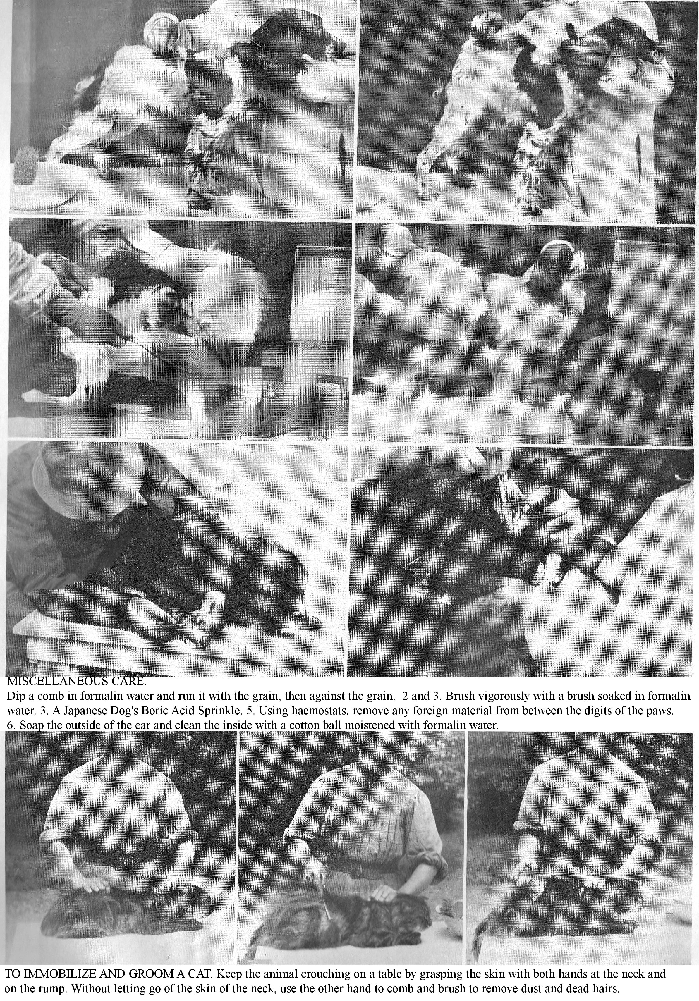
MISCELLANEOUS CARE.
Dip a comb in formalin water and run it with the grain, then against the grain.
2 and 3. Brush vigorously with a brush soaked in formalin water.
3. A Japanese Dog's Boric Acid Sprinkle.
5. Using haemostats, remove any foreign material from between the digits of the paws.
6. Soap the outside of the ear and clean the inside with a cotton ball moistened with formalin water.
TO IMMOBILIZE AND GROOM A CAT. Keep the animal crouching on a table by grasping the skin with both hands at the neck and on the rump. Without letting go of the skin of the neck, use the other hand to comb and brush to remove dust and dead hairs.
A VISIT TO THE VETERINARIAN. Alsatian Shepherd Dog presented by Miss Geneviève Spitz for consultation with our collaborator Mr. Taskin. The Dog, lying on the operating table, is carefully examined by the veterinarian.
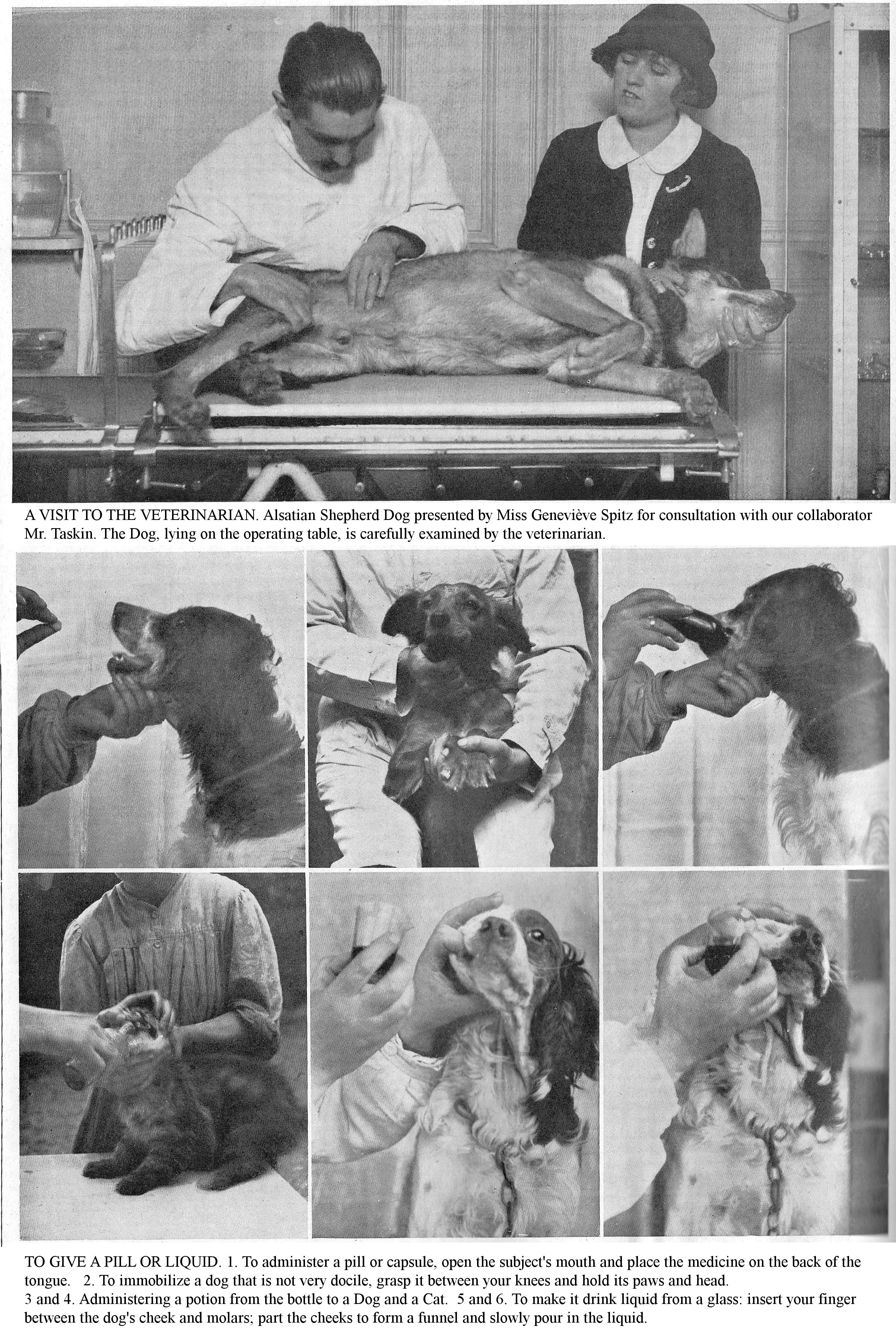
TO GIVE A PILL OR LIQUID. 1. To administer a pill or capsule, open the subject's mouth and place the medicine on the back of the tongue.
2. To immobilize a dog that is not very docile, grasp it between your knees and hold its paws and head.
3 and 4. Administering a potion from the bottle to a Dog and a Cat.
5 and 6. To make it drink liquid from a glass: insert your finger between the dog's cheek and molars; part the cheeks to form a funnel and slowly pour in the liquid.
INDIGESTION.
Indigestion is a temporary acute disorder of the digestive function, which usually occurs a few hours after ingesting too much food or poor-quality food, sometimes under the influence of an external cause such as cold, fatigue. Indigestion can also be the result of microbial self-poisoning, following paralysis or weakness of the digestive viscera. Dogs and cats that consume all kinds of food, foreign bodies or rotting matter, are very prone to stomach or intestinal indigestion; but the ease with which these animals vomit makes such digestive accidents mild.
Symptoms: The patient is depressed, worried, agitated, shows signs of mild colic, paces, lies down, gets up, complains; you sometimes notice salivation and nervous disorders.
Treatment: Induce vomiting by administering syrup of ipecac, or emetic, 3 teaspoons per day. Subsequently, if signs of depression persist, institute a milk diet for a few days. Prophylaxis: Through sensible feeding (sufficient number of meals varying with the age of the animals) avoid gluttony, ingestion of excess food, coarse or spoiled food, toxic substances or foreign bodies.
ACUTE GASTRITIS.
A most common disease, particularly frequent in young subjects, where it is a major cause of mortality. Gastritis, acute or chronic inflammation, has a complex aetiology, ingestion of coarse or indigestible food, too cold or too hot food, water tainted with irritating or caustic substances, foreign bodies, overeating, meals containing excessive fat, etc. Cold and dyspepsia are predisposing causes. This inflammation is rarely localized and often spreads to the intestinal lining (gastroenteritis).
Symptoms: Apart from the general symptoms (prostration, depression, febrile reaction), this disease is characterized by intense thirst, nausea and vomiting first of food, then mucous or bilious matter. The mouth is dry, furry, the tongue coated, the breath foul; palpation in the epigastric region indicates abnormal tenderness. The patient looks for cool places. From the start there is constipation which usually persists for the duration of the condition. You may observe jaundice as a complication during the course of the disease.
Treatment: Protect patients from cold and damp; give them a milk or water diet: milk diluted with Vais water, Vichy water or water with a little baking soda. Combat frequent vomiting with opium extract (2 cgr.), Sydenham's laudanum (50 cgr. to 1 gr.), or chloroformed water. Treat constipation with simple warm water enemas, or mucilaginous ones, or with a laxative (5 to 50 grams of castor oil). Stimulate the appetite with stomachics (tincture of gentian, cinchona, cola, etc.). After recovery, monitor the diet and health-care to avoid relapses; add a small dose of baking soda to its drink.
Prophylaxis: Avoid cold, ingestion of coarse or indigestible foods; observe great regularity in the number and interval of meals (especially with youngsters), use clean water.
ACUTE ENTERITIS.
Symptoms: While vomiting is the specific symptom of gastritis, diarrhoea is the hallmark of Enteritis; stools are frequent, semi-liquid, greyish, oily, often foetid; sometimes they contain small greyish-white flakes or bloody mucus. The febrile reaction is marked in proportion to the degree of infection; palpation of the abdomen indicates abnormal tenderness. You quite commonly see signs of mild colic and tenesmus [the need to defecate even though the bowel is empty]. In severe forms of acute enteritis, there is intense fever, the temperature can reach 41 centigrade, there is complete prostration, watery and profuse diarrhoea. Patients lose weight quickly; poison absorbed from the intestine can cause fatal disorders.
Lesions: Lesions are usually localized in the small intestine; the swollen, reddish mucosa, devoid of its epithelium, is covered with thick, yellowish-white mucus; it is sometimes softened in a few points; the mesenteric nodes are infiltrated.
Treatment: Apply the same health care as for patients with gastritis. According to the degree of illness, institute a water diet for 24 to 48 hours (boiled water, Vais water, Vichy water) or give in small quantities good quality food at the same time: a little meat, milk with added lime water and milk preparations (milk soup, rice, semolina and vermicelli soups). In severe forms of Enteritis, use a wet stomach wrap or emollient poultices. At first, achieve intestinal antisepsis by using a purgative; then calm the diarrhoea with the use of astringents: bismuth sub-nitrate (1 to 5 gr.), tannin (10 to 25 cgr.); opiates: tincture of opium, laudanum (I to VIII drops); intestinal antiseptics: calomel (0.25 to 1 gr.), salol (0.25 to 1 gr.), bismuth salicylate (0.5 gr.), benzonaphthol (5 to 50 cgr.), lactic acid (2 to 5 gr.). Use enemas of warm water with the addition of starch, potassium permanganate or laudanum (IV to X drops per enema).
Prophylaxis: Prophylaxis lies entirely in food hygiene: avoid the use of coarse, indigestible foods, and irritating or caustic substances, over-abundant meals, or mash containing excessive fat. Suppress, by a well-regulated diet, the gluttony and voracity of patients; give unpolluted water; reject empirical procedures (such as giving a large dose of coarse sea salt); avoid the harmful effects of cold and damp.
CHRONIC GASTROENTERITIS.
Chronic Gastroenteritis, a common ending to acute Gastroenteritis, can result from repeated ingestion of irritating, greasy or spoiled foods.
Symptoms: When chronic stomach catarrh predominates, you will notice disturbances in appetite, pale coating in the mouth, vomiting, first of food and then of mucus and bilious matter, constipation or diarrhoea. In the case of Chronic Enteritis, you will notice a decrease in appetite, and foul diarrhoea alternating with constipation. These two forms are frequently associated.
Treatment: In the case of chronic gastritis, institute the following diet: boiled milk, whey, broth with added meat juice, light soups, eggs; dilute the milk with 1/3 Vichy or Vais water or a small dose of baking soda. Calm the vomiting and stimulate the appetite using the treatment indicated for Acute gastritis. Gradually re-feed the patient to avoid relapses. Use the preparations indicated for the treatment of acute enteritis.
Individuals with chronic Gastroenteritis, especially in miniature breeds, exhibit sluggish digestion which often resists medical treatment. Combat this inappetence with healthy feeding: increase the number of meals, reduce their size, and above all vary the diet. Here are the foods and culinary preparations that can enter the menu of these delicate patients, a. Milk and milk preparations: solid creams made from flour, sweetened milk and eggs, liquid creams (sweetened milk and eggs), pasta pudding (semolina, vermicelli, noodles, tapioca), b. Broth, broth with added meat juice or eggs, c. Light toast soups, d. Minced or grated raw meat, e. Mashed well cooked vegetables. f. Dry cakes, biscuits, cookies, sugar, chocolate, etc., in moderate quantities.
TOXIC-INFECTIOUS GASTROENTERITIS [ACUTE FOOD POISONING].
This form of foodborne enteritis, with a very severe prognosis, is caused by ingesting rotten, spoiled meat. The bacteria and ptomains contained in these materials cause severe inflammation of the gastrointestinal mucosa and general disorders. Wandering and stray animals, and Shepherd Dogs are predisposed to it.
Symptoms: This poisoning is characterized by vomiting, signs of abdominal pain, profuse, fetid, bloody diarrhoea, intense fever, nervous phenomena, weakness, prostration, and coma. Death can occur within hours.
Treatment: Induce expulsion of toxic foods by vomiting (5 to 50 cgr. Ipecac); perform intestinal antisepsis (calomel 1 to 5 cgr.), benzonaphthol, salol, etc. Combat debility with infusions of caffeine, camphor oil; combat diarrhoea with astringents, opiates. (See "Treatment of Acute Enteritis.")
DYSENTERY.
Dysentery in dogs, characterized by frequent bloody evacuations and signs of colic, is generally a symptom of haemorrhagic gastroenteritis or various toxins.
Lesions: The disruption of the mucosa is sometimes very advanced (swelling, epithelial desquamation [shedding of lining], ulcerations, etc.).
Treatment: Subject the patient to a water or milk diet. Perform intestinal antisepsis by using a purgative (castor oil), calomel, salicylic acid, naphthol, etc.; relieve pain with laudanum, emollient enemas; calm diarrhoea with astringents: bismuth sub-nitrate, tannin, iron perchloride (5 to 15 cgr.). Combat fever with antipyretics (quinine, acetanilide); combat weakness, debility, with coffee, lightly alcoholic tea, injections of caffeine, camphor oil, etc.
GASTROENTERITIS IN PUPPIES [DISTEMPER].
Symptoms: This disease, of which simple diarrhoea is often the first stage, is characterized by lack of appetite, great thirst, signs of colic and tenderness of the stomach to pressure. Soon food vomiting occurs, then bilious or haemorrhagic vomit, and diarrhoea; faeces depending on the degree of intestinal infection are frequent, abundant and mucus, greenish yellow, streaked with blood, often foetid. Profuse diarrhoea and fever quickly lead to emaciation and exhaustion in young patients.
Treatment: Protect young Dogs and Cats with simple diarrhoea or gastroenteritis from cold and damp; in severe cases, perform a padded stomach wrap. If the wet nurse is ill or her milk is insufficient or poor quality, wean the young prematurely, and feed them with boiled cow's milk, given pure or diluted with rice water or lime water. If the diarrhoea is profuse, foetid, institute a water diet: boiled water, by the teaspoonful or dessertspoonful, for 24 hours. Combat vomiting with tincture of opium or belladonna (I to VI drops in a spoonful of cold water, every two hours); calm diarrhoea with lime water, astringents, intestinal antiseptics.
Prophylaxis: Gastroenteritis of young age [distemper] is the disease which plays the greatest role in mortality; the poor innate resistance of the subjects makes the results of treatment more uncertain, therefore, prophylaxis is of the greatest importance. During nursing, feed mothers sensibly. If they are sick, wean the young prematurely and resort to artificial nursing; when performing the latter, use sterilized milk; observe great regularity in the number and intervals of meals. Use disinfected containers; practice late and gradually weaning; shield the young from the harmful effects of cold and damp.
SIMPLE DIARRHOEA IN PUPPIES.
Simple diarrhoea, which is the passing of frequent, abnormal, soft and watery faeces, can be seen during nursing, but is more common in artificial feeding. It is distinguished from distemper by the absence of serious general symptoms.
The mother's constitutional unhealthy states - anaemia, cachexia [wasting], digestive ailments or nutritional disorders - which are always accompanied by deterioration of milk, adversely affect newborns. Remember that the nature of the diet of nursing females is of considerable importance, because the mammary gland is a secretory organ through which toxic or irritant substances introduced into the body can be expelled. Although not affecting the mother’s health, the milk can therefore be harmful to her young. Diarrhoea seen during the period of artificial breastfeeding indicates a fault in food hygiene: use of unsterilized milk, irregularity in the number and interval of meals, use of non-disinfected containers, etc. Apart from faulty food hygiene, cold, damp, and poor kennel cleanliness, are important etiological factors.
INFECTIOUS DIARRHEA IN PUPPIES
Enteritis in newborns is the result of umbilical infection, and most often results in death within 2 or 3 days, sometimes in less than 24 hours. Differentiate Simple Diarrhoea from Infectious Diarrhoea by examining the umbilical region; in the latter case, whether the umbilicus is inflamed or suppurating.
Prophylaxis: From birth, disinfect the umbilical region (tincture of iodine, hydrogen peroxide, collodion iodoform); use clean litter when whelping; renew it frequently.
CONSTIPATION.
Constipation, common in apartment dogs and house cats, and in the elderly, is often the result of poor hygiene or food: insufficient exercise, excess meat, or an exclusively meat diet, or overuse of sugar and pastries by luxury animals. It can be caused by digestive ailments, by foreign bodies, by lesions, the pelvic organs, or by febrile illnesses.
Symptoms: Constipation is the symptom that catches your eye; the patient makes violent and painful efforts to open the bowels; it manages to expel small, dry, hard, earthy, whitish stools, with bloody streaks. Gradually, defecation becomes impossible, and you observe painful bloating on percussion. On palpation, you perceive a bulky roll extending from the rectum to the caecum and the colon; this hard, depressible mass sometimes reaches the size of the fist. Anorexia is complete, and the animal shows general signs of poisoning due to food stasis: hyperthermia, dry nostrils, foetid breath, depression, difficult and painful gait, hind legs apart, sparse dark urine, visible mucous membranes, slight jaundice, faeces-like vomit. If the intestinal blockage cannot be released, the disease results in death due to peritonitis or perforation of the intestine.
Treatment: You must first find the cause of constipation to combat it. Change the diet, institute a refreshing diet: milk, white meat, herbal soup, cooked vegetables. Do not use biscuit-based food preparations, which are generally very heating. Induce the softening of the material and its evacuation by laxatives: olive oil, castor oil, magnesia, manna and enemas (based on glycerine or sodium sulphate). Add exercise to this treatment, massage the abdomen, and apply hot, wet compresses.
Treat obstinate constipation, resistant coprostasis, by administering small doses of calomel (1 to 5 cgr. per day). Use hot rectal douches of decoctions of marshmallow with the addition of glycerine and sodium sulphate. Generally this method is not sufficient; you will need to curettage the rectum using a long-handled, blunt-edged spoon. Lay the patient down on a table, squeeze the intestinal roll with one hand, avoid puncturing the duct and extract the blockage using twisting movements. If this is unsuccessful, resort to laparotomy, the results of which are most uncertain.
Prophylaxis: The prophylaxis of Constipation and coprostasis lies entirely in hygiene and diet: avoid insufficient exercise; avoid overeating of meat, the use of bones, sugar or too much pastry; during febrile illnesses, put together a refreshing diet, based on milk, white meat, herbal soup and cooked vegetables,
FOREIGN BODIES.
The swallowing of foreign bodies or bone fragments is a common accident in dogs, especially in fox terriers, used to retrieving objects thrown by their masters (pebbles, rubber balls, lead balls, glass balls, buttons, coins, pieces of wire, spinning tops, pieces of leather, sponges, stoppers, rags, straw, etc.); moreover, you must not encourage them to do this. The migration of foreign bodies is highly variable: when they do not stop at a point in the oesophagus, they enter the stomach; many are expelled by vomiting. Some remain there without causing serious symptoms, owing to this organ’s tolerance of foreign bodies; others enter the gut. If the dimension of the object is less than the diameter of the duct, they can be expelled; otherwise, they stop a short distance from the pylorus and cause bowel obstruction disorders. Palpation of the abdomen, methodical exploration of the intestinal mass, the patient being first examined standing, then resting on its back, often make it possible to identify the presence of foreign bodies.
X-rays are a valuable means of diagnosing foreign bodies in the stomach and intestines; they are essential when surgery (gastrotomy, laparotomy) is indicated. In addition to local symptoms, repeated vomiting should make you suspect the presence of foreign bodies.
Treatment: Induce vomiting within hours of ingestion of a foreign body, by administration of ipecac (5 to 50 cgr.), emetic (0.5 cgr. To 5 cgr.). If this has no result, aid its progression through the intestine with a special diet (clear mash, milk soups, mashed potatoes). If you notice signs of intestinal obstruction, give olive oil, by the spoonful, every 2 or 3 hours, or a purgative (10 to 50 gr. Of castor oil). If unsuccessful, surgical intervention (coeliotomy) is indicated.
POISONING.
This is very diverse in nature and severity, and is frequently observed in Dogs, more often during the opening period of the hunt when the victim encounters bait intended for vermin. It is not uncommon, especially in the countryside, where intimacies and sometimes hatred are tenacious, to view the death of a Dog as revenge. Finally, administration of too high or too prolonged drug doses to a patient, absorption through the skin or by licking a toxic drug applied topically can give the same result.
The roaming character of the Dog, its voracity and its special taste for spoiled food, explain the relative frequency of poisoning. A common cause of cat poisoning is the ingestion of mice poisoned by arsenic or phosphorus.
Symptoms: Poisoning can be acute, rapid with fatal termination; super-acute, with slow or chronic course. Poisoning is manifested by symptoms common to many poisons, and often by special symptoms, dependent on the properties of the poison.
A sudden onset of gastrointestinal disturbance should arouse the suspicion of acute intoxication. You will usually notice signs of colic, crying out, vomiting, first of food, then of mucus and blood-stained, then serous or bloody diarrhoea. From a medico-legal point of view, only, in view of the uncertainty of the diagnosis, examination of the vomited matter and faeces can confirm the poisoning.
General treatment: Rid the body of any unabsorbed poison, induce vomiting by administration of ipecac, emetic, apomorphine; administer oily, gummy, albuminous (4 egg whites per litre of water) or mucilaginous (60 gr. of flaxseed per litre) enemas and beverages. Neutralize the effect of the poison absorbed by the administration of chemical antidotes. While waiting for specific antidotes, give plenty of milk, albuminous water, flaxseed water. Give skimmed milk, because the fat can serve as a vehicle for several poisons, such as cantharides, phosphorus, etc.; administer cooked food, to coat the poison. Use multiple antidotes which have the property of combatting poisoning even when the nature of the toxic agent is uncertain.
Multiple antidote of ferric hydrate: 100 gr of 40% ferric sulphate solution; 110 gr. iron perchloride; 120 gr. hydrated magnesia; 40gr. washed animal charcoal; 100 gr water. Store the ferric sulphate solution separately; and the other part, animal charcoal in a flask with the water. When needed, pour ferric solution (Jeannet) into this vial. This formula comprises, with ferric hydrate, hydrate of magnesia, sulphate of magnesia, and animal charcoal, and suits a large number of poisons: poisoning by acids, arsenical preparations, metal salts, mineral acids, alkaloids and their salts. Administer 50 to 100 gr. of this antidote according to the size of the Dog to promote the expulsion of the absorbed poison, and use the following symptomatic medication: combat vomiting with cold or iced herbal teas, supplemented with tincture of opium, laudanum, chloroform, chloral syrup.
Calm excitability with narcotics or hypodermic injections of morphine. Combat debility and nervous depression by coffee or tea, or in severe cases, use hypodermic injections of ether, camphor oil or caffeine.
SPECIFIC POISONS.
To help you apply first aid while waiting for your veterinarian to arrive, here are the symptoms and treatment of Poisoning most commonly seen in Dogs.
CAUSTIC ALKALIS (lime, soda, potash). Main symptoms: Salivation, inflammation and blackish sores of the oral mucosa. Treatment: Give, by teaspoonfuls, dessertspoonfuls or into the mouth, a mucilaginous beverage (decoction of linseed) acidified by the addition of 3 p. 100 vinegar, 1 p. 100 of citric or tartaric acid.
CAUSTIC ACIDS. Poisoning in this group is usually caused by sulphuric, nitric or hydrochloric acid. Main symptoms: Severe swelling of the lips, blackish, greyish or yellowish sores. Treatment: Give soapy water by spoonful; lime water (second) and, 2 to 4 gr. of calcined magnesia in a little milk in several batches.
ARSENIC. Acute poisoning, which is most common, is often produced by ingesting arsenic which forms the basis of rodent poisons. Main symptoms: Salivation, colic, albuminuria, signs of great weakness and paralysis. Treatment: Every quarter of an hour give a spoonful of one of the following solutions: magnesia, 10 gr.; albumin water, 200 gr.; iron sesquioxide [ferric oxide] hydrate, 5 gr.; mucilaginous decoction, 200 gr.
PHOSPHORUS. This poisoning has the same sources as arsenic. Main symptoms: Swelling of the oral mucosa, intense thirst, vomiting of material which may be phosphorescent in the dark, onion-breath, jaundice and albuminuria, great weakness, tremor. Treatment: Administer spoonfuls of a solution of iron sulphate at 1 p. 200.
MERCURY. This poisoning can be caused by the use of a mercurial ointment as an antiparasitic, of calomel as a purgative, of sublimate in baths, in extensive lotions. Main symptoms: Salivation, foetid breath, gingivitis, loosening of the teeth, ulcerations on the inside of the lips and cheeks, rash, stupor, tremors, paralysis. Treatment: Every two hours give spoonfuls of an albuminous preparation: milk or herbal tea, 200 gr., 3 egg whites.
STRYCHNINE. This poisoning most often results from ingesting food or bait intended for the destruction of vermin. Symptoms: Signs of sickness and anxiety, hyperesthesia and hyperexcitability, general stiffness, stiff ears and limbs, falls, convulsions, tetaniform contractions. Treatment: Subcutaneous injection of atropomorphine; every hour administer spoonfuls of syrup of chloral.
ACUTE AND CHRONIC PERITONITIS.
Peritonitis, inflammation of the peritoneum, always derives from infection: common pyogenic microbes, streptococci, staphylococci, colibacilli being the usual agents. They can enter the peritoneal cavity by the bloodstream, by trauma, by perforation of organs, or by the uterine, bladder or intestinal tracts. Chilling is only as an occasional cause.
Symptoms: The main symptoms of acute peritonitis are: severe depression, colic, vomiting, a sharp tenderness of the stomach to pressure and a high fever: 40 – 41 centigrade. If the effusion is abundant, you will notice a swollen belly, especially in its lower regions, and dyspnoea. An exploratory puncture produces a sero-fibrinous fluid, haemorrhagic or purulent. In chronic peritonitis, the effusion is usually abundant, but fever is always moderate.
Lesions: At the opening of the abdominal cavity you will find a serous exudate, reddish yellow, clear or cloudy, more or less abundant; the peritoneum is covered with false fibrinous membranes which join the layers of the serosa and the various parts of the intestine, either to each other or to the liver, diaphragm, and abdominal walls.
Treatment: To start with, apply large mustard poultices, on the abdominal walls, wet and hot wraps; then, subsequently, maintain the superficial inflammation treatment by brushing tincture of iodine on the skin of the stomach, and cover the trunk with a cotton bandage; over a few days, administer III to XXX drops of tincture of opium, morning and evening, in a little milk. Activate the absorption of exudate by diuretics (tincture of seille, V to XV drops); prevent constipation with oily or glycerine enemas, lukewarm and scanty. When the effusion is abundant and the dyspnoea is intense, perform a belly puncture aseptically to remove most of the fluid. Since chronic peritonitis is often tuberculous in nature, have this checked by tuberculin injection. During treatment, institute the milk diet; support the patient by feeding them small amounts of liquid food: broth with the addition of meat juice or minced meat.
Prophylaxis: Avoid chills or staying in damp kennels.
ASCITES.
Dropsy of the peritoneum, particularly common in dogs and cats, is the accumulation of serum in the abdominal cavity. Ascites is not a disease, but a symptom common to various organic changes: chronic conditions of the pericardium, endocardium, lung, pleura or liver. In the vast majority of cases, it is of tuberculous origin.
Symptoms: Ascites usually develops gradually and is detected by the increasing the volume of the belly, especially in the most sloping region; the thoracic and abdominal subcutaneous veins have dilated, and percussion gives a dull sound. Later, the fluid compresses the abdominal viscera and makes it breathing difficult. In the final stages, the mucous membranes are pale and infiltrated, and the patient succumbs to cachexia [wasting]. An exploratory puncture produces a serous, straw-coloured liquid.
Diagnosis: Do not confuse Ascites with obesity or advanced pregnancy; percussion of the abdomen and rectal exploration will prevent this error through the following observations and differences: displacement of the area of dullness when the patient is made to change position and the dull sound in response to percussion characterize Ascites. Palpation of the belly and rectal exploration detect any state of gestation.
Treatment: Before any treatment, have the patient tested with tuberculin. Treat the causative condition. Overfeed the patient; promote absorption of fluid by administering diuretics. If breathing is difficult, have paracentesis performed to remove most of the fluid.
LIVER DISEASES.
The liver of the Dog and the Cat, which is not very bulky, is located in the right diaphragmatic region, between the 12th and 13th ribs. Explore it by palpation in the last intercostal spaces and posterior to the 10th rib. When it is normal, fingers engaged under the circle of the hypochondrium can hardly feel it; in morbid hypertrophy, it extends beyond the last rib, and palpation often reveals abnormal tenderness. Percussion, through variations in the dull zone, allows you to circumscribe the large or small space occupied by the liver. Due to its multiple functions, the liver is the seat of frequent ailments and various lesions of toxic, parasitic, infectious origin, etc.
LIVER CONGESTION.
Congestion of the liver is quite common in dogs and is often unrecognized.
Aetiology: A bloated state and high external temperature are predisposing causes of active congestion, which appears as a result of violent muscular exertion (draught Dogs) or fast chases soon after a meal (Hunting dogs); it can be consequential to traumas or falls.
Symptoms: The appetite is finicky; the animals are dejected and present with dull colic, quite mild, but persistent, and above all with stubborn constipation, accompanied by slight bloating; the stomach is often sensitive to pressure, especially in the area of the right hypochondrium. The urine is dark brown and sometimes contains bile pigments.
Lesions: The shape of a hyperaemic liver is unchanged, but it has become larger; it is engorged with blood, darker in colour; its tissue is friable.
Treatment: Keep the patient well covered, in a warm room; put it on the milk diet. Dilute the milk with Vais water or add baking soda, 4 to 5 gr. per litre. Induce a slight intestinal bypass, by administering a laxative (castor oil or calomel).
Prophylaxis: Avoid, through diet and sensible health care, bloating, violent muscular efforts and rapid runs after meals.
LIVER CIRRHOSIS.
Cirrhosis of the liver, induration or Sclerosis of this organ are quite common in older dogs. The liver may be enlarged or reduced in size. These lesions are manifested, along with chronic gastroenteritis disorders, by rapid panting during fast walking, ascites, oedema of the limbs and trunk, and sometimes by jaundice. Patients gradually lose weight and get weaker.
Treatment: Institute the milk diet as food, give milk soups, white meats, well-cooked vegetables, etc. For drink, give water with the addition of 4 to 5 gr. baking soda per litre; promote the absorption of oedemas by diuretics (potassium nitrate, 10 cgr. to 2 gr.).
JAUNDICE.
Catarrhal jaundice of Dogs is an infectious disease caused by Gastro-Duodenitis, with inflammation (cholangitis) of the bile ducts and obstruction of the mucous membrane of the excretory ducts. Boschetti found the principal agent of this disease to be Baccillus coli communis, a short microbe, with rounded ends, very mobile, which gives this hepatic affection its very serious form.
Aetiology: The main causes of jaundice infection have been reported: 1st childhood illness e.g. distemper; 2nd violent emotions; sudden chills (soakings during the hunt); 3rd intestinal ailments: Enteritis, Gastro-Duodenitis, overuse of purgatives; 4th Tumours and parasites; 5th external influences: season (Autumn). Piroplasmosis [Babesia] is implicated as cause of jaundice.
Symptoms: At first, you notice digestive disturbances, lack of appetite, sharp, intense thirst, with vomiting, often bilious, sometimes bloody, and severe constipation. There is marked fever of 39 – 40 centigrade; dark urine with a reddish-brown, or even bloody tinge; the patient is lying in a coma, it has chills. In mild cases, characterizing the pre-yellowing phase, these symptoms disappear, the appetite returns, and the animal seems to wake up. Most often, after two or three days, the visible mucous membranes and then the skin, especially where it is white, develop a yellow colour which characterizes the icteric phase. Then comes grey or brownish, foul-smelling diarrhoea. The patient loses weight, becomes prostrate, his temperature drops to 36 degrees and death occurs.
Diagnosis: At first, establish the diagnosis by digestive disturbances, vomiting, intensity of fever and prostration. As soon as the mucous membranes and skin take on an yellowish tinge, diagnosis is easy.
Lesions: Intense gastro-duodenitis; the lining of the bile ducts is very inflamed. intussusception of the intestine is often seen. The liver is often damaged. Kidneys are congested and friable.
Treatment: Isolate the patient; protect it from cold and damp; institute an exclusive milk diet: boiled milk diluted with Vichy or Vais water or simply boiled water with the addition of 4 gr. of baking soda per litre, and given by teaspoonful every hour if possible. Carry out intestinal antisepsis by using calomel (0.25 to 1 gr.), Benzoate or sodium salicylate (0.30 to 2 gr.). Carry out the elimination of bile poisons with hypodermic injections of artificial serum. Rancilla recommends making the patient swallow three 16 gauge lead bullets; give a little castor oil and walk the dog. This treatment would aim to mechanically unclog the biliary system. Piroplasmic jaundice [Babesia] involves arsenical treatment.
INTERNAL PARASITIC DISEASES
BY THEIR FREQUENCY, THEIR ENZOOTIC FORM [seasonal or localised], AND THEIR SEVERITY, THESE CONDITIONS CAUSE SERIOUS GROWTH DELAYS AND SOMETIMES HIGH LOSSES IN YOUNG ANIMALS.
Rare during the lactation period, because the young do not yet take food from outside and which contains the parasitic germs, parasitic diseases become very frequent after weaning. Young age, deficient nursing, premature and sudden weaning, poor hygiene, crowding, and dirty kennels are all predisposing causes of parasitism.
Of all domestic animals, the Dog is by far the most prone to parasites in the gut. Of the Dogs Krabb examined in Copenhagen, 336, or 67%, carried parasites. The following statistic concerning the degree of parasitism in several canine varieties is most instructive: Butchers’ dogs, 65%; Watch dogs, 40%; Harness dogs, 72%; Sheepdogs, 57%; Luxury dogs, 70%.
Infection: Thanks to advances in parasitology, the aetiology of parasitic diseases is familiar. The spread of these diseases is dependent on the conditions of the parasites. The eggs laid by the worms in the intestinal tract are expelled with the excremental contents of the organs. Those of the respiratory tract are expelled by coughing and snorting. Often it is whole worms or fragments of worms which are expelled this way and which, by natural disintegration, release the eggs they contain to external agents.
Helminths always come from embryonic eggs or larvae, taken by Dogs in the outdoor environment, with food or drinking water; a few of these parasites produce eggs by the million! The latter need an intermediate host to complete their lifecycle. Maternal infestation, often unrecognized, plays a major role in parasitism of the young and requires special prophylaxis.
Symptoms: Symptoms are usually non-specific. You observe an irregular appetite, sometimes voracious, sometimes apathetic; depraved tastes, depression, weight loss despite an abundant diet, dry, sticking skin, prickly and dull hair; the flanks are either bloated or pulled in; the belly is large and contrasts with the general thinness of the subject. Yawning, nausea, foul breath, constipation or diarrhoea, spasmodic movements of the upper lip, anal pruritus are observed. The many symptoms reported with entero-helminthiasis are seldom combined, and this clinical picture is often incomplete.
Parasitic diseases can cause nervous disorders: convulsions, epileptiform seizures, rabies-like signs, muscle contractions, paralysis. You will see the convulsions at mealtimes; when the seizure is imminent, the animal utters high-pitched cries, walks backwards, falls backwards, remains lying on its side; then all the main muscles of his body are agitated by convulsive tremors. The duration of the attack is variable, from a few seconds to a few minutes; breathing is rapid, the eyelid reflex disappears; there is profuse salivation. When the fit is over, the breathless Puppy gets up and staggers towards the trough to continue eating.
Diagnosis: The diagnosis of parasitic diseases involves physical examination and testing for parasites.
Search for Parasites: Pinpoint the clinical diagnosis of parasitism, which is often vague, by looking for parasites; perform macroscopic or microscopic examination of vomit and excrement; there you will find the kind of parasite which is the cause, or, in the absence of the parasite, find its eggs or larva. In autopsies, examine the entire digestive tract from the oesophagus to the rectum; worms often reveal their presence by wave movements in liquid media; to reveal them, immerse the digestive tract, after opening it, in a solution of lukewarm water.
Examine this with a magnifying glass or microscope (magnification from 100 to 200 diameters); dim the lighting to highlight any transparent worms. Also test with de-wormer, which also gives useful information; put the suspected subject on a diet and administer a vigorous de-wormer; if the result is positive, then diagnosis is easy; if it is negative (absence of parasites), do not come to a conclusion until after a second dose of de-wormer.
Prognosis: The affect of parasites on dog health has always been the subject of debate. Some regard most of the parasites as harmless to their host and even beneficial to its health. Certainly, at autopsy of adult subjects who appeared perfectly healthy, you can find the intestines filled with a considerable number of worms. The prognosis for parasitism in young animals is severe; it causes, in addition to serious pathological disorders, markedly stunted growth and predisposes them to morbidity at a young age.
The seriousness of seasonal or localised parasitic diseases prevalent in the Kennels is related to the changes caused by the parasites. Insignificant in some Dogs, it can range from simple inconvenience to fatal disease. Often adults with a mild infestation show no sign of disease, but they constitute permanent foci of contamination for the puppies.
STOMACH PARASITES.
The most frequently encountered parasite is spiroptera sanguinolenta, a reddish thread-like worm 3 to 5 cm. long; it is found in tumours of the stomach and oesophagus and, in the latter case, can induce compulsive vomiting. This worm has cockroaches as an intermediate host; Dogs infest themselves by eating them.
HELMINTHIASIS.
The presence of Helminths in the intestines of Dogs is extremely frequent; three quarters of subjects are carriers; one distinguishes the flat or ribboned worms – Tapeworms - and the cylindrical or filiform round worms - Ascarids, the Uncinaries, or Ankylostomes (hookworms).
Tapeworms. Tapeworms are the most common and least offensive parasites; their larval phase takes place in a primary organism (pig, rabbit, flea, etc.) and their development ends in the intestine of the Dog or Cat, where they reproduce.
These are the most common tapeworms: 1. Taenia serrata (length 50 cm. to 2 m.): The dog contracts this tapeworm by eating the viscera of rabbits or hares loaded with Cysticerci. 2. Taenia marginata (length 1.50 to 2 m.) Comes from Cysticercus taenicolis, found in the peritoneum of various domestic ruminant animals. 3. Taenia cenurus (rarely exceeding 1 m.) Is the Coenure cerebralis, which develops in the brain of sheep and causes the disease called "spinning". The Dog contracts this tapeworm by eating the heads of parasitized sheep. 4. Taenia serialis (length 45 to 75 cm.); you see this in Dogs that have ingested Coenurus serialis, which you find in the connective tissue of various parts of the body in Rabbits. 5. The Taenia echinoccus often goes unnoticed due to its small size (4 to 5 mm.); it appears in the form of small yellowish filaments, swollen at their posterior end. Its hydatid form is found in most organs of various herbivores (liver and lungs of ruminants, pigs and even humans). Small luxury dogs who share their master's home and especially their mistress's bed are dangerous when they harbour Echinococcus tapeworms. It is through accidental ingestion of this parasite’s eggs that echinococcal cysts develop in humans, usually located in the liver. 6. Taenia cucumerin (length 10 to 40 cm.) spends its larval life in the dog flea, and infests it when it ingests eggs found on its skin, the Dog infests and reinfests reinfesting itself by ingesting a parasitized flea while washing itself. 7. Taenia bothriocephalus (length 2 to 7 m., can reach up to 20 m.), is greyish in colour; the head is oblong, lanceolate and spineless. During its intestinal life, Bothriocephalus occasionally sheds fragments of variable length that are somewhat shrivelled or twisted.
You often find several Tapeworms together in the gut of the same Dog; it is sufficient for the latter to eat various carcase scraps which give him several cysts from which these ribbon worms develop.
ASCARIDES.
Ascarids, the most common roundworm (whitish or reddish body, curved head, length 5 to 10 cm., curved tail) are particularly common in young dogs, which are often infected in the first weeks of their life by ingesting eggs either from the mother’s skin, mainly from her teats, or from the straw litter. It is especially around the age of 2 or 3 months that Ascarids are particularly abundant. These parasites do not have an intermediate host and develop directly; they inhabit the small intestine and often the stomach, causing vomiting of mucus in which you find them.
The most common type of helminthiasis in dogs is caused by Ascarids; their presence in adults often goes unnoticed; however they can give rise to various digestive disorders: thinness, bad hair, enlarged belly, irregular appetite, constipation, diarrhoea. By curling up in the intestine they can be the starting point for deadly bowel obstruction and intussusception. You also observe epileptiform symptoms and rabies-like signs. At autopsy, apart from the presence of parasites, you will find all the signs of Anaemia and irritative Gastroenteritis.
PERNICIOUS ANAEMIA.
Hookworms cause serious illness in Dogs, Pernicious Anaemia or nosebleeds. Ankylostoma communis attaches itself to the intestinal lining and feeds on blood. Pernicious anaemia is only rife in Hunting Dogs, especially Pack Hounds, but you can find it in all breeds. Moisture is an important aetiological factor, favouring the spread of the disease, which occurs seasonally and locally and destroys or seriously compromises valuable packs.
Infection: Animals are usually infected by lapping water from ponds and streams contaminated with the faeces of sick individuals carrying Ankylostoma larvae. Once introduced into the kennel, the disease spreads quickly and persists there, spread by the droppings of infected animals. Overwork during the hunt promotes infection.
Infection is most commonly through the digestive tract, from water or ingested food, but the infection can have an external origin; Dogs with skin wounds, especially in the lower trunk areas and limbs, can become infected by lying on damp ground where parasite larvae exist.
Packs with anaemia often present with hookworms (Uncinaria stenocephala) which play an active role in the development of the disease. These parasites attach themselves to the intestinal lining and feed by sucking blood, causing Dog Pack Pernicious Anaemia.
Symptoms: The early signs are those of profound anaemia; you see weakness, progressive weight loss despite a good appetite. Dogs are less eager to hunt, even morose; the hair becomes spiky, the skin becomes scaly, especially at the point of the buttocks and the nose; the latter becomes swollen, cracked, rough, excoriated and there is a mucopurulent or bloody discharge from both nostrils. Later, true nosebleeds occur; the blood is bright red or simply pinkish, and the Dog can lose a decilitre of blood each time. These nosebleeds occur at intervals of a few days to several weeks. When anaemia has developed rapidly, it is sometimes accompanied by convulsions. Another important symptom is oedema and flaccid engorgement of the limbs; this is intermittent at first and then becomes chronic. Subsequently, diarrhoea sets in, giving way to dysentery; weakness and thinness worsens, and subjects die emaciated, in physiological wretchedness, coma, or with convulsions. The disease is perpetuated in the Kennels if you do not vigorously fight it.
Diagnosis: Make the diagnosis based on the seasonal nature of the condition and on finding hookworms or their eggs in the faeces.
Treatment: The treatment of parasitic diseases has some peculiarities in the choice of drugs and how they are administered. a. Put the Dog on a diet the day before and give him a light purgative or an enema, b. Purge animal 2-3 hours after administering a de-wormer. c. Repeat the treatment twice a week, for two or three weeks. d. Prevent vomiting by holding the Dog upright on its hind legs e. Avoid re-infestation by de-fleaing and de-lousing the Dog. f. Check that the parasite’s head is rejected. g. Check for new segments in the faeces, h. In an infested environment, treat all animals to avoid successive reinfection. i. Isolate the Dogs for 48 hours to collect the expelled parasites and burn them.
TAPEWORMS. These are the de-wormers from which you can expect the best results: use the tapeworm-expelling properties of chloroform: chloroform: 4 gr.; simple syrup: 30 gr.; distilled water: 120 gr. Or chloroform: 1 to 4 gr.; castor oil: 20 to 50 gr. Give 4 times, 1 hour apart.
Cresyl (0.5 to 0.8 gr.) to be given in pill form.
Kamala powder (0.2 to 0.4 gr.): two doses in a little milk, 1 or 2 hours apart. As this powder is both deworming and laxative, there is no need to administer a purgative.
Fresh areca nut (0.25 to 3 gr.): 1 dose in the morning in milk. Repeat the medication 2 or 3 times a few days apart.
Fresh pomegranate bark (5 to 50 gr.): Macerate in 200 or 300 gr. of water, reduce by boiling to 1/3 and filter. Give twice, 1/2 hour apart. Two hours later, administer 5 to 50 gr. castor oil.
Pelletierine tannate: 0.15 to 1.5 gr.; powdered sugar: 1 to 2 gr. Give on an empty stomach in a little milk.
[Pelleterine is a poisonous alkaloid obtained from the bark of the pomegranate tree.]
Male Fern Powder: This is a slow-acting, de-wormer; give preference to ethereal extract of male fern (milk, in pills, capsules, potions or syrups). For a large dog: 2 to 5 gr.; for a small Dog: 0.5 to 2 gr.
Thymol (Pills, packets, or suspended in syrup); administer repeated doses of 0.5 to 0.75 gr. every hour, for 4 hours.
ASCARIDES. Semen Contra: 15 cgr. Given as 3 gr doses, for 3 days, in milk or jam.
Santonin constitutes the best de-wormer for young Dogs and Cats; Give it as a biscuit or tablet, or in sweetened milk, syrup or olive oil. As the absorption of santonin can cause poisoning at too high a dose, it is best to give it in combination with calomel, divided into doses of 1/2 to 5 cgr. of each substance, and continue dosing for 3 to 6 days. For the Cat, do not exceed 2 to 3 cgr.
Pinworms: Add to Semen Contra or Santonin treatment enemas of cold soapy water or vinegar wate at 20%; calm anal itching with applications of zinc oxide petroleum jelly.
ANKYLOSTOMES. Give calomel alone (20 to 80 cgr.) or mixed with scammoy (40 cgr. of each); 24 hours later, administer thymol (2 to 6 gr., in 4 doses an hour apart), in water or milk. Repeat the treatment 8 days later if necessary. Give specific treatment for Anaemia (ferruginous and arsenical preparations; sodium cacodylate); cod liver oil; small doses of stimulants (coffee, alcohol, caffeine, injections of pure or caffeinated artificial serum). Give substantial food. The drug doses are indicated to you for information purposes; due to the toxicity of some de-wormers, use only those specified by your veterinarian.
Prophylaxis: Due to the frequency and severity of parasitic diseases in dogs, attach great importance to preventive treatment and care. In nursing young, avoid organ infestation by de-worming the bitches in a timely manner and disinfect the nipples by washing them twice a day with salicylic acid solution; prevent self-infection of the Dog, by de-fleaing and de-lousing it using an external antiparasitic lotion (See formulas in the chapter "External parasitic diseases").
Cook the intestines of rabbits, pork scraps, mutton brains, parasitized meats, etc., used as food; prevent the Dog from drinking from streams or ponds; give boiled or filtered water to drink; do not give food on the floor; clean the feeding bowls; perform weekly disinfection of the Kennels; disinfect the kennel manure and fresh bedding; burn all parasitized organs.
Do not limit yourself to treatment of individual patients, treat all adult animals systematically to avoid subsequent re-infections. By applying all these prophylactic measures wisely, you will also avoid stunted growth and high mortality from parasitic diseases, which often is greater than that caused by childhood diseases.
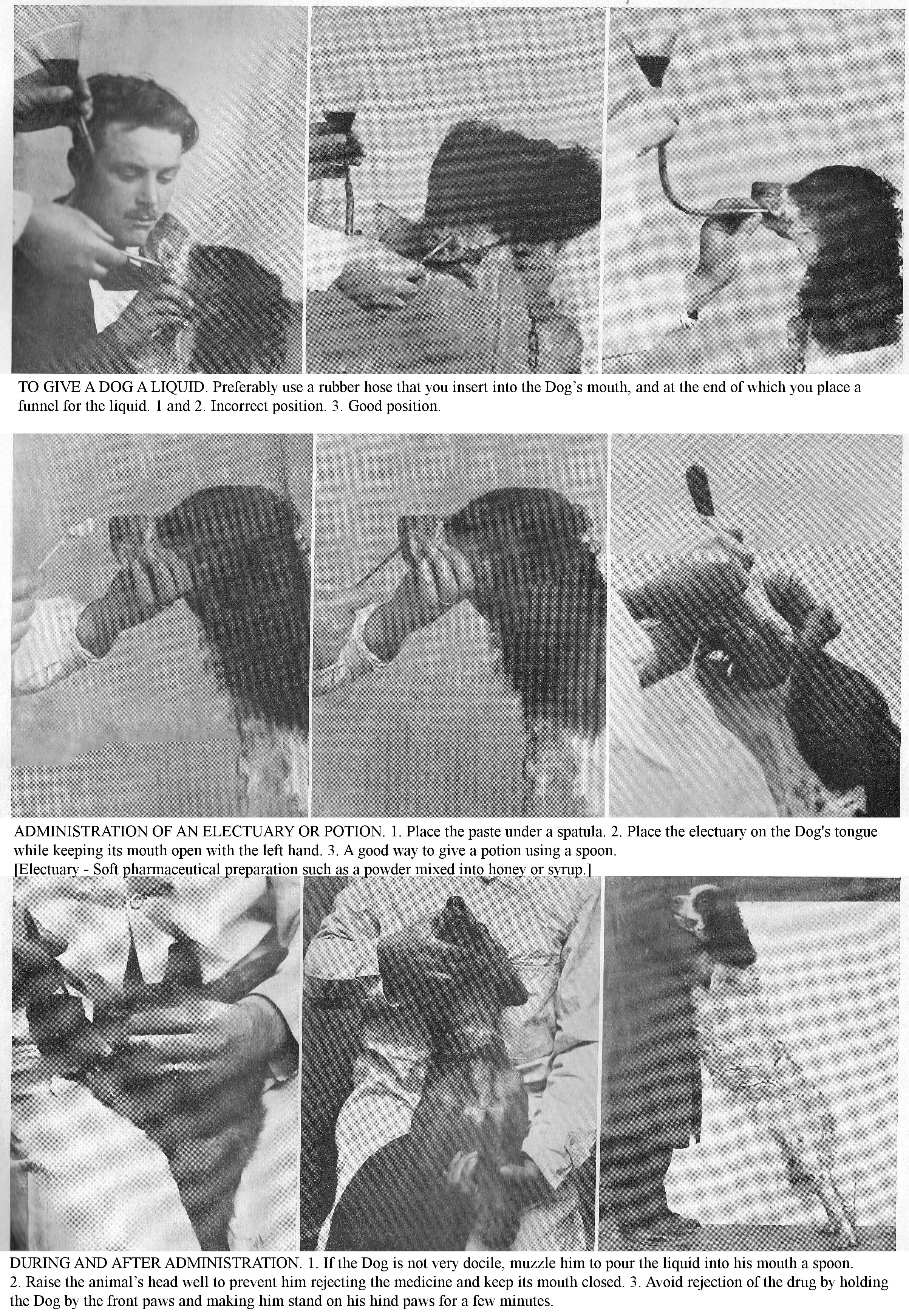
TO GIVE A DOG A LIQUID. Preferably use a rubber hose that you insert into the Dog’s mouth, and at the end of which you place a funnel for the liquid. 1 and 2. Incorrect position. 3. Good position.
ADMINISTRATION OF AN ELECTUARY OR POTION. 1. Place the paste under a spatula. 2. Place the electuary on the Dog's tongue while keeping its mouth open with the left hand. 3. A good way to give a potion using a spoon.
[Electuary - Soft pharmaceutical preparation such as a powder mixed into honey or syrup.]
DURING AND AFTER ADMINISTRATION. 1. If the Dog is not very docile, muzzle him to pour the liquid into his mouth a spoon. 2. Raise the animal’s head well to prevent him rejecting the medicine and keep its mouth closed. 3. Avoid rejection of the drug by holding the Dog by the front paws and making him stand on his hind paws for a few minutes.
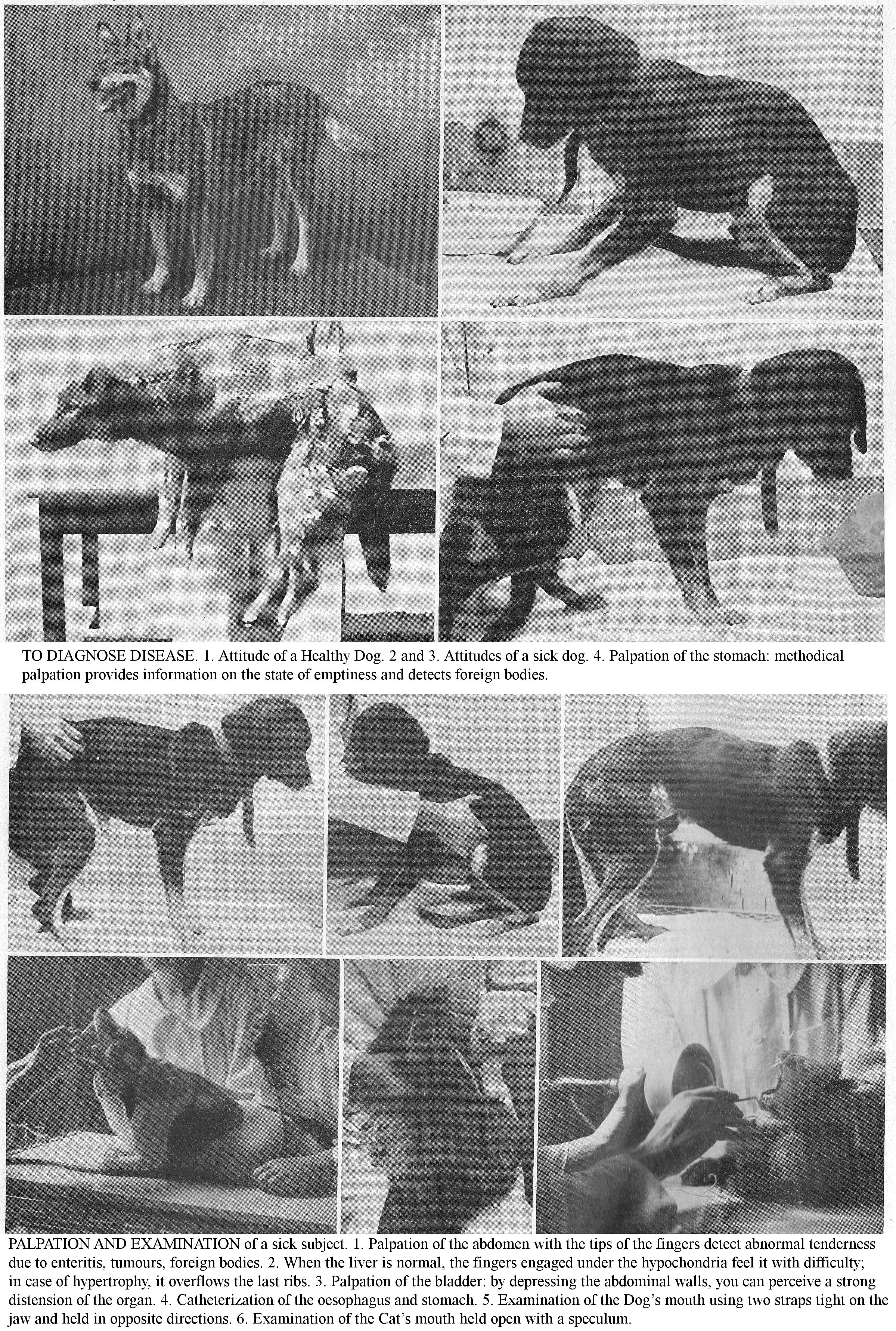
TO DIAGNOSE DISEASE. 1. Attitude of a Healthy Dog. 2 and 3. Attitudes of a sick dog. 4. Palpation of the stomach: methodical palpation provides information on the state of emptiness and detects foreign bodies.
PALPATION AND EXAMINATION of a sick subject. 1. Palpation of the abdomen with the tips of the fingers detect abnormal tenderness due to enteritis, tumours, foreign bodies. 2. When the liver is normal, the fingers engaged under the hypochondria feel it with difficulty; in case of hypertrophy, it overflows the last ribs. 3. Palpation of the bladder: by depressing the abdominal walls, you can perceive a strong distension of the organ. 4. Catheterization of the oesophagus and stomach. 5. Examination of the Dog’s mouth using two straps tight on the jaw and held in opposite directions. 6. Examination of the Cat’s mouth held open with a speculum.
PARASITIC DISEASES OF THE CAT.
The cat's stomach may contain Ascarids, Tapeworms, especially Taenia crassicalis, but the specific parasite is represented by the three-pointed Allulan strongyle [a nematode]; in adulthood, it lives inside the gastric mucosa and is often found there in such quantities that this membrane is soft, red and bruised.
The intestinal parasites are represented by three species of Tapeworm, including the thick-necked tapeworm (length 15 to 60 cm.) which lives in the liver of common rats, Norway rats, mice, voles and water rats in its vesicular state. The symptoms of tapeworm are not distinctive; you notice gradual decline, then loss of appetite; the abdomen is tucked up; afterwards there is profuse salivation, vomiting, and nervous phenomena may also appear.
Ascarids (4 to 6 cm. in length) are found in the small intestine and sometimes in the stomach of young cats; their pathogenicity is low.
At autopsy, you find the parasites widespread from the stomach to the middle of the small intestine, and you will observe chronic enteritis and severe gastric catarrh. Prophylaxis and therapy are identical to those for parasitic illnesses of the Dog.
PARASITES OF THE URINARY SYSTEM.
The Giant Strongyle is a parasite of the Dog’s kidneys. The first visible symptoms are progressive weight loss, complaining, screaming at night; at times the urine is bloody or purulent.
Diagnosis is difficult; on its own, the presence of parasite eggs in the urine is symptomatic of this parasitic illness. Sometimes Strongyles can cause a subcutaneous tumour to form; in this case, have a puncture performed.
PARASITES OF THE RESPIRATORY SYSTEM.
Linguatules [tongueworms]: Quite common in Dogs, in several regions, Linguatules can occupy various parts of the nasal cavities.
Aetiology: Dogs are infected by consuming the viscera of herbivores, usually rabbits and sheep, if they contain larvae and linguatules; the larvae enter the nasal cavities through the throat openings.
Symptoms: Frequent sneezing, more or less marked respiratory discomfort, pruritus which causes scratching of the nose, and mucopurulent discharge, characterize this affliction. Diagnosis: Establish the diagnosis by microscopic examination of the discharge; it will contain eggs of Linguatules.
Treatment: Give nasal cavity injections every 2-3 days with weak cresyl solution, or give sternutatory powder [powder to cause sneezing], or use inhalations of ammonia, chloroform. When Linguatules cause alarming phenomena, have sinus and nasal cavity trepanation performed.
CIRCULATORY SYSTEM PARASITES.
In dogs, haematozoa (filariae worms) are quite frequent; their pathogenicity is extremely variable.
Heartworm: The grievous heartworm lives as an adult in the right chambers of the heart and pulmonary artery. When the parasites are numerous, they cause severe symptoms, dyspepsia, pulmonary congestion, epileptiform phenomena, etc. They can cause an enlarged heart, more or less marked endocarditis, and sometimes the rupture of the myocardium.
The aetiology of this helminthiasis is rather obscure; infection is believed to be through drinking water. In infected pregnant bitches, Galob and Pourquier found embryos of this worm in the blood of the young as well as in that of the mother.
Strongyles: The strongyle of the blood vessels resides in the right chamber and atrium of the heart, in small pea-sized bundles, which can obstruct the opening of the pulmonary artery. Symptoms consist of marked tightness in breathing, coughing fits, irregular heartbeat, and dilation of the jugular veins coinciding with normal appetite and absence of fever. The mode of infection is in drinking water; filtration of the latter, recommended as a preventive means, is not practical. There is no medical treatment because the diagnosis of parasitic diseases of the circulatory system is tricky, if not impossible.
DISEASES OF THE RESPIRATORY SYSTEM
MANY DOGS AND CATS FALL VICTIM TO COLD AND DAMP; AVOID THE CONDITIONS WHICH THEY CAUSE BY GIVING EVERY CARE TO THE HOUSING AND HYGIENE OF YOUR ANIMALS.
By their frequency and severity, respiratory diseases are one of the dominant issues in Canine and Feline pathology. Cold, damp and drafts play a very important aetiological role; therefore plan a hygienic setup for your Kennel; avoid long-haired dogs getting chills from hunting in swamps; dry them completely before returning them to the Kennel. In Luxury Dogs, because of their extreme delicacy, prevent the organic repercussions a cold by using protective coats (wool, felt, etc.). The application of these simple hygienic measures decreases the high losses caused by respiratory diseases to a notable extent.
Examination of the respiratory system includes examination of the nostrils, nasal cavities, larynx, trachea and chest. The narrowness of the nasal cavities in dogs, and particularly in cats, does not allow direct inspection. On the other hand, palpation and percussion of the path of the chambers can inform you about possible deformations caused by tumours, exostoses [bony nodules], etc.
Examine the larynx in Dogs and Cats by holding the jaws apart and lowering the tongue with a spatula. You can see the entrance to the larynx, the movements of the glottis. By causing a sharp sensitivity, external palpation signals the presence and position of foreign bodies (needles, pins, fragments of wire, etc.).
The methods of examining the thorax are: inspection, palpation, percussion and auscultation. Inspection allows you to appreciate the whole conformation of the thorax, congenital or acquired deformities, and respiratory rate. The average number of breaths varies from 12 to 20 in the young dog; to 16 to 18 in adults; to 14 to 16 in the elderly.
By palpation, you can recognize the degree of sensitivity of the chest wall, by percussion, you appreciate the sound of the chest in its different regions. Practice this directly with your hand, or by using a plessimeter.
Auscultation, that is, examination by hearing, is the most valuable method of investigation, but like percussion it requires technical knowledge. It plays a major role in the diagnosis of pulmonary, pleural or cardiac lesions. When you apply your ear to the chest of a healthy animal, you should hear a light, soft, even sound: this is the murmur or vesicular noise. The decrease or weakening of this respiratory murmur may be general or partial, depending on the state of disease. The modalities of inspiration noise are variable: heavy, harsh, gravelly, painful breathing; the character of the exhalation sound are: tubal murmur, pleuritic murmur, crackling, sub-crackling, mucous, cavernous, etc.
CORYZA [COMMON COLD]
Acute Coryza or nasal catarrh is very common in Dogs due to the narrowness of their nostrils and its easy infection when exploring the rubbish of the street; it is often caused by cold and damp, by moving from a warm room to a cold atmosphere. In packs, due to its contagiousness, coryza can take the enzootic form. Secondary Coryzas, acute or chronic, are manifested in the course of various infectious diseases, especially in distemper; some are due to the presence of a tumour or tongueworms in the nasal cavities.
Symptoms: Coryza manifests as frequent sneezing, rubbing and scratching at the nose, through nasal discharge that is first serous, then mucous, finally mucopurulent. When it is abundant, breathing is restricted, sometimes noisy, and, if the nasal cavities are blocked, air enters and exits through the mouth, lifting the lips with each exhale (mouth breathing).
Prognosis: Not serious; symptoms usually subside and healing occurs after a few days. In chronic Coryza, the mucopurulent discharge persists, it may be foetid or blood-streaked. Apart from parasites and tumours of the nose, this form is rare, except in breeds with a short muzzle, where it sometimes results in the pus collecting in the turbinates and sinuses.
Treatment: Clean the nostrils several times a day; coat the nasal cavities with the following preparations: sulphurated petroleum jelly, boric glycerine, mentholated petroleum jelly, etc. In the case of respiratory discomfort, use fumigations of simple water or water containing some eucalyptus leaves, menthol, cresyl, etc. Give internally: sodium benzoate (0.80 to 4 gr.) with diacode syrup. For chronic Coryza, use a few drops of menthol oil, at 1 part in 50.
Prophylaxis: When establishing the Kennels, provide a set-up that prevents the action of cold or damp on young animals. In built-up areas, where contagion is always possible, isolate the patient and disinfect the room.
Tumours of the nasal cavities and sinuses: The nasal cavities and sinuses of dogs can be the site of polyps, epitheliomas, and sarcomas. Surgical intervention (removal after making a gap in the upper wall of the nasal cavities) is indicated in different cases.
ACUTE LARYNGITIS.
Laryngitis, an acute inflammation of the laryngeal mucosa, is often confused with Pharyngitis under the name "Angina."
Aetiology: The predisposing causes are: young age, debility, staying in too hot kennels; mor occasional causes are cooling, caused by drafts, sudden changes in temperature, cold rain showers, trauma to the laryngeal region, irritation of the laryngeal mucosa, by road dust, localised strain following prolonged barking. In winter, you frequently observe cold-induced laryngitis, in luxury dogs sensitive to bad weather. Laryngitis is particularly common among the King's Charles breed.
The determining cause is infection by nonspecific microbes that inhabit the back of the throat or that get there by exudates and become pathogenic when the integrity of the laryngeal mucosa is impaired (frequent erosions caused by swallowing bone fragments).
Secondary laryngitis occurs in the course of various infectious diseases (childhood illnesses, rabies, etc.), or results from the extension of neighbouring inflammation to the mucous membrane.
In large Kennels, Laryngitis can exceptionally take on an enzootic character, either by contagion, or because the causes which provoke it affect many animals at the same time.
Symptoms: The first symptom that comes to your attention is a frequent, nagging cough, first dry and hoarse, then greasy, easily caused by the feeling of cold air. Examination of patients reveals abnormal tenderness in the laryngeal region. After a few days, you will observe a light bilateral foamy discharge. The barking is dull or hoarse; in a few cases, the disease is accompanied by general disturbances (depression, prostration, fever, inappetence) and dyspepsia.
Treatment: Protect the patient from cold, give him hot drinks. Practice revulsion [provoke blood flow to the area] with tincture of iodine, ammoniacal liniment, or use wet and warm compresses with a flannel bandage renewed every two hours. Calm the cough by fumigating with simple steam, or steam with the addition of a few eucalyptus leaves, a few drops of menthol or carbolic acid; internally, administer diacode syrup by the teaspoonful; terpine (0.1 to 1 gr.), sodium benzoate are indicated.
Prophylaxis: Especially for young dogs, avoid sudden changes in temperature, cold rains and staying in damp kennels.
CHRONIC LARYNGITIS.
Aetiology: Termination of the acute form which persists in attenuated form. You will observe chronic laryngitis especially when the acute condition has not been treated or when the patient remains in poor hygienic conditions (cold, damp kennels). The flattened conformation of the short, crushed noses like that of Bull Mastiffs, Pugs, Brussels Griffons, Pekingese, Chow-Chows, various tumours, predispose animals with these anomalies to chronic laryngitis.
Symptoms: The main symptom is heavy, nagging, dry, or slightly oily cough, easily provoked by light pressure exerted on the throat. The Voice is often changed; discharge, especially mucous, is scarce, intermittent or absent.
Treatment: Keep the patient out of inclement weather, do not take him out unless the atmospheric conditions are favourable. Practice revulsion on the throat with tincture of iodine; calm the cough by fumigating and by administering calming potions based on diacode syrup, terpine, sodium benzoate. Use iodized medications (potassium iodide (0.50 to 2 gr.) and arsenicals (Fowler’s liqueur, II to X drops) on alternate days for 10 days. Also use Mont-Dore water, La Bourboule water (1 dessert spoonful to 1 glass per day).
LARYNGEAL PARALYSIS.
This condition, symptomatic in the majority of rabies cases, gives rise to voice and breathing disorders: barking is muffled, breathing is difficult and wheezy, and can cause attacks of suffocation.
ACUTE BRONCHITIS.
Acute bronchitis, inflammation of the mucous membrane of the bronchi, is caused by sudden cooling, especially if damp (rain, sweat, drafts) on young animals and during the seasons with sudden atmospheric variations (Spring, Autumn). In addition to the first disorders caused in the mucosa by the aforementioned environmental factors, is added self-infection caused by bacteria deposited by the air on this membrane. In Kennels, acute bronchitis can become enzootic: when the inflammatory causes act on many subjects at the same time, or by contagion, when the bacteria swarming in the discharge of the first patient become pathogenic.
Symptoms: The patient is sad, depressed, refuses all food and seeks cold drinks; its temperature rises by one degree on average; rapid respiration and pulse, the mucous membranes protrude. You first observe a dry, hard cough, which, after a few days, becomes oily and is accompanied by bilateral discharge, mucous or mucopurulent. Auscultation of the chest indicates a harsh, breathy murmur. Sometimes it resolves itself; the symptoms improve and appetite returns. In severe forms, these signs accompany an intense fever: 39.5 centigrade to 40.5 centigrade, with anorexia and prostration. The coughing period is longer, and the flank is pulled in. In capillary bronchitis, there is marked fever, the dyspnoea is strong, continuous and there is a painful cough. There is abundant discharge, breathing is through the mouth, causing labial breath. The prognosis is particularly severe in young dogs and in small breeds due to the smallness of the bronchial ducts.
Lesions: Inflammatory lesions are seen on the large and medium bronchi; sometimes the mucous membrane is desquamated, ulcerated in places. The bronchial nodes are enlarged and indurated if the disease has progressed slowly.
Treatment: Keep patients resting, away from cold and damp; if indicated, cover the chest with a wadded bandage; give as a diet hot milk, milk preparations, raw or cooked meat. Practice the revulsion on the thorax by sinapized friction [counter-irritation with mustard plasters]. Calm the cough by administering narcotic substances: tincture of belladonna, tincture of opium at a dose of I to X drops, 3 to 4 times a day, diacode syrup, codeine, by the spoonful.
Promote expectoration by using mineral Kermes (0.20 to 1 gr.) [kermesite, a mix of antimony oxides and sulphides] or ipecac syrup, benzoic acid, terpine (0.10 to 1 gr.), etc. Give simple, mildly antiseptic steam fumigations; fight weakness and debility, with hot infusions of coffee or lightly alcoholic tea.
Prophylaxis: Avoid cold, damp, drafts; in cold and wet weather provide delicate dogs of small breeds with protective coats.
CHRONIC BRONCHITIS.
This ailment is one of the end results of acute bronchitis. It can occur as a result of repeated colds, but it is often a consequence of a predisposition to acne or herpes infections.
Symptoms: The cough is greasy, nagging, and accompanied by mucopurulent discharge. Brisk exercise quickly causes shortness of breath and fatigue. On auscultation of the lung, you will notice mucous and wheezing groans. The thoracic sound is abnormal on percussion. When coughing fits are frequent and violent, Bronchitis becomes complicated by emphysema. As chronic bronchitis is often of tuberculous origin (Cadiot), have tuberculination performed.
Treatment: Over-nourish the patient (mash containing a lot of meat, raw meat, dairy products). Facilitate expectoration at the beginning with mineral Kermes (0.20 to 1 gr.). Modify bronchial secretions with tar, creosote (0.20 to 1 gr.), Terpine (0.0 to 1 gr.), potassium iodide (0.50 to 2 gr.); calm frequent and painful coughs with aqueous extract of belladonna (0.15 to 0.20), aconitine granules, etc. Modify the general condition with Fowler's liquor (II to X drops); cod liver oil (1 to 2 tablespoons).
Prophylaxis; Protect the Dog from the effects of cold and damp. Treat acute bronchitis right away to prevent it becoming chronic.
BRONCHO-PNEUMONIA.
This disease, which plays a large role in canine mortality, most often follows Bronchitis and is also due to cold. Watchdogs, Dogs hunting in winter, on the water or in the marshes, Dogs traveling by rail, Apartment dogs that you take out in cold, wet weather, or that you bathe in cold weather, etc., are prone to Broncho-pneumonia. Foreign bodies (drugs, food), accidentally entering the windpipe, bronchi and lungs, cause Broncho-pneumonia. Often secondary broncho-pneumonia is a childhood disease. This condition results, in reality, from an increased virulence of saprophytic microbes due to either physical predisposition or to contagion. Sporadic broncho-pneumonia quickly becomes contagious to other animals in the Kennels; always consider it as such in a built-up area or in a breeding kennels.
Symptoms: At first you notice depression, weakness, decreased appetite, strong thirst and infrequent dry cough; respiration and pulse are rapid; the nose is dry and hot, there is a marked fever, 38.5 to 39.5 centigrade. These symptoms worsen rapidly, the breathing becomes dyspnoeic, the labial murmur is very marked, the Dog remains seated on its hindquarters, the forelimbs spread apart to facilitate breathing; a viscous, greyish or bloody discharge flows from the nostrils. Percussion of the chest denotes decreased sound, sometimes dullness; auscultation detects mucous rattles, crackling rattles, and often a tubal murmur. Complications soon appear in the liver (jaundice) and intestine (enteritis with vomiting and diarrhoea). The disease can progress very quickly, and death from asphyxiation, toxaemia and uraemia occurs within days.
Lesions: Lesions are most often limited to a single lung and generally sit on the lower or deeper parts of the lung. The diseased lung tissue forms a homogeneous, compact mass, very friable, reddish-brown in colour; caseous islets (purulent foci) or gangrenous islets may be observed. Prognosis: Very serious, death is the usual end-result.
Treatment: Attach great importance to health care during winter, spring and in wet and cold weather; isolate and put any Dog or Puppy which coughs or has nasal discharge (Coryza) in a warm and ventilated room; coat his nostrils with gomenolated [essential oil of niaouli] or mentholated petroleum jelly, put him on a milk diet, give him plenty of clean, dust-free straw. Strictly observe these precautions in packs, because any sporadic broncho-pneumonia can quickly become contagious and infectious; the virus is present in discharge, phlegm, sputum, faeces and urine.
Chest revulsion forms the basis of treatment; achieve this by prolonged rubbing with mustard flour, tincture of iodine, or have your veterinarian make a fixation abscess in the sternal region; do not use antimony ointment rubs for revulsion due to their toxicity when licked. Calm the cough with the appropriate preparations for treating Bronchitis; facilitate expectoration with mineral kermes, sodium benzoate; combat cardiac weakness with subcutaneous injections of ether, camphor oil, or caffeinated camphor oil (1 to 4 cm), Lower the temperature with febrifuges, antipyrine (1 to 2 gr.), quinine (0.5 to 2 gr.); combat physical infection with colloidotherapy (collargol, electrargol, etc.). Use serotherapy with injections of polyvalent anti-streptococcal serum.
The recovery period is slow; reduce it by good health care (progressive exercise in the sun); support the patient with raw meat and give them spoonfuls of hot sweet milk, simple broth or broth with the addition of minced meat; if necessary, use food enemas; use the following formula: a cup of milk, a tablespoon of peptone, an egg yolk and a few drops (V to XV drops) of laudanum to prevent diarrhoea.
Prophylaxis: The frequency and severity of this disease, the high losses it causes (75% to 80%) in kennels, emphasise the importance of planned prophylaxis. Isolate the patient and those suspected of having illness; disinfect the Kennels. Avoid cold and damp; in cold weather, use coats (in woollen fabrics) which protect the chest, especially for luxury Dogs. Administer drugs without getting them into the airways.
ACUTE PNEUMONIA.
This condition, rare in dogs, is almost always caused by cold, so you usually see it in late autumn or during winter, especially in dogs hunting in swamps. Almost all cases of Pneumonia are a manifestation of distemper.
Symptoms: Lobar pneumonia is initially characterized by signs of depression and weakness, fever, anorexia, sharp thirst, rapid breathing and a troublesome, painful cough. Subsequently, mucous discharge appears, blood-streaked or rusty. Auscultation indicates a crackling rattle in one or both lungs, followed by tubal murmur. Percussion first reveals reduced sound, then dullness. You may observe complications of pleurisy, pericarditis, and nephritis during this period. When close to recovery, the symptoms subside on the 6th to 8th day and appetite returns. Treatment: The therapeutic prophylactics are those indicated for Broncho-pneumonia.
CHRONIC PNEUMONIA.
Chronic uncomplicated pneumonia is very rare. In dogs, almost all chronic lung lesions are associated with Tuberculosis (Cadiot). Symptoms: This condition is characterized by cough, a little discharge, particular physical signs (dullness, reduced sound, rattles, etc.), short, rapid breathing, disturbed appetite, weakness and progressive weight loss. Have the patient subjected to the tuberculin test; if there is discharge, have a bacteriological examination of the discharge done. Treatment: Over-nourish the patient, lots of raw meat; use iodized or creosoted cod liver oil (1 to 2 tablespoons).
PLEURISY.
Pleurisy, or inflammation of the pleura, is quite common in dogs; the main predisposing and occasional causes are: prolonged chilling of sweaty, wet animals exposed to drafts; cold and wet seasons; previous infectious illnesses (rheumatic diseases, childhood diseases, etc.); infected chest wounds (gunshot wounds, injuries from horns of cattle, deer, wild boar, etc.). The causal agent is a microbial infection (staphylococci, streptococci, diplococci, pasteurellas, etc.), acting sometimes by physical predisposition, sometimes by local penetration directly into the thorax.
Symptoms: Pleurisy is announced by signs of depression and weakness, fever, chills, sharp thirst, inappetence, difficulty breathing, with rapid movements of the ribs, by a small, dry, painful cough, abnormal tenderness of the chest walls in response to percussion. Depending on whether the pleurisy is bilateral or unilateral, the percussion produces very clear dullness on both sides or on the diseased side only. By holding the patient in various attitudes, the physical signs are not modified by displacement of the effusion.
Diagnosis: To confirm diagnosis, have an exploratory puncture performed; to identify the nature of the causal illness, have the tuberculin test performed. Prognosis: Pleurisy may end with recovery, death, or transition to a chronic condition. Recovery occurs when the effusion is scarce; it is heralded by the gradual return of appetite and by an improvement in general condition. The convalescence is long, often lasts 2 or 3 weeks, during which relapses are to be feared. Death is the most frequent ending; either quickly due to asphyxiation or late on due to exhaustion.
Lesions: The liquid effusion is variable in colour, density and abundance, is sometimes amber, lemon yellow, whitish, purulent, holding in suspension the debris of false membranes, soft and spongy and pale yellow in colour. The heart, liver and kidneys show asphyxic lesions.
Treatment: Keep the patient resting, away from cold and damp. Feed it raw or cooked meat. If he refuses meat, support him with liquid foods: milk, broth with the addition of minced meat. Practice chest revulsion, using a revulsive friction (antimony ointment, tincture of iodine, mustard flour), or use a shunt with the aid of fixation abscesses (injection of turpentine); use the specific action on the serous membranes of calomel (0.25 to 1 gr.) and sodium salicylate (0.3 to 1.5 gr). Combat fever with antipyrine (1 to 2 gr.); combat cardiac weakness with tincture of digitalis (XX to L drops,). If there is severe dyspnoea, have thoracentesis performed aseptically.
Two new therapeutic methods, aerotherapy (injection into the pleura of a quantity of air approximately equal to that of the extracted exudate) and autoserotherapy (injection into the subcutaneous connective tissue of 1 to 10 cubic cm fluid extracted from the pleura) can be used.
Prophylaxis: Avoid the effects of prolonged cold on sweaty, wet animals exposed to drafts; treat chest wounds aseptically.
CHRONIC PLEURISY.
Chronic Pleurisy can be an end-result of Acute Pleurisy, but it is also observed following repeated chills (unhealthy kennels) affecting animals with a predisposition.
Symptoms: Inflammation affecting the pleura slowly becomes apparent; you first notice only a small dry cough, erratic appetite and dull, prickly coat. The patient becomes breathless following the slightest exercise; he loses weight, his mucous membranes turn pale. His temperature remains more or less normal while Bacillary pleurisy is accompanied by frequent bouts of fever. Before starting treatment, test the patient for tuberculin; perform a bacteriological examination of the exudate to determine whether or not the pleurisy is tuberculous. Prognosis: Very serious, recovery is uncertain, often incomplete, and requires a long convalescence.
Treatment: Support the patient with a tonic diet; give plenty of raw or cooked meat. Use the same treatment as that indicated for Acute pleurisy; use the same prophylaxis.
EMPHYSEMA AND ASTHMA.
Pulmonary emphysema is seen quite frequently in older dogs; this condition is characterized by rapid and difficult breathing, rapid panting, and mouth breathing when the animal is forced to run, often also by fits of dry coughing.
Basic asthma presents with attacks of dyspepsia consisting mainly of great difficulty in exhaling.
Treatment: Ensure good hygiene and a good diet for emphysema patients. Give them iodized and arsenical medications; ten days a month, give a teaspoonful to a dessert-spoon or soup-spoon of the following solution: potassium iodide, 1 to 5 gr.; distilled water, 100 to 200 cc. On the other 10 days, give 1 to 6 drops of Fowler's liquor. Combat asthma attacks with sedatives (ether, chloral, diacode syrup, etc.) or hypodermic injections of morphine hydrochloride.
PNEUMOTHORAX.
Pneumothorax is produced by a gas effusion in the thoracic cavity, caused by puncture of the lung or parietal pleura (penetrating wound of the chest, rib fracture with torn lung, poorly performed thoracentesis, etc.).
Symptoms: From the start, dyspepsia is intense; percussion on the affected side gives a tympanic sound; auscultation reveals amphoric breathing [low pitch bronchial breath with high pitch overtones].
Treatment: In the case of traumatic pneumothorax, perform aseptic closure of the chest wound; calm dyspnoea with subcutaneous injections of morphine; combat weakness by administering a mildly alcoholic tea or coffee infusion.
THE MAIN NUTRITIONAL DISEASES
A POORLY BALANCED DIET CAUSES DISORDERS THAT RESULT IN FUNDAMENTAL DECLINE OF THE ANIMAL
IT is necessary to give your animals a nutritionally balanced diet to provide the essential elements for maintaining the body. Modern studies have made immense progress in nutritional matters, and the name of deficiency diseases is currently reserved for conditions caused adults and young people by a deficient diet, that is to say, a diet lacking, more or less completely, various basic principles required for maintain the body and health.
DEFICIENCY DISEASES.
Protein Deficiency: The body constantly needs protein because living matter is in a continual state of destruction; this constant need for albumin is so imperative that an animal subjected to a protein fast, succumbs as if you were imposing an absolute fast; it resists for longer, but death is always the end result. Protein deficiencies in the diet of Dogs and Cats can relate either to the amount of albuminoids distributed, or to the chemical composition. Amide acids [proteins & amino acids], derived from prolifics, must be supplied in sufficient quantities in food; if there is a deficiency in a youngster, growth stops and death may result.
Mineral deficiency: The mineral content of various organic tissues implies that minerals play an essential part in the constitution and the formation of all organs, and that minerals are essential for life activity. Mineral requirements, especially in the young, are an imperative physiological need. Mineral deficiency diseases cause arrested growth and rickets.
Symptoms: Gauntness, despite providing copious meals, the appearance of nervous disorders, poor general condition despite the conservation of appetite, signs of diarrhoea, and paralysis, are signs of mineral deficiency. Poor hygiene and care conditions cause such deficiencies to evolve more quickly; they place the organisms in a reduced state of resistance, facilitate infections and parasitic infestations. Deficiency disorders (protein and mineral deficiency, lack of vitamins) have a negative effect on fertility.
Prophylaxis: Prophylaxis lies entirely in correct feeding: provide the body, through a correct diet, with all the materials necessary for its maintenance and growth; remember that protein and mineral requirements and the vitamin content are physiological essentials.
VITAMIN DEFICIENCY.
Recent research has shown the important role played in the body by ill-defined substances – vitamins - which are found in the plant kingdom (fresh herbs, roots, tubers, etc.) and in the animal kingdom; (cod liver oil, egg yolk, milk, etc.). Current experience tends to admit the need for nutrition to contain, in addition to the ordinary principles (albumin, fat, carbohydrates, salts, etc.) "fermenting elements" which allow these to be used by the body. Vitamin deficiency quite quickly leads to nervous or paralytic disorders. The prevention of vitamin deficiency is through nutrition; give a mixed animal and vegetable diet, and give youngsters eggs, milk, and cod liver oil; foods rich in vitamins.
ANAEMIA, LYMPHADENIA AND LEUKAEMIA.
Anaemia is characterized by a decrease in blood cells, with a marked increase in the amount of serum; there is both Anaemia and Hydroaemia.
Lymphadenia and Leukaemia are rare conditions characterized by alterations in lymph organs and the blood.
Aetiology. Among the causes of Anaemia, a fairly frequent morbid condition in Dogs and Cats, we can cite incorrect diet - too uniform, insufficient or lacking in vitamins - copious haemorrhages, chronic diseases, parasitic diseases, poor hygiene conditions, etc.
Symptoms. Pale mucous membranes, weakness, and rapid fatigue caused by the slightest effort, are the main symptoms of Anaemia. Appetite is decreased and irregular, digestion is disturbed. On examining the circulatory system, you will notice a weak pulse, often palpitations, sometimes a slight heart murmur. Prognosis: The severity of the prognosis varies with the causes, the degree of blood changes and the amount of blood loss.
Treatment: Investigate the causes of Anaemia and combat it with the appropriate treatment. Treat the subjects with good hygiene and boost their diet; institute a mixed diet especially meat: raw meat, meat pate. In case of inappetence, stimulate the appetite with stomachic preparations (gentian tincture (0.05 gr. to 2 gr.); Cinchona (3 to 15 gr.); kola (0.05 gr to 0.2 gr.), etc. Use iron tonic medications (iron protoiodide, citrate, lactate, 10 to 50 gr., etc.) and arsenical preparations (Fowler's liquor); to young dogs, preferably administer cod liver oil and phosphate of lime syrup (1 teaspoonful, dessert-spoon or tablespoon). In the case of profound anaemia, give a subcutaneous injection of 10 to 100 g of lukewarm salt water every two or three days at 9 parts per 1000. Prophylaxis: Prevent Aanemia by giving a highly digestible vitamin-rich diet, by good hygiene and by regular exercise in the open air.
RICKETS.
Rickets is a bone disease seen in young Dogs and Cats, characterized by physiological ossification disorders leading to transient or lasting skeletal deformities.
Aetiology: Primarily a childhood condition, occurring either while in the womb or during nursing and weaning; breed also plays an important aetiological role. Observation shows that certain subjects, particularly Great Danes and Mastiffs, are predisposed to Rickets. Heredity also has a very clear role, especially when perpetuated a female with osteomalacia nursing [see note], as does faulty hygiene of the premises (staying in narrow, damp kennels). Mineral deficiency, food that is insufficient, too uniform, or too low in nitrogen compounds. Dietary deficiency, due to stinginess or lack of vitamins in food, leads to Rickets.
[Translator’s note: osteomalacia is bone decalcification caused by poor mineralization/demineralization (lack of calcium and phosphorus) in the protein matrix of the skeleton. It is the adult equivalent of childhood rickets]
Symptoms: Rickets proceeds somewhat slowly. Usually the Puppy is sad and dejected, and has difficulty moving. Its digestive system functions poorly: the appetite is irregular and sometimes depraved [pica]; gastritis and enteritis are not unusual. Then bone deformities occur; the joints are deformed, and palpation reveals the existence of breaks and irregular growths. The forelimbs deviate severely from the vertical and can resemble very disparate figures: K. D. X. In exceptional cases the spine may curve upwards (Kyphosis), in the opposite direction (Lordosis) or laterally (Scoliosis). When the rib joints with their cartilaginous extensions are affected, you will see nodules which gradually increase in volume (rickets).
The rickety gait is characteristically hesitant and irregular; and patients often walk on the carpus or tarsus, kneeling or squatting. It is not uncommon to see spontaneous fractures that heal quite easily, but which leave permanent deformities. Left untreated, the disease ends in consumption, stagnation and death.
Lesions: Rickets lesions show as disordered ossification in the epiphyseal cartilages. All the bone tissues undergo profound nutritional modifications which make the skeleton a fragile, light framework; the marrow becomes diffluent [mushy], pinkish and gelatinous, the periosteum is inflamed. On chemical analysis, the minerals in the bones, mainly phosphates, were half of what they should be.
Diagnosis: Diagnosis is easy once the bony deviations appear. Do not confuse Rickets with Infectious Polyarthritis or Rheumatic Arthritis, which have rapid onset with symptomatic inflammation and joint cavity swelling. The manifestation of Rickets, on the contrary, is symptomised by a slow course, absence of inflammation or joint congestion; the clinical picture is dominated by bone lesions. Prognosis: If treated from the start, Rickets quite frequently resolves, but the subject remains small, sickly, and weak.
Treatment: Good feeding is the basis of treatment; build up the patient with organic phosphates, particularly a decoction of cereals and bone powder, or use lime hydrochlorophosphate syrup (2 to 3 tablespoons per day), cod liver oil (1 to 2 tablespoons per day), lecithinated or phosphorated cod liver oil (phosphorus, 1 mgr.). For Luxury Dogs, use thyroid opotherapy; take half a lobe (about 75 cgr.) of fresh sheep thyroid; finely chop in slightly warm milk or broth, and give this to the patient for 10 days on an empty stomach; give this treatment and repeat the cycle for 6 weeks. Local treatment is not important: rub the limbs daily with dry flannel, or with one soaked in a topical stimulant (brandy or camphorated alcohol). Prevent limb deformation by using bandages.
Prophylaxis: The prevention of Rickets, deduced from its aetiology, is very complex; it concerns both the mother and the offspring. Feed the mother intensively during pregnancy and lactation; give an easily digested and varied diet ensuring she consumes mineral protein and vitamins. Leave the mother with a number of offspring commensurate with her strength and milk yield; ensure the young suckle copiously and wean them late and progressively. Increase the mineral content of the diet by adding phosphate flour; avoid undernutrition, insufficient exercise, and damp, narrow kennels. Do not breed from animals that are too old or show signs of vital decline, and especially not from those suffering from a tendency to soft bones. You will thus remove the aetiological causes of Rickets, a condition which often compromises the future of a particular line of purebred dogs.
DIABETES MELLITUS, DIABETES INSIPIDUS.
Diabetes, a relatively common disease in dogs, have the common features of eating disorders and excessive urination, polyuria. In diabetes proper, or Diabetes Mellitus, the urine contains varying amounts of sugar. In Diabetes Insipidus, the urine is qualitatively altered, but does not contain sugar.
Diabetes Mellitus: Aetiology. This ailment is found mainly in luxury breeds and elderly animals. Its main causes are eating too much food or too rich food, the overuse of treats, sweets, lack of exercise, strong emotions, head trauma, liver and pancreatic damage.
Symptoms: Due to its insidious onset, diabetes is usually not diagnosed until late. Glycosuria is a fundamental symptom; normally the blood sugar varies between 1.5 gr. per 100 to 7.5 gr. In glycosuria, the sugar excreted in urine can reach up to 100 g. per litre; this is yellowish, stringy, syrupy, and the Feehling reaction (cupro-potassic liquor) gives a clearly reddish precipitate. Polyuria is characteristic: the Dog urinates everywhere and often, even if it is house-trained. The patient has a raging thirst. Finally, weight loss and sluggishness quickly become extreme (diabetic wasting), but sometimes there are Diabetics who remain overweight (fatty diabetes). During the course of the disease, most people develop double cataracts, which sometimes leads to blindness within a few weeks. You also observe digestive disorders, vomiting, alternating constipation and diarrhoea, eczematic rashes, persistent bronchitis, paralytic or epileptiform incidents. Wounds heal badly; they suppurate and develop an ulcerative appearance. Suspect the following as signs of Diabetes: polyuria, extreme thirst and weight loss; confirm the diagnosis by urine analysis. Prognosis: Very serious; the duration of the illness varies between 4 and 8 months and ends in diabetic coma.
Treatment: Diet is the basis of treatment: remove starchy and sugary foods; cut down on cooked meat. Give small amounts of raw meat, milk and especially herbal soups. Distribute Vittel, Vais, or Vichy water or a baking soda solution (4 to 5 gr. per litre). Combat diabetic coma with lightly alcoholic coffee and tea, injections of caffeine, ether or camphor oil (1 to 4 cc); use sodium salicylate, 10 gr. Antipyrine in 300 gr. water at the rate of 3 to 4 tablespoons per day. Only perform urgent operations and in conditions of rigorous asepsis, watch carefully for wounds.
Prophylaxis: Avoid, through sensible feeding, overeating, overuse of treats and sweets; give the Dogs regular exercise.
OBESITY.
Obesity is a morbid condition characterized by excessive overweight. More common in female dogs, during adulthood or old age, it is caused by: over-feeding, the overuse of starchy and sugary foods, insufficient exercise. The obese dog has slow, painful digestion; he is sluggish, feverish and quickly becomes breathless from the slightest effort. Obesity promotes a tendency to arthritis and often results in impotence and relative sterility in breeding stock. Treatment: Treatment is in the realm of care and feeding. Avoid sugar and pastries, eliminate or reduce starchy foods, reduce the rations, and make the obese dog take long walks on an empty stomach. Speed up under-nutrition using alkalis (baking soda, 5 cgr. to 4 gr.), weekly purging (5 to 50 gr. Of castor oil) and iodine medication (tincture of iodine, X drops in a little milk before the meal), for periods of 10 days, separated by 10 day intervals. Prophylaxis: Avoid overeating, sweets and lack of exercise.
MUSCULAR RHEUMATISM.
Muscular rheumatism mainly affects older and obese dogs, those who stay in kennels or in damp kennels or who hunt in the marshes. It is quite common during the cold season. In all subjects, an initial bout of the disease favours its return, and sometimes muscular rheumatism alternates with articular rheumatism.
Symptoms: Muscular rheumatism can progress in acute, subacute or chronic forms. It is rarely generalized; most often it is localized to a muscle group (neck, back, lower back and upper regions of the limbs). The affected muscles are slack, very painful on pressure; patients avoid contracting them, and the corresponding joint is immobilized. There is intense lameness, the animals have great difficulty moving around, and when lying down they have great difficulty getting up. You do not see any localised swelling or inflammation, only great sensitivity to palpation. The pain is sometimes moderated by atmospheric influences; it increases in cold, damp weather and is worse at night. The condition, unlike rheumatoid arthritis, usually progresses without fever.
Prognosis: The course, duration and termination of muscular rheumatism are very variable. Complications of the serous membranes (pleura, pericardium, endocardium) can occur. When the disease takes the chronic course, the muscles atrophy and you may observe paralysis.
Treatment: Protect the patient from cold and damp; give them a mixed diet or milk diet; add Vichy water, Vais water, or baking soda to the milk; calm the sharp pains using bromide medication, or give two to fifteen drops of colchicum seed tincture in milk. Lightly massage the affected areas with flannel or calming ointments.
Prophylaxis: Avoid cold and damp in the premises (kennels and sleeping boxes). Before bringing them in, thoroughly dry any Dogs that hunt in swamps or in rainy weather.
DISEASES OF THE CAT.
FELINE PATHOLOGY is very similar to canine pathology; the most frequently observed ailments are as follows:
DIGESTIVE SYSTEM: Gastroenteritis enteritis, jaundice, cirrhosis, ascites, helminthiasis.
RESPIRATORY SYSTEM: angina, coryza, bronchitis.
CIRCULATORY SYSTEM: anaemia.
LOCOMOTOR SYSTEM: trauma, fractures.
GENITOURINARY SYSTEM: testicular tumours, metritis, pyometritis.
MICROBIAL AND MISCELLANEOUS DISORDERS: tuberculosis, rabies; tumours: adenoma, carcinoma, epithelioma, fibromyoma, etc.
Refer to the different chapters of this Issue.
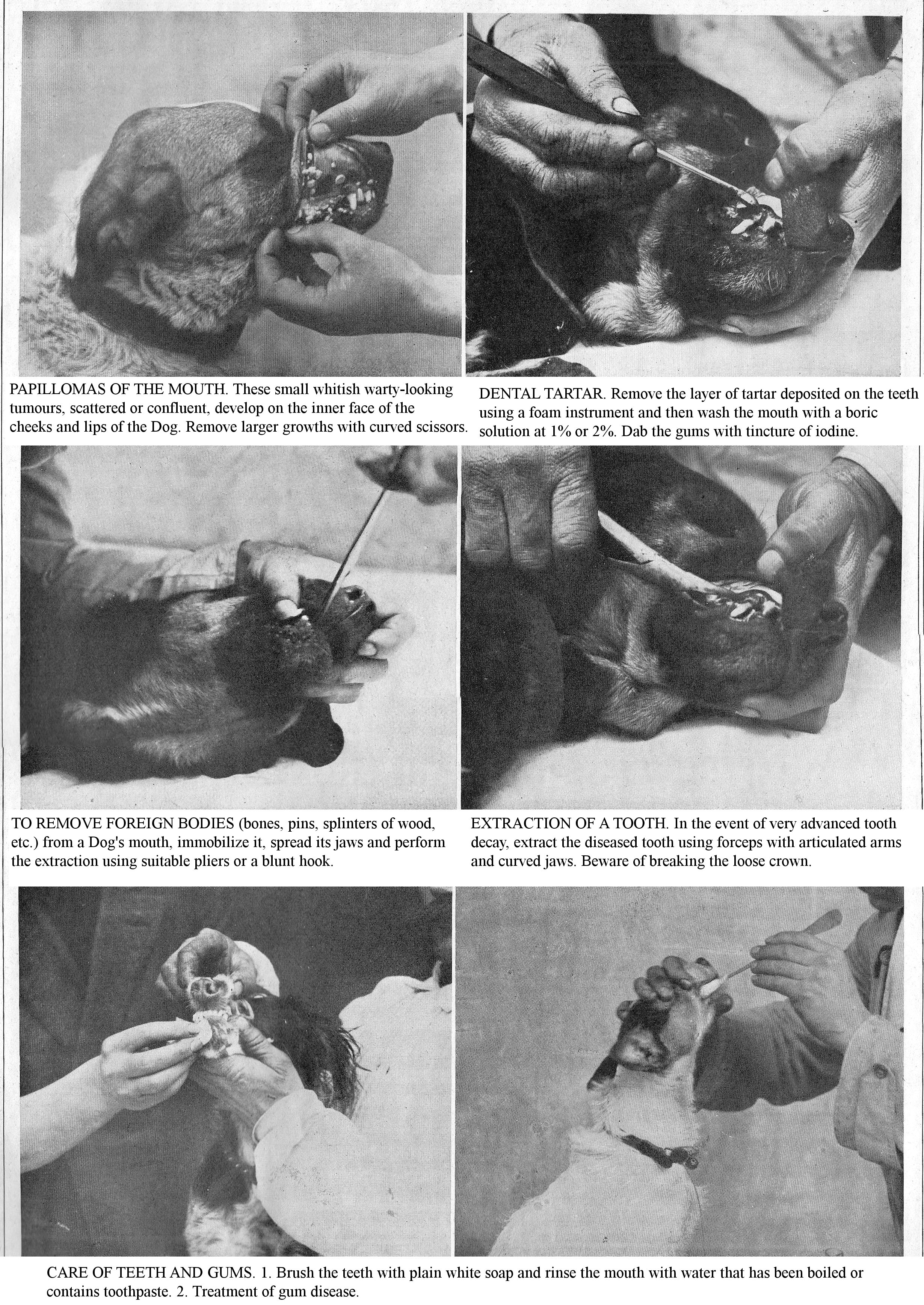
PAPILLOMAS OF THE MOUTH. These small whitish warty-looking tumours, scattered or confluent, develop on the inner face of the cheeks and lips of the Dog. Remove larger growths with curved scissors.
DENTAL TARTAR. Remove the layer of tartar deposited on the teeth using a foam instrument and then wash the mouth with a boric solution at 1% or 2%. Dab the gums with tincture of iodine.
TO REMOVE FOREIGN BODIES (bones, pins, splinters of wood, etc.) from a Dog's mouth, immobilize it, spread its jaws and perform the extraction using suitable pliers or a blunt hook.
EXTRACTION OF A TOOTH. In the event of very advanced tooth decay, extract the diseased tooth using forceps with articulated arms and curved jaws. Beware of breaking the loose crown.
CARE OF TEETH AND GUMS. 1. Brush the teeth with plain white soap and rinse the mouth with water that has been boiled or contains toothpaste. 2. Treatment of gum disease.
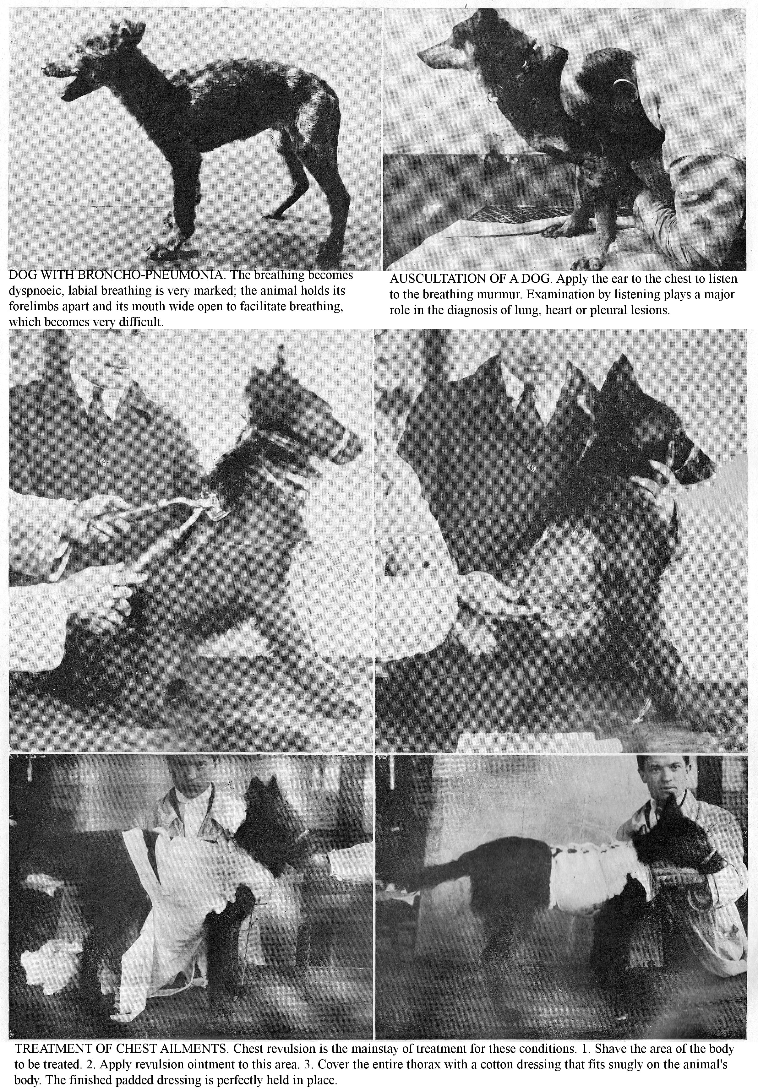
DOG WITH BRONCHO-PNEUMONIA. The breathing becomes dyspnoeic, labial breathing is very marked; the animal holds its forelimbs apart and its mouth wide open to facilitate breathing, which becomes very difficult.
AUSCULTATION OF A DOG. Apply the ear to the chest to listen to the breathing murmur. Examination by listening plays a major role in the diagnosis of lung, heart or pleural lesions.
TREATMENT OF CHEST AILMENTS. Chest revulsion is the mainstay of treatment for these conditions. 1. Shave the area of the body to be treated. 2. Apply revulsion ointment to this area. 3. Cover the entire thorax with a cotton dressing that fits snugly on the animal's body. The finished padded dressing is perfectly held in place.
DISEASES OF THE CIRCULATORY SYSTEM
THROUGH PALPATION, FINGER PERCUSSION, AUSCULTATION AND EXPLORATION OF THE PULSE, ESTABLISH AN EARLY DIAGNOSIS OF THESE CONDITIONS TO AVOID INCURABLE CHRONIC FORMS.
IN CANINE PATHOLOGY, heart ailments often go unnoticed; thus, various significant heart lesions are found at autopsy. The aetiology of heart disease can be classified into four broad groups: 1, diatheses; 2, infection; 3, overwork; 4, miscellaneous accidents. Heredity, arthritis and rheumatism are predisposing tendencies. Infectious diseases (distemper, contagious bronchopneumonia, tuberculosis, septicaemia, etc.) play an important role as underlying causes. Colds, bad weather, shocks, bruises, and traumas, all predispose an individual to cardiac afflictions. "Cardiacs" are particularly observed in those where overwork is a factor [Hunting, coursing (greyhound), draught dogs].
Consider rapid panting, swelling of the lower parts, mainly of the limbs, as suspicious signs of heart damage; have the integral functional of the heart checked by your veterinarian. Specific treatment given from the start can halt the gradual progress of these conditions, which significantly reduce longevity and often undermine the economic usefulness of animals.
Semiology: The heart is located in the thoracic cavity roughly in the median plane. Examining the circulatory system includes clinical exploration of the heart, examining the pulse, and examining the blood. Practice palpation by pressing your hand flat in the heart zone; it will tell you the intensity and regularity of the heartbeat. Use percussion with a finger or plessimeter, to determine the extent of physiological cardiac dullness, as well as variations in disease states. Practice auscultation, which, like percussion, requires professional knowledge, either directly or using a stethoscopes. It tells you about normal or pathological heart sounds, and about the intensity and rhythm of the heartbeat. The heart auscultation zone in Dogs is found on the left, between the 4th and 6th ribs, and on the right between the 4th and 7th ribs.
The normal heart sounds are two in number: a first noise or large noise, a little heavy, which lasts quite a long time, and a second noise, or small noise, which is clear, metallic, and of shorter duration. These noises can be altered in their location, extent, intensity, timbre and rhythm. Pathological noises include: a breathy sound comparable to that of wind coming out of a bellows; this noise varies in terms of intensity and duration (systolic, diastolic, presystolic murmur) and caused by the failure of the mitral, tricuspid, sigmoid valves, etc.
The number of heartbeats in a given time, and the regular order in which the movements follow one another, may be disturbed by illness (intermittence, doubling).
Examine the pulse on the upper part of the inner side of the thigh. It has enormous clinical importance, allowing the frequency, quality, and regularity of the pulse to be recorded. The pulse rate varies in the young dog from 110 to 120; in adults, 90 to 110; in the old dog, from 70 to 80. In the cat, it varies from 110 to 140 per minute.
Examine the vascular condition of the visible mucous membranes (conjunctiva, mouth, vulva, etc.); this varies with states of congestion, anaemia, etc. Paleness mucous membranes indicate Anaemia, Cachexia; flushed coloration indicates an overly congestive state.
Blood examination is sometimes essential for the diagnosis of some diseases; the external appearance, shade, degree of colouring, and speed of coagulation provide valuable information. Microscopic examination is sometimes necessary to get an accurate diagnosis.
ACUTE PERICARDITIS.
Pericarditis, the inflammation of the serosa surrounding the heart, occurs in the course of various infectious diseases, pneumonia, distemper age and especially tuberculosis (Cadiot). Dogs that hunt in marshes, as a result of prolonged immersion in cold water, are predisposed to this condition.
Symptoms: The disease is marked by signs of depression, weakness, inappetence, rapid breathing, anxiety, dyspnoea, weakness, fever or disappearance of cardiac shock, by a large area of dullness corresponding to the distended pericardium, by the presence of a marked venous pulse. Percussion denotes a marked sensitivity of the region. Death is due to asphyxiation resulting from pulmonary engorgement, fatigue and heart failure; it is preceded by extreme dyspnoea; often the disease becomes chronic.
Diagnosis: Very tricky at first, the slight rise in temperature, general condition of the patient, frequency and irregularity of the heartbeat suggest heart disease or pleurisy. After the exudative period, diagnosis is easier; dullness, localized to the cardiac zone, the disappearance or attenuation of cardiac shock and especially the presence of the venous pulse characterize acute pericarditis.
Prognosis: Severe in all cases.
Lesions: The pericardium is distended by sero-fibrinous exudate; false membranes line the layers of the serosa; the effused fluid is very albuminous, generally yellowish and clear, sometimes bloody or even purulent; the lungs and liver are congested and infiltrated.
Treatment: Use revulsions and vesicants applied to the entire extent of the dull area; internally, administer very low doses of calomel (10 to 15 cgr.); combat cardiac weakness by administering of digitalis, caffeine, diffusible stimulants, subcutaneous ether injections; address dyspnoea with opiates, subcutaneous injections of morphine; when the effusion is very abundant and the symptoms alarming, have paracentesis of the pericardium performed.
CHRONIC PERICARDITIS.
This ailment, quite frequent in dogs, is the consequence of tuberculous localizations; it rarely begins in the chronic form from the outset; often it follows the acute inflammation.
Symptoms: The symptoms at the start are not very characteristic and often go unnoticed (inappetence, weight loss, seeping and pale mucous membranes, rapid panting; fast, irregular breathing; small, weak pulse). Heart palpation allows you to see the absence or remoteness of the cardiac shock; percussion denotes a fairly extensive zone of dullness; auscultation reveals irregular heart sounds. In addition, there is dilation of the jugulars with a venous pulse and oedematous infiltration of the lower parts, especially the limbs; afterwards, chronic diarrhoea appears, and the animal succumbs exhausted and wasting.
Lesions: The pericardium contains a variable amount of serous fluid, clear, transparent, yellowish, albuminous; the serosa is thickened, indurated, and covered with false membranes; the heart is pale, atrophied, its surface is wrinkled; there are almost always serous effusions into the pleura and peritoneum.
Treatment: Before starting treatment, have your veterinarian inject tuberculin to find out whether or not the pericarditis is tuberculous in origin. Treatment can only delay the progress of the disease: vesicants on the chest; internally, mercurials, digitalis, diuretics, stimulants. Very moderate exercise. Support the patient with raw or cooked meat, milk and formula.
La Vie à la Campagne will soon devote in its monthly edition an Important study by M.J. Taskin, Doctor of veterinary medicine U.S.A., graduate of Alfort and radiological veterinarian: WHAT WE CAN EXPECT FROM CANINE RADIOLOGY illustrated by superb demonstrative photographs.
ACUTE ENDOCARDITIS.
Acute endocarditis, the inflammation of the serosa lining the chambers of the heart, is very common in canine disease. According to Cadiot, a fifth of Dogs are affected; it generally occurs in the course of infectious diseases (pneumonia, distemper, rheumatism, tuberculosis, septicaemia, etc.).
Chilling is only an occasional cause of endocarditis in animals predisposed by rheumatic tendencies, or already having changes in their valves. Overwork is an important predisposing cause in some breeds (draught Dogs, Hunting, Pointing, Coursing Dogs, etc.). Secondary endocarditis follows Pericarditis, Myocarditis, Infectious Pneumonia, and Bronchopneumonia.
Symptoms: The illness begins suddenly with dullness, depression, lack of appetite and hyperthermia; the heartbeat is strong and noisy; that of the artery is strained and rolling; the pulse is strong and pulsating, or is or weak, fleeting and often irregular; sometimes breathing is difficult, and nosebleeds and mild colic may be observed. Auscultation reveals a continuous rolling noise, due to doubled noises and the existence of a heart murmur. Diagnosis: tricky. Prognosis: Severe.
Lesions: Lesions are mainly localized to the left side of the heart and most often at the level of the valves. The chambers of the heart contain a firm, white fibrinous clot; after removal of this clot, the endocardium appears dark red in colour, dotted with small ecchymoses. The inflamed valves are swollen and increased in size; they are covered with soft, reddish-grey granulations, which make their surface rough and irritated. Secondary lesions include enlarged liver, congested kidneys, and effusions from the pleura, pericardium and peritoneum.
Treatment: Impose absolute rest and support the patient with milk and broth. Practice revulsion (stibiated ointment, mustard flour) on the precordial region. Depending on the state of the pulse, use cardiac tranquilizers (potassium and sodium bromide, 0.5 to 2 gr.) or cardiotonics (digitalis, caffeine, etc.).
At a more advanced stage of the disease, treat the symptoms: treat pulmonary complications with strong revulsion, ammonia acetate; intestinal accidents, by saline purgatives; renal disorders, by diuretics, etc. Prevent valve complications by using iodine medication during and after recovery.
Prophylaxis: Avoid heart disease by not breeding from animals with arthritic and rheumatic tendencies; remove the Dogs from the effects of chilling and inclement weather. Prevent the effects of overwork in those used in hunting.
CHRONIC ENDOCARDITIS.
This condition, commonly seen in Hunting and Draught Dogs, is sometimes the result of Acute Endocarditis, but the disease usually begins in the chronic form. Advanced age, cold, staying in damp kennels are predisposing causes; in young animals, it follows localised rheumatics and tuberculosis.
Symptoms: The main signs are weakness, feelings of suffocation, and rapid dyspnoea under the influence of even moderate exertion; decreased appetite and periods of anorexia and ascites. A murmur is perceived on auscultation of the precordial region. The characteristics of a strong and regular pulse, or a weak and fleeting one, vary according to the type of mitral involvement.
Lesions: Lesions of chronic endocarditis are almost exclusively seated on the valves; they are characterized by fibrous growths, thickenings, and indurations. Secondary lesions consist of myocardial hypertrophy, congestion of various organs (lung, liver, kidneys), and effusions in the serous membranes, oedema, etc.
Diagnosis: Rapid panting, weight loss, irregular breathing, loss of appetite; the presence of a murmur on auscultation suggest the existence of the condition.
Prognosis: Chronic endocarditis is incurable; the condition can be stopped at the beginning by subjecting the sick to a good healthy diet.
Treatment: Treat the symptoms; give digitalis whenever the heartbeat is weak or irregular; suppress it when blood pressure rises; use potassium iodide (0.50 to 2 gr.), sodium salicylate (0.30 to 1.50 gr.), diuretics, etc. For Dogs who are very attached to their owners or who are very impressionable, avoid strong morale emotions, isolation or hospitalization.
MYOCARDITIS.
Acute or chronic inflammation of the myocardium, Myocarditis, is a secondary condition caused by states of toxic-infectious disease (distemper, pneumonia, tuberculosis, etc.); sometimes it coexists with Endocarditis and Pericarditis.
Symptoms: The symptoms are easily unrecognized amid the disturbances caused by the primary disease. At first, cardiac palpitations appear, then come irreguarities, weakened pulse, dyspnoea. Cardiac asphyxia and syncope cause death, or the disease progresses to a chronic state.
Lesions: The heart is enlarged, yellowish in colour; it is soft, crumbly and infiltrated; sometimes abscesses are found in the walls of the heart, where the suppuration is disseminated.
Chronic myocarditis causes rapid panting, noisy heartbeat, palpitations and abnormal sounds on auscultation. The lesions gradually worsen and eventually lead to death from cachexia, syncope or rupture of the heart.
Treatment: Stimulate the heart with diffusible stimulants: wine, coffee, alcohol, ammonia acetate; regulate it with caffeine, digitalis; eliminate toxins from the body with diuretics; reduce valve damage, myocardial degeneration with potassium iodide and sodium iodide (10 days each month).
OUR CONSULTATION DEPARTMENT Our Employees are at your disposal to answer any questions you may ask them. Send us each request for information on a separate sheet: attach 2 fr. with your subscription band (5 fr. if you are not a subscriber). Under these conditions you will be answered by letter.
DISEASES OF THE URINARY TRACT AND GENITAL ORGANS
THE KIDNEY, BLADDER, PROSTATE AND VARIOUS REPRODUCTIVE ORGANS ARE THE SITE OF CONDITIONS THAT YOU MUST PREVENT OR MUST TREAT FROM THE START TO AVOID SERIOUS COMPLICATIONS.
The PHYSIOLOGICAL INTEGRITY of the urinary system is essential: you must ensure it through perfect hygiene and nutrition: on the other hand, know how to recognize any symptoms of disorders in order to treat your animals without delay. Cases of infertility are almost always caused by chronic localised diseases affecting the genitals: learn to diagnose and treat these before the disease opens the door to serious complications.
DISEASES OF THE URINARY SYSTEM.
Anatomy: The kidneys are the essential organs of urinary purification; they are responsible for removing excess water, nitrogenous or excremental products, urea, uric acid, etc., from the blood. These arise from the vital processes and are found in the urine. The kidneys are two glandular organs, located in the abdominal cavity, to the right and left of the lumbar region. Their average weight in dogs is 50 gr. and in the Cat is 25 gr.
Physiology: The kidneys are the organs of urine secretion; the urine then goes to the bladder through the ureters. Urinary secretion is a simple filtration of the elements of urine contained in the blood; this secretion is continuous; if it stops, the urea, uric acid, creatinine, etc., found in the urine, remain in the blood and poison the body (Uraemia).
Semiology: Exploration of the kidney includes inspection and palpation. Examination of the lower back in the costoiliac region may, in the case of renal suppuration, show deformation of the area. To practice external exploration, keep the patient supine or lateral; engage the hand in the false ribs to examine the situation, volume and mobility of the kidney. Exploration of bladder disease is based on logical symptoms and local symptoms. Frequent urination often indicates a bladder stone or bladder inflammation; difficult urination indicates a decrease in the excretory duct (narrowing, calculus, enlarged prostate) as the cause. Abdominal palpation gives precise information; lay your hand flat in front of the pubis and depress the abdominal wall, you will feel great distension, indicating a sore bladder. In males, use the index finger to explore the rectum, and in females, use vaginal exploration. In the Dog, perform catheterization in the dorsal position using a rubber probe, 30 to 35 cm long. and 2 to 3 mm. diameter; this exploration specifies the site of any stones and indicates strictures. However, requiring anatomical knowledge means these data can only be used by a veterinarian.
CONCEPTS OF UROLOGY.
Here we give you some urological data (characteristics and composition of normal urine, urine examination, abnormal elements, etc.), which are essential for diagnosing urinary tract disorders.
Characteristics of Normal Urine: In Dogs, urine varies in colour from yellow to reddish-yellow; it is clear and gives off an odour of broth. The cat's urine appears identical to that of the Dog, with a more unpleasant odour. The amount of urine emitted in 24 hours for a 25 kg. Dog ranges between 500 and 1500 cc. This amount varies when there is illness: it decreases during febrile illnesses, and increases when fever declines, and with diabetes mellitus or chronic nephritis; it becomes excessive in polyuria. A dark yellow tint, turning green in the air, indicates the presence of bile; a pink or red tint indicates the presence of blood or simply of haemoglobin; urine is whitish and milky when it contains fat. During acute illness, urine often has a high density; in chronic diseases it is generally below average. In Dogs, the density varies between 1010 and 1015. The reaction is acidic in Dogs and Cats.
The urine of carnivores contains dissolved organic and mineral substances, the nature and quality of which vary with disease states. In the normal state, these substances are represented by: urea, uric acid and urates, creatinine, traces of hippuric acid, colouring pigments, chlorides, sulphates, alkaline and alkaline earth phosphates.
Abnormal Items: Albumin: Albuminuria is a morbid symptom; it is particularly seen in kidney disease. Sugar (glucose and lactose): Glucosuria is a symptom of diabetes mellitus, nervous disorders, chorea, rabies. Elements of bile: Urine that contains bile is said to be jaundiced.
Urinary stones: These are sediments which, if not expelled with the urine, settle in the urinary tract and grow through the continual deposition of substances of the same or different nature. Variable in size and shape, they may be rounded and smooth, or rough and spiky. Microscopic examination of the urine can detect the presence of urine sediment, pus (Pyuria), or blood (Haematuria).
KIDNEY ABSCESSES.
Purulent nephritis and kidney abscesses are complications of a pre-existing disease: Sepsis, Pneumonia, Distemper, etc. The symptoms are not specific; they are almost always masked by the signs of the primary disease. Apart from serious general symptoms (high fever, abdominal pain, etc.), on palpation of the lumbar region you may detect symmetrical pain. The urine contains pus (Pyuria). The disease usually ends in death.
KIDNEY CONGESTION.
Congestion of the kidneys, the first stage of nephritis, occurs mainly in young animals, particularly during infectious diseases; it may be due to trauma (contusions, falls, etc.); chilling is a strong causal factor.
Symptoms: Renal congestion is manifested by discomfort during locomotion; the animal walks with its hind legs apart and its back arched, it has an anxious expression and rapid breathing. Often the patient shows signs of mild colic. After a few hours, it expels, with violent efforts, a copious amount of clear urine of lower than average density; sometimes it is tinged with blood. In exceptional cases, the symptoms worsen, the colic persists, the weakness increases, the pulse weakens and the animal succumbs. More usually, symptoms quickly resolve, the appetite returns, and urination becomes easy.
Lesions: The kidneys are enlarged; they are more highly coloured and have haemorrhagic spots on their surfaces; they are soft, swollen, infiltrated; sometimes the organ is torn.
Diagnosis: Quite difficult at first, base it on the coexistence of colic, polyuria due to the absence of fever, and the abnormal symmetrical tenderness revealed on palpation of the lumbar region. Prognosis: The incident is generally not serious.
Treatment: Keep the patient rested and protected from the cold; give it a milk diet: milk with added Vais or Vichy water or small doses of baking soda, given by the spoonful. Calm kidney pain and induce revulsion by applying hot compresses to the lumbar region that are left in place. Perform an intestinal diversion by administering a purgative (5 to 50 gr. of castor oil, buckthorn syrup). If the pain is severe, use painkillers and sedatives: potassium bromide (0.50 to 2 gr.), Camphor (5 to 20 cgr.), etc.
ACUTE NEPHRITIS.
Nephritis, or acute inflammation of the kidney, has a complex aetiology: infectious diseases (canine distemper), bronchopneumonia, ingestion of spoiled food, cold, blows, trauma in the lumbar region, etc. Nephritis a frigore [idiopathic nephritis] is seen in young dogs subjected to external chills, exposure, rain, etc., in those that travel in the cold kennels of rail wagons.
Symptoms: The disease begins suddenly, the animal is dejected, anxious, does not eat and has dull colic. Its movements are painful, especially those of the hind quarters; in some patients, the lumbar region is tender on palpation. At the start, urinary secretion is reduced; urination is frequent, painful and scant; the urine is sometimes reddish (haematuria), thick, mucilaginous, strongly albuminous. Microscopic examination reveals the presence of numerous rolls of the uriniferous tubes, epithelial cells, and blood cells. In severe cases, kidney function may be suspended, and complete anuria may be observed. Uraemia, intoxication by urine poisons, is shown by vomiting, dyspnoea, nervous disorders (convulsions, muscle weakness, comatose state) and by odour of urine in exhaled breath.
General symptoms consist of rapid breathing, fast and pounding heartbeat; the stomach is tight and painful. Later on, weakness and weight loss increase; constipation becomes marked, oedemas appear in the lower parts, and you will see a state of extreme prostration. Resolution is heralded by the disappearance of general symptoms, returning appetite, reduced tenderness of the lower back and abundant urination. Death occurs in half of all cases; it is the result of complete disruption of kidney function and uraemia.
Lesions: When the inflammation is localized, the kidney usually retains its normal size; when inflammation is diffuse, the kidney is soft, crumbly, and increased in size. The organ is dark brown in colour, with haemorrhagic spots; later, it undergoes fatty degeneration and turns pale grey in colour. In infectious nephritis, there are often foci of suppuration. Prognosis: very serious.
Treatment: Keep the patient resting and protected from the cold; give it a milk diet. Calm kidney pain by applying warm, moist compresses to the lumbar region, renewed every 3 hours; induce intestinal diversion with purgatives (5 to 50 gr. of buckthorn syrup, castor oil).
Eliminate urine toxins by blood-letting 30 to 250 gr. and give hypodermic injections of physiological saline; combat debility, cardiac weakness with stimulants (coffee, alcohol, injections of caffeine, ether, etc.). During convalescence, continue the milk diet and give alkalis in small doses; gradually return the subject to his normal diet.
Prophylaxis: Avoid chilling in all its forms, avoid staying in damp kennels and do not use spoiled food (toxic nephritis).
CHRONIC NEPHRITIS.
This frequent dog ailment can follow the acute disease; it is generally linked to tuberculosis, cardiac or pulmonary illness (Cadiot and Breton); exceptionally, it can be caused by the presence of a calculus in the pelvis.
Symptoms: The onset is insidious, the appetite is capricious, the mucous membranes are infiltrated and pale, breathing is shallow and rapid, weight loss is marked and oedema, due to heart problems, appears in the lower part of the trunk and limbs. The disease progresses slowly, especially if the animal is in good hygienic conditions. Over time, albuminuria exhausts the patient; you observe profuse diarrhoea, bronchial catarrh, heart trouble, and death occurs from exhaustion or uraemic intoxication. Prognosis: Very serious.
Lesions: Lesions vary depending on whether the disease results in renal enlargement or renal atrophy; this last form is the most frequent; the kidney has shrunk; its surface is pale and grainy; the tissue is resistant and dotted with whitish streaks. Secondary lesions consist of an enlarged left heart and engorgement of the lung.
Treatment: Before starting treatment, have the dog tested with tuberculin to determine the causal condition. Carefully avoid cold and damp conditions for the patient; give him a milk diet; encourage the disappearance of oedemas by using diuretics (baking soda); combat cardiac weakness with caffeine, digitalis tincture (I to V drops).
URINARY LITHIASIS, CALCULUS.
Calculi [stones] are concretions that form in an animal’s body through the precipitation of saline elements dissolved in the liquids of the bodily system. Aetiology: Lack of exercise, arthritic tendencies, overeating, advanced age, all predispose dogs and cats to urinary lithiasis.
Symptoms: The mechanical effects of Stones vary according to their location, volume, weight, shape and number. Developing slowly, some attain a rather considerable size without causing serious symptoms. On the contrary, the majority act as foreign bodies and become harmful, irritating the walls of the channels and reservoirs that contain them; they cause inflammation, suppuration, gangrene or often fatal obstructions.
According to their location, the Calculi cause chronic Nephritis or Cystitis; vaginal or rectal examination makes it possible to recognize those in the bladder. Emitting bloody urine only after walking should make you suspect a bladder stone. Sand and gravel, usually carried away by urine, are released during urination, in the male they stop behind the penile bone, obstructing the urethra and causing urinary stasis.
Treatment: Institute a mixed diet, not very nutritious with milk, light soups, white meat and well-cooked vegetables; add 3 gr. baking soda or benzoate or lithium salicylate to the drinking water. In the case of urine retention due to the accumulation of gravel in the urethra, the treatment must be surgical (Urethrotomy).
Prophylaxis: Through correct health care and diet (vegetable-and-meat diet), avoid overeating, obesity, arthritic tendencies, lack of exercise, mainly in sedentary dogs, which are factors that encourage the production of urinary stones.
HAEMATURIA.
Haematuria, the emission of haemorrhagic urine, has, apart from trauma, various causes: kidney congestion, acute nephritis and cystitis, renal tuberculosis, kidney and bladder tumours, etc. Depending on the amount of bleeding, the urine may be pink, red, brownish, or blackish.
Treatment: Treat traumatic haematuria with rest, milk diet, barley herbal teas and the use of haemostats: iron perchloride (5 to 15 cgr.). The other varieties of haematuria are amenable to symptomatic treatment, and the veterinarian's intervention is essential to determine the causal lesion.
URETHRITIS AND PROSTATITIS.
Urethritis, inflammation of the urethral lining, is caused by injuries to the duct caused by a stone, or by bruising of the penis during mating.
Symptoms: This condition is defined by mucopurulent urethral discharge, redness and swelling of the meatus; urination is rather painful.
Treatment: Institute a milk diet, give decoctions of quackgrass or cereals; calm local inflammation with salol, baking soda, drugs that are eliminated through urine; dry up the flow by injections containing boric acid, salicylic acid, potassium permanganate, etc.
Prostatitis: Found especially in older dogs, this is marked by frequent, painful, scant urination and stubborn constipation. Rectal exploration shows the size and tenderness of the prostate.
Treatment: Institute a refreshing diet; milk, herbal soups, cooked vegetables, meat broth, barley tea; combat constipation with emollient enemas (marshmallow, flaxseed) and purgatives. Against the enlarged prostate, use iodine medication (0.50 to 2 gr.).
PROLAPSED BLADDER.
This accident occurs due to repeated efforts to defecate in patients with stubborn constipation; the bladder forms a cyst-like protrusion on the side of the anus, more or less voluminous. This abnormality hinders urination; in the latter case, you will see fruitless efforts to urinate, colic and a marked tension of the protrusion which can lead to it rupturing.
Treatment: Address constipation with enemas and the administration of purgatives (olive oil, castor oil, 5 to 50 gr.). Have the herniated organ reduced, using the technique described in the "Surgery" chapter.
Prophylaxis: Avoid constipation, a common cause of bladder prolapse, through rational health care and nutrition.
CYSTITIS.
Cystitis, inflammation of the bladder lining, result from congestion or inflammation of the kidneys, due to the ingestion of irritating substances (cantharid preparations, turpentine), the presence of stones, the retention of urine when the Dog is kept in for a long time, trauma or the spread of nearby inflammation (peritonitis, metritis).
Symptoms: The animal is dejected, anxious, refuses to eat; urination is difficult and painful; urine is thick, coloured and contains epithelial debris, white and red blood cells, sometimes pus cells and always some albumin; the kidneys are unresponsive; rectal or vaginal exploration shows fullness and abnormal tenderness of the bladder; defecation is always painful and constipation is the rule.
Prognosis: Severe, due to possible complications (rupture, gangrene, transition to a chronic condition). Lesions: The bladder is full of thick, dark-coloured urine containing fibrinous false membranes; the bladder lining is inflamed, dark red, thickened; sometimes it is denuded or gangrenous in places.
Treatment: Give the patient a refreshing diet: milk, herbal soup, cooked vegetables, meat broth, barley or quackgrass tea; modify the qualities of the urine using diuretics, baking soda (1 to 5 gr.); calm painful urination with potassium bromide, camphor bromide; combat bladder infection with bladder antiseptics: salol (0.25 to 1 gr.); combat constipation with purgatives, hot water, oil or glycerine enemas. If the cystitis is intractable, especially if the urine is purulent, carry out catheterization with a rubber probe, previously sanitized and coated with boric petroleum jelly; then empty the urine, irrigate the bladder with a lukewarm solution of boric acid or salicylic acid. Before performing this procedure, compress the bladder slightly rectally to facilitate the flow of urine. Prophylaxis: Avoid colds, ingestion of irritants and urine retention due to prolonged impounding.
DISEASES OF THE GENITAL ORGANS.
In dogs, the penis is long and terminates in a point; the anterior half is supported by a long bone, intended to facilitate the intromission of the organ. Almost all of the penile bone forms the supporting structure of the entire portion of the penis held in the sheath; In addition, this part has two distinct erectile bulges. Each is erected separately during copulation; they then grow considerably, and the large volume of the posterior bulge necessarily prolongs the duration of copulation until flaccidity returns. In the cat, the penis is short and backward pointing; but when erect it points forward for mating. The free part of the penis is covered with an integument bristling with small, rather rough papillae, which may straighten during erection. The glands annexed to the organs are made up of two oval masses of 15 mm. approximately, located on the sides of the rectal opening; the secretion is brownish and foetid. The empirical practice (emptying these anal glands) is falsely considered to have a preventive and curative role in distemper.
Semiology: Palpation of the genitals in the female dog is external or internal; the latter is vaginal or rectal. First introduce the oiled and disinfected index finger into the vagina as far as the cervix, whose volume, consistency and surface damage you will feel. Complete the examination by placing the animal on its back and depressing the abdominal wall between the umbilicus and the pubis with the free hand (bimanual exploration); your hands will also detect the volume, consistency and sensitivity of the uterus.
CONGESTED MAMMARY GLANDS.
A physiological issue towards the end of gestation, congested mammary glands may be seen in virgin or unfertilized female dogs at the time of heat and especially after the total removal of new-born infants.
Symptoms: The female dog usually appears completely healthy; breathing and circulation are normal; there is no fever, appetite is preserved. The expressed milk does not contain fibrin clots or microorganisms; it is coloured pink or red. The absence of inflammation helps distinguish breast congestion from mastitis. Locally, you observe a hot swelling, not very painful, non-oedematous, more or less marked, of the mammary gland.
Treatment: Institute a diet or half-diet (lean soups and vegetables, boiled milk). Calm congestion by using purges: castor oil (10 to 15 gr.), buckthorn syrup; locally decongest the mammary glands by applying an astringent poultice made from Spanish white [purified calcium carbonate], vinegar and cold water.
Prophylaxis: Pay strict attention to preventing mammary gland ailments, because congestion, the starting point of mastitis, or tumours, can permanently reduce or compromise the maternal milk yield, which directly affects the future of the young. Chronic lesions frequent cause the need for artificial nursing.
ACUTE MASTITIS.
Acute mastitis, the acute inflammation of the mammary glands, always infectious, is caused by mammary congestion, injuries to the mammary glands or repeated irritation produced by sucking.
Symptoms: The onset of acute mastitis is always sudden; mammary glands and teats become bulky, turgid; the skin is bright red. On palpation, they are hard, hot, tender, oedematous; the female dog tries to avoid them being touched. Instead of milk it expresses a yellow or reddish yellow serous fluid, curdled and purulent. When Mastitis is severe; the milk secretion, due to inflammation, is also dried up in the healthy mammary glands. In a few cases, suppuration occurs and is heralded by the formation of abscesses; in others, the disease becomes chronic and the glandular tissue in diseased areas is atrophied.
Treatment: If the female dog with Acute Mastitis is nursing, depending on the age of the pups, wean them or nurse them artificially. Tackle local inflammation by applying moist compresses to the mammary glands soaked in a light, warm antiseptic solution. In the case of suppurative mastitis, have the abscess punctured; disinfect the cavity and protect the area with a bandage. The treatment of chronic mastitis, which is of questionable effectiveness, involves daily applications of iodine and potassium iodide ointment; bandage or muzzle the female dog to prevent licking.
Prophylaxis: The notion of the infectious nature of mastitis demands preventive measures; carry out breast hygiene by washing with boric water; avoid engorgement of the nipples; renew the litter frequently.
MAMMARY TUMOURS.
Mammary tumours are common in the female dog, especially during the second half of normal life. They are classified as benign (cysts, fibroids, myxomas, papillomas, lipomas); malignant (sarcomas, epitheliomas, chondromas); and mixed, the last comprising elements of the two preceding classes. Benign tumours grow slowly, are not painful, are not accompanied by fever, and do not spread. Malignant tumours, on the contrary, spread to the lymph nodes, are painful, ulcerate, affect nutrition and often cause fatal wasting. Early and complete ablation is the only effective intervention, except for benign tumours, which cause no pain or discomfort and don’t spread. For clearly limited tumours, the operation is usually easy, and with antiseptic care, complications are unlikely. But tumours surrounded by an area of infiltration, with neoplasia in the connective membranes, require extensive extirpation, to avoid immediate or imminent recurrence. The operating technique is indicated in the "Surgery" chapter.
VAGINITIS-VULVITIS.
Inflammation of the vagina and vulva coexist with Metritis, or follow mating, or manoeuvres in obstructed parts.
Symptoms: The lips of the vulva are swollen, sore, soiled with a mucous, mucopurulent or purulent discharge. When the condition is chronic, you notice the presence of a discharge which clumps the hairs on the lower corner.
Treatment: At the beginning, give vaginal emollient injections (marshmallow root, flax seed water); then use antiseptic solutions (3% boric acid, 1 part per 1000-2000 potassium permanganate).
VAGINAL PROLAPSE.
This condition is quite common in young bitches and occurs towards the end of heat. You observe, through the vulvar orifice, a rounded, reddish tumour, the surface of which is sometimes ulcerated in places.
Treatment: After washing the tumour with hot boric water, or a 1 part per 1,000 solution of potassium permanganate, gently reduce the tumour with fingers smeared with boric petroleum jelly; then, to contain it, insert a cotton ball into the vagina. If the mucous membrane is heavily swollen, make small incisions to decongest it. If this treatment is insufficient, induce mortification of the tumour by applying an elastic ligature leaving the urethra free.
Tumours of the vagina and vulva: Polyps of the vagina and vulva are common and are transmitted by mating. Treatment: Have it removed and curetted.
ACUTE AND CHRONIC MERITS.
Metritis, inflammation of the uterine lining, may be acute, chronic or septic in form.
Acute metritis is caused by infection of the mucous membrane from trauma, obstetrics, abortion, non-delivery, or accidental infection of vaginal origin.
Symptoms: Local symptoms consist of a mucous, mucopurulent, bloody or foetid discharge, usually scanty. Speculum examination shows vaginitis of varying intensity. There are marked general symptoms; anorexia, fever, depression. Prognosis: It is an always serious disease which temporarily, and often permanently, prevents reproduction.
Chronic metritis is a frequent outcome of acute metritis. Symptoms: A fairly profuse purulent fluid flows from the vulva; the animal loses weight. Sometimes during the disease, the cervix closes and the muco-pus collects in the womb; this purulent pocket empties at occasional intervals. This is how Hydrometra and Pyometra develop, which can resemble pregnancy or ascites. Diagnosis: Based on the evacuation of pus through the genital tract and by two-handed palpation of the abdomen.
Septic metritis: Septic metritis or puerperal sepsis, quite common, is a very serious disease resulting from parturition or abortion.
Aetiology: Polymicrobial infection is the main cause: streptococci, staphylococci, para and colibacilli, putrefactive bacteria enter the body through uterine breaches. Lack of asepsis in surgical practice (dirty instruments) plays an important role.
Symptoms: The first symptoms usually appear 1 to 3 days after the birth; in cases of retained foetuses, you observe them later. General symptoms will attract your attention; the patient suddenly becomes dejected, there is a high fever (40 to 41 centigrade); the stomach is painful if pressure is applied. Soon local signs appear, swelling and infiltration of the external genitalia, which are stained by purulent, bloody fluid with a foul odour. Often death occurs within 24 to 36 hours.
Treatment of Metritis: From the start, apply damp, warm compresses to the abdomen, held in place by an appropriate bandage, and renewed every two hours. Disinfect the uterus by large irrigations, with a rubber probe, suitable for a glass containing boiled water, and at a temperature of 40 centigrade; when the liquid comes out clear, give a copious injection of 1/4 or 1/5 hydrogen peroxide or 1 part per two-thousand potassium permanganate. Combat fever with daily administration of 15 cgr. to 1 gr. quinine sulphate or antipyrine; address weakness, debility, by infusions of tea or coffee, with the addition of a little alcohol or by injections of caffeine, camphor oil, etc.
Prophylaxis; Isolate bitches that are about to give birth; ensure the cleanliness of the room assigned to them; during obstructed labour, perform asepsis of hands and instruments; after birth, disinfect the soiled genital tract by a large irrigation with antiseptic liquid. In the case of puerperal septicaemia, disinfect the premises.
NON-DELIVERY [RETAINED FOETUS].
Non-delivery, or retained foetus, relatively rare in bitches, is marked by depression, lack of appetite, maternal indifference to the offspring, and intermittent expulsive efforts. Subsequently, the depression worsens, thee is high fever (39.5 to 40 centigrade); the mammary glands are flabby, milk secretion has almost dried up; the vulva discharges a cloudy, seropurulent fluid and produces the odour of the putrefying foetus.
Treatment: Do not extract the foetus with forceps; restrict yourself to disinfecting the uterus with hot antiseptic irrigations (boric acid solution at 3%, permanganate at 1 part per 1000; etc.), apply warm, wet compresses to the stomach, renewed every two hours.
PHIMOSIS, PARAPHIMOSIS AND POLYPS.
Phimosis, the narrowing of the foreskin, interferes with urine flow, prevents mating and is accompanied by inflammation of the lining of the sheath. Paraphimosis, the constriction of the organ behind its erectile bulge, by the preputial ring, is usually an accident of mating. Treatment: Phimosis and Paraphimosis require surgery (debridement of the anterior part of the sheath).
PENILE AND FORESKIN POLYPS.
These are very common in Dogs. Symptoms: The most constant symptom is a discharge of yellowish or bloody material through the opening of the sheath. More rarely, it is overwhelmed by the Polyps that you see protruding outside. To recognize the presence of these lesions, pull the penis out of the sheath, you will usually find them at the point where the lining of the penis folds up to form the foreskin, around the bulge. They are granules, initially the size of a pinhead, later developing into a fairly soft and crumbly sort of cauliflower at the top, but firm and fibrous at the base. These polyps can remain static for a long time and, by their presence, cause only a little local irritation of suppuration. A few cases are contagious, transmissible during mating.
Treatment: Make an incision and cauterize the base of these tumours with silver nitrate or iron perchloride; wash the area frequently with a weak antiseptic solution. Carcinomas and Epitheliomas can be sited on the sheath and the glans. These malignant tumours ulcerate quickly and interfere with urination. Have them removed early on. Prophylaxis: Due to the possible transmission of penile and sheath polyps by mating, do not breed from affected individuals.
ORCHITIS AND TUMUORS.
Orchitis, inflammation of the testis, is characterized by swelling of the gland and scrotum, engorgement of the area, and pain on palpation. This condition is accompanied by feverish disorders, more or less marked. Testicular tumours are seen mostly in older animals; the testicle is enlarged, hard, painless and usually lumpy; often it is a malignant tumour.
Treatment: Combat Orchitis by applying wet compresses (boric water, white water), or by anointing with guaiacolated Vaseline. For testicular tumours, have castration done.
[Guaiacol - 2-Methoxyphenol - is found in essential oils from celery seeds, tobacco leaves, orange leaves, and lemon peels.]
BLENNORRHEA OR BALANITIS
Canine blennorrhoea is chronic catarrh giving rise to a generally weak mucopurulent discharge from the orifice in the sheath which agglutinates in the hairs lining its opening. Affected animals seem to experience n itchy sensation which prompts them to lick the end of the sheath.
Aetiology: Recognised causes of this condition are: distemper, eczema, foreign bodies in the sheath, injuries of the penis, phimosis and paraphimosis. The duration of mating, the frequency of its repetition, especially between large males and small females, can be a predisposing cause. Contagiousness has not been proven.
Treatment: Clean the sheath and the organ daily with a hot boric solution, then make injections with permanganate, sublimate (1 part per 2000), zinc sulphate (1% to 2%); if the flow persists, instil a few drops of a 1% solution of silver nitrate into the foreskin.
ANIMAL TEMPERATURES.
ASSESS the animal’s internal body temperature using a verified thermometer inserted into the rectum for at least 5 minutes. This average temperature, apart from pathological states, varies with a large number of circumstances: climate, season. It is higher in the evening than in the morning by at least a half degree. The normal temperature of canines and felines fluctuates between 38.5 and 39 centigrade.
EYE DISORDERS, 1. Inflammation of a Dog's eye following trauma. 2. Asepsis of the eye. Regardless of the ailment affecting the eye, immediately proceed to its asepsis by washing thoroughly with boiled water or with a solution of sublimate. 3. Cat with an enucleated eye. Suppuration of the middle of the eye frequently requires removal of the visual organ.
TREATMENT OF EYE DISORDERS IN CATS. 1. Examination of the eye. With the Cat kept on a table, the operator uses a mirror to direct a ray of light from an electric lamp into the diseased eye. This allows him to examine the entire visual organ in detail. 2. Washing a cat’s eyes with a cotton ball soaked in antiseptic solution.
INSTILLATION AND APPLICATION OF OINTMENT. 1. Secure the Dog on your lap; hold its head with her left hand, and with one finger of that hand, widely open the eye to be treated. 2. Apply yellow mercury oxide ointment using a small cotton ball rolled around a thin piece of wood. Gently spread the ointment on the part of the diseased eye; with your left hand, hold the Dog's muzzle to keep it still.
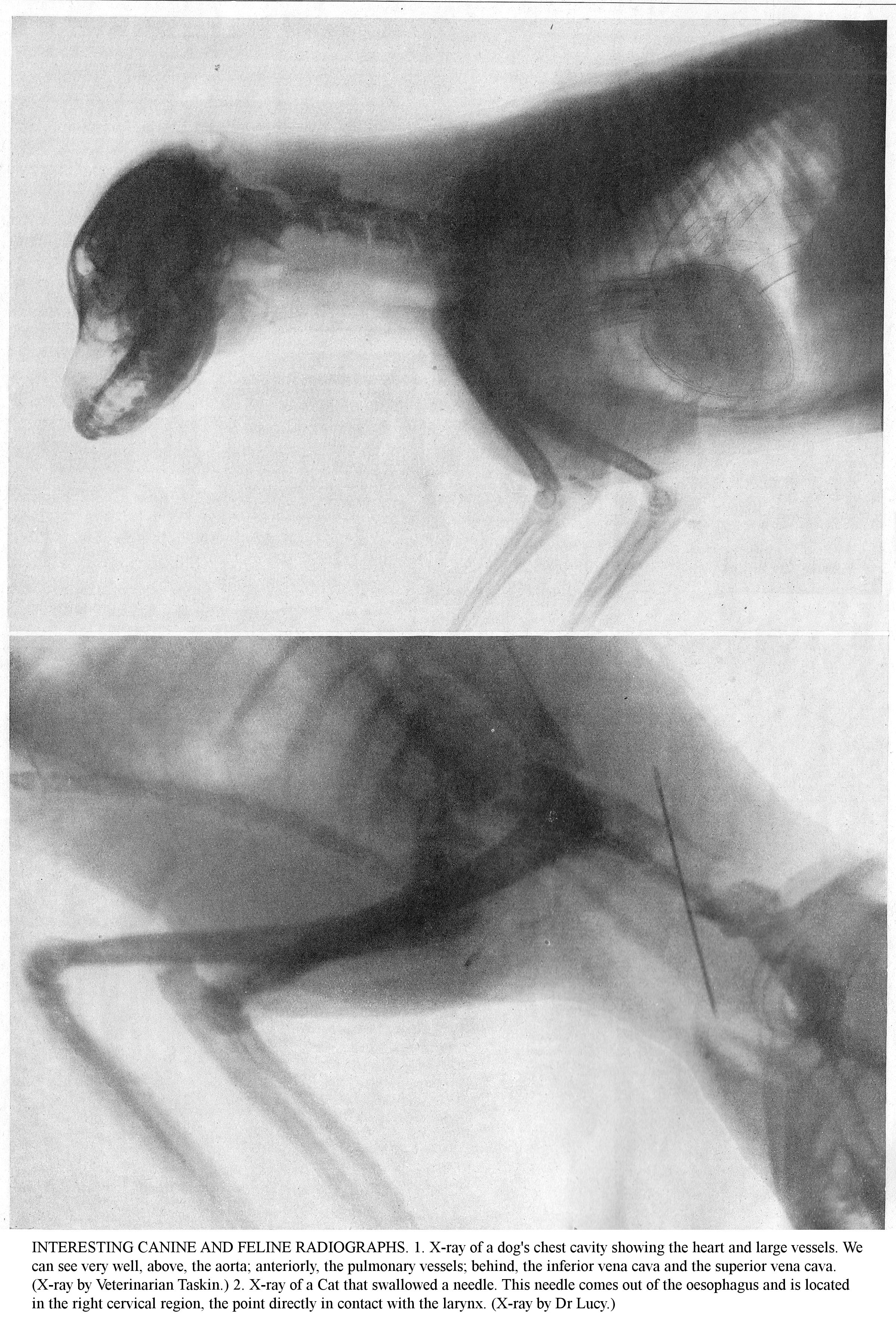
INTERESTING CANINE AND FELINE RADIOGRAPHS. 1. X-ray of a dog's chest cavity showing the heart and large vessels. We can see very well, above, the aorta; anteriorly, the pulmonary vessels; behind, the inferior vena cava and the superior vena cava. (X-ray by Veterinarian Taskin.) 2. X-ray of a Cat that swallowed a needle. This needle comes out of the oesophagus and is located in the right cervical region, the point directly in contact with the larynx. (X-ray by Dr Lucy.)
RADIOTHERAPY TREATMENT OF A DOG. With the animal lying on the operating table, the bulb holder is attached to it, the operator checks the bulb output with the milliammeter and the ray penetration using the spintermetre. (Clinic of veterinary surgeon Taskin.)
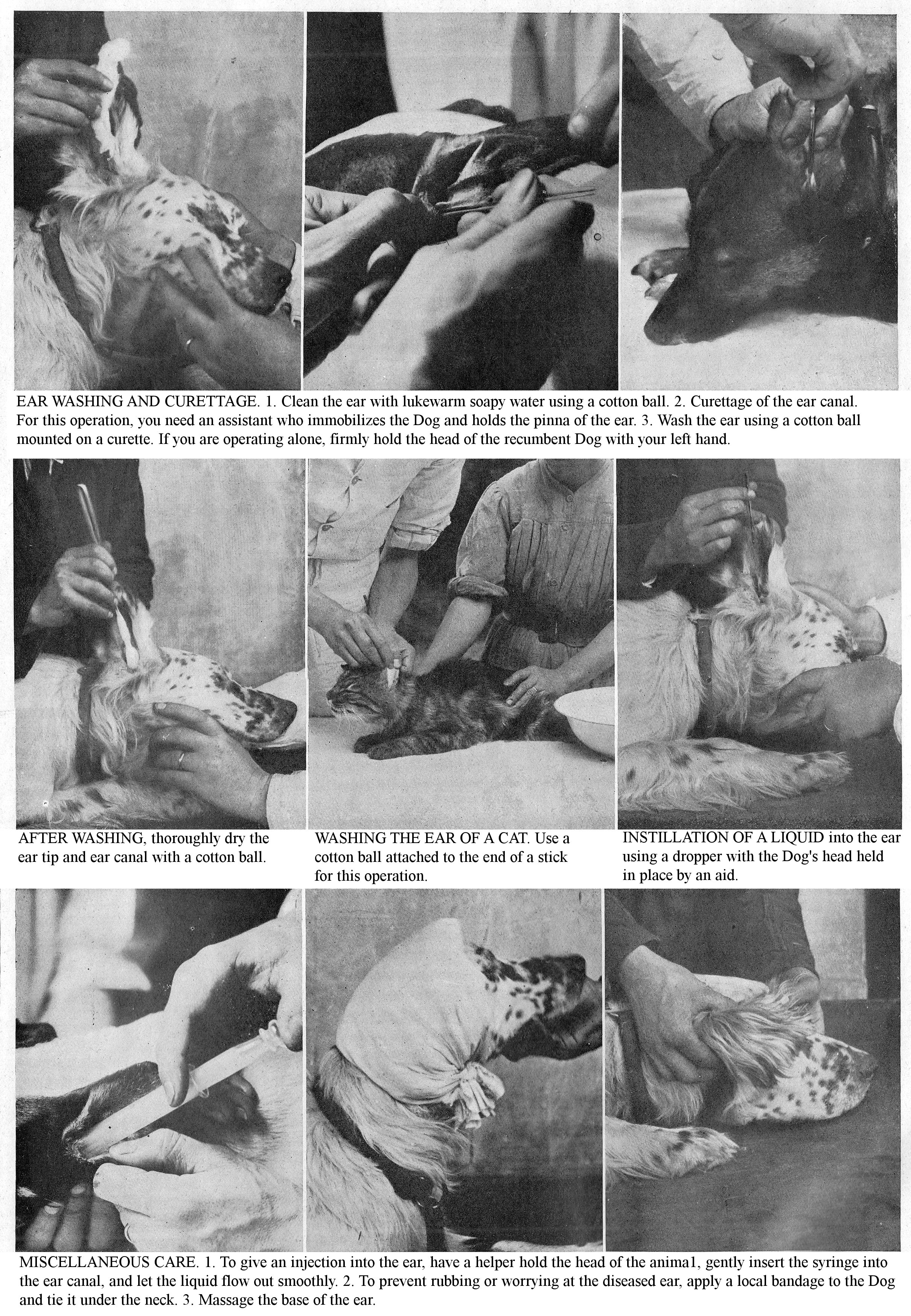
EAR WASHING AND CURETTAGE. 1. Clean the ear with lukewarm soapy water using a cotton ball. 2. Curettage of the ear canal. For this operation, you need an assistant who immobilizes the Dog and holds the pinna of the ear. 3. Wash the ear using a cotton ball mounted on a curette. If you are operating alone, firmly hold the head of the recumbent Dog with your left hand.
AFTER WASHING, thoroughly dry the ear tip and ear canal with a cotton ball.
WASHING THE EAR OF A CAT. Use a cotton ball attached to the end of a stick for this operation.
INSTILLATION OF A LIQUID into the ear using a dropper with the Dog's head held in place by an aid.
MISCELLANEOUS CARE. 1. To give an injection into the ear, have a helper hold the head of the anima1, gently insert the syringe into the ear canal, and let the liquid flow out smoothly. 2. To prevent rubbing or worrying at the diseased ear, apply a local bandage to the Dog and tie it under the neck. 3. Massage the base of the ear.
DISEASES OF THE NERVOUS SYSTEM
THESE CONDITIONS ARE VERY OFTEN COMPLICATIONS DISTEMPER, PREVENT THEM BY TREATING THE LATTER DISEASE RATIONALLY.
The Dog’s Nervous System is the seat of several diseases with various causes: they are sometimes incurable and are often difficult to cure. To these diseases are connected the compression of the medulla/spinal cord, osteomas of the meninges, ossifying pachymeningitis. The diagnosis of these lesions is very tricky and can only be made by the veterinarian.
BRAIN CONGESTION.
Head traumas, high ambient temperatures, forced exposure to hot sun, violent or prolonged effect of cold, distemper, are all major causes of Cerebral congestion.
Symptoms: The patient is initially anxious, overexcited; he screams, howls, snaps at the air, tries to bite and hide away. Do not confuse these short-lived rabies-like attacks with real rabies. Sometimes there is vomiting and convulsions; locally, the skull is warmer than normal; circulation and respiration become faster; the pupils become narrow. Signs of coma are observed later: dizziness, drowsiness or stupefaction. The gait is shaky; the animal makes involuntary movements or collapses and remains prostrate out on the ground.
Treatment: Keep the patient at absolute rest in a cool place and away from all excitement. Combat Congestion by bleeding (20 to 200 gr.); place the head in an elevated position and spray it with water or cover it with cold compresses; use ice packs if possible. Induce intestinal diversion by administering a strong purgative (5 to 50 g of buckthorn syrup). Give a cold enema every two hours. Treat the symptoms of depression with light coffee and tea, to which are added a little alcohol, caffeine or ether.
CEREBRAL HAEMORRHAGE [STROKE].
Cerebral haemorrhage, characterized by the rush of blood into the medulla, can affect either the meninges or the medullary/spinal cord tissue.
Aetiology: Various traumas, fractures, falls, blows with a stick, being run over by vehicles, shotgun wounds during hunting, are common causes of medulla/spinal cord haemorrhages in dogs.
Symptoms: The sudden onset of the following symptoms is characteristic of Cerebral Haemorrhage: tilting, lateral bending or twisting of the head on the neck; rolling eyes, weak pulse, slow breathing. Depending on the site of the lesion, you may see various forms of paralysis and especially paraplegia which is more pronounced on one side than on the other. Appetite and intelligence are unaffected; the temperature is normal. Sometimes you will see poor coordination of movements and locomotor ataxia.
Lesions: Haemorrhages can be multiple and have their seat in the spinal cord or envelopes; the pith is often softened, reduced to a mush. Prognosis: Very serious.
Treatment: Use ice packs, local revulsion (a seton at the back of the neck); but the treatment is completely random. Treat resulting paralysis with massage, exercise, strychnine, and electricity. Subsequently, promote resorption of the effused blood with iodine medication. Prophylaxis: Avoid overeating, give the dog regular exercise, and above all, do not expose it to blazing sun, or to the violent or prolonged effect of cold.
[seton: a piece of surgical thread through a fistula, left for several weeks to keep it open, allowing it to drain and heal.]
EPILEPSY.
Epilepsy, a very common condition in dogs, is a chronic disease of the brain, characterized by seizures, with loss of consciousness and sensitivity. It has no fixed seat of disease; the nervous system is subject to over-arousal which manifests as attacks produced under the influence of a wide variety of causes.
Aetiology: Apart from heredity, the basic causes of epilepsy are unknown; predisposing causes include young age and improved breeds; it can be a result of skull asymmetry, fright, the development of brain tumours, ear parasites, brain lesions of the brain or spinal cord, to intestinal worms, digestive disorders, constipation, foreign bodies, etc.
Symptoms: Epilepsy manifests as attacks at varying intervals, which occur without prodromas. The animal is suddenly seized with tremors and dizziness, accompanied by the sudden loss of sensory functions; it experiences convulsive agitation which soon causes a fall; it staggers, suddenly collapses, makes confused and convulsive movements; first clonic, then tonic, convulsions are noticed in the head, neck and extremities. Some patients, however, remain standing: they then present general stiffness in all the muscles of the limbs and trunk, convulsive agitated jaw movements and profuse frothy salivation. The animal on the ground is unconscious; the eyes are protruding, fixed, wild and roll in their orbits; the pupils are generally dilated and fixed. The patient grinds its teeth. The Dog sometimes bites its tongue while clenching it between the teeth; the patient moans, utters plaintive cries, breathes anxiously. The limbs become stiff, tense and are seized by convulsions; urine and faeces are sometimes expelled involuntarily. The pulse is small, slow, and irregular. Little by little the convulsive movements diminish, the animals recover consciousness and sensibility, they get up and recover gradually, remaining, however, for some time sleepy, weak and dejected. Subsequently, the patient shakes itself, urinates, resumes its usual calm and tries to eat and drink. Usually, episodes only last 3, 4 to 5 minutes; sometimes they go on for a quarter of an hour, or even longer. The duration of the attacks, their frequency and severity, become more pronounced as the already old and established disease, through numerous attacks, has increasingly debilitated the body. In the interval between two attacks, there are no characteristic symptoms.
Diagnosis: This is easy, due to the sudden onset of symptoms, their violence and also their intermittent nature. Prognosis: It varies with the nature of the underlying disorder. Symptomatic epilepsy, caused by auricular acariasis or intestinal helminthiasis, unlike true epilepsy, is curable. Clinically the vast majority of cases, considered to be epilepsy, involve epileptiform seizures.
Treatment: Investigate the causes of the attacks. Pseudo-epilepsy, due to auricular acariasis and intestinal helminthiasis, require specific treatment (disinfection of the ear canal, anthelmintics). Apart from these cases, there is almost no treatment. Use sedatives, mainly potassium bromides, sodium (0.50 to 2 gr.).
ECLAMPSIA.
Eclampsia is an acute, feverish disease affecting female dogs before or after parturition, characterized by convulsive contractions and coma, with complete loss of sensibility; it is particularly observed in apartment-living female dogs, guard dogs, and overweight dogs.
Aetiology: Eclampsia is caused by pregnancy poisoning the body, nephritis, albuminuria.
Symptoms: The onset is abrupt, marked by signs of disquiet, anxiety, acceleration of major functions and neuro-muscular disorders (stiffness of the limbs, uncoordinated movements). Soon the patient collapses and makes vain efforts to get up; convulsions shake the whole body, the limbs are stiff, the head stretches out on the neck, the lips become soiled with foamy saliva. Breathing and circulation are rapid, seizures usually persist for several hours, sometimes for a whole day, more severe at various times than others. Left on its own, eclampsia can lead to death; but it can be cured easily.
Diagnosis: Do not confuse this malady with Epilepsy in which the symptoms are more acute (loss of consciousness, abolition of sensitivity), and the crisis lasts only a few minutes. You can easily tell the difference between Eclampsia and Rabies.
Treatment: Place the patient in an isolated place away from various causes of excitement. Combat seizures using nervous system sedatives: chloral, chloroform or ether syrup (5, 10 to 15 gr.). In severe cases, use artificial serum injections; during the course of treatment and a few days after, give the patient a milk diet.
PARALYSIS.
Paralysis is the loss of muscle contractility and tenderness. Clinically, you can observe local or isolated paralysis, involving one or more muscles or several muscle groups; monoplegia or extensive paralysis of a single limb; hemiplegia or paralysis of a lateral half of the body; paraplegia or paralysis of the posterior half of the body.
Aetiology: Paralyses follow lesions of the brain, spinal cord or nerves. The causes are varied: anaemia, congestion, haemorrhage, softening, acute or chronic inflammation, tumours of the nervous centres, infection and intoxication with microbial toxins (childhood diseases). Traumatic paralysis is caused by shocks, muscle tears, fractures, bruises (animal run over by a car) as the cause.
Symptoms: The symptoms of Paralysis necessarily vary with the degree of paralysis, the regions affected and especially with its origin. Sometimes the muscles are paralyzed, relaxed, limp and soft; other times they are hard, stiff, and contracted. Tenderness may be imperceptible, or there may be severe lameness in a limb, or the affected area may be deformed. Sensitivity is increased, suppressed, or perverted. In acute lesions of the brain (haemorrhage, softening), the paralysis begins suddenly; it is limited to the lateral half of the body, and hemiplegia occurs on the side opposite the lesion. In lesions of the spinal cord, the usual site of the paralysis is in the abdominal limbs, with extension to the thoracic limbs. Paralysis of peripheral origin is usually limited to a single nerve and rarely involves a large number of muscles. Let us mention, among the most frequent forms in dogs, those of the facial, lower jaw, radial, crural and greater sciatic nerves.
Treatment: Use massage, dry or lively rubs: brandy, camphorated alcohol, subcutaneous, irritating injections of a saturated solution of sea salt. As internal treatment, administer sodium salicylate and potassium iodide (0.50 to 2 gr.). In severe cases, use subcutaneous injections of a strychnine salt, an effective treatment but which, due to its toxicity, requires the intervention of a veterinarian. Electrotherapy (electricity, galvanic and faradic current), by the subsequent ionization of the muscles, acts positively on the degenerative process which leads to the destruction of nervous elements, and it causes contractions in the muscles which prevent their atrophy.
CHOREA.
Chorea, a disease characterized by irregular and involuntary contractions of one or more muscle groups of the musculoskeletal system, is most often seen as a toxic-infectious complication of distemper. Purebred dogs are particularly exposed to it.
Symptoms: The dominant symptoms are involuntary muscle contractions, true clonic convulsions which can be general or local; in the latter case, they extend to the lateral part of the body or are confined to one region. The disease is usually continuous, and the tremors appear at equal intervals; at other times, remission or intermittence may be observed. During sleep, the contractions stop or decrease in intensity. Often walking is confused, vegetative functions are normal, sensibility is preserved. In ordinary cases, patients appear to remain healthy for a long time, then they lose weight, lose their appetite and die. The disease lasts for months or years, and is resistant to various treatments.
Lesions: Lesions, sometimes inappreciable, account for the changes of meningitis, meningoencephalitis, resulting in vascular inflammatory foci, mainly located in the grey matter. The cerebellum, medulla, meninges, and the entire cerebrospinal axis show inflammatory lesions of the vessels and perimyelitis. The predominance of lesions is especially frequent and obvious in the lumbar cord. Prognosis: Very serious.
Treatment: Build up the patient, give him plenty of raw meat and tonics, especially cod liver oil. Let it run free or take it for walks in the sun; protect itfrom cold and damp. Remember that diet is more effective than the therapy. Treat paralytic disorders of the hind limbs with revulsive frictions (stibiated ointment) on the dorso-lumbar region; combat lesions of the nervous centres using arsenico-iodide medication; reduce convulsions with bromides (0.50 to 2 gr.), chloral (0.30 to 2 gr.), sulphonal [sulphonmethane] (1 to 2 gr.). Balneotherapy is sometimes recommended; immerse the animal abruptly and several times in a tub of cold water, then perform the other actions (rubbing with hot cloths, running).
MENINGOENCEPHALITIS.
Meningoencephalitis, inflammation of the cranial meninges, brain, can be caused by head trauma, sunstroke, parasites of the nerve centres; but it is seen mainly as a condition secondary to distemper.
Symptoms: Initially the illness is characterized by anxiousness, restlessness, complaints, screaming; the eyes are wild; the cranial region is warmer than usual. Convulsions, contractions and sometimes rabiform or epileptiform symptoms may be observed. In the latter, you will see coma and various types of paralysis. The chronic form results in mobility problems (dizziness, convulsions, contractures).
Treatment: Place the patient in a dark room, feed him liquid food: milk, broth. Combat congestion by cooling the skull, bleeding; perform bowel bypass by administering a purgative; calm the phenomena of excitement using sulphonal sedatives (potassium bromide); fight coma with low-alcohol coffee, tea, caffeine or ether injections.
MENINGO-MYELITIS.
Infections, poisoning, bruises, traumas, colds and especially complications of distemper are the common causes of meningo-myelitis.
Symptoms: The disease is characterized by abnormal sensitivity of the spine to pressure, symptoms of excitement, tremors, contractures, paralysis. The prognosis is serious; recovery is rare and almost always incomplete. Chronic meningo-myelitis follows the acute form. Overworked muscles (hunting dogs, working dogs), excessive pregnancies, are predisposing causes.
Treatment: Calm congestive phenomena by applying cold compresses to the spine; practice revulsion by rubbing stibiated ointment on the dorso-lumbar region; perform internal bypass with the administration of a strong purgative. Moderate the phenomena of excitement by using bromides. Give strychnine injections under the skin aginst the paralysis. Chronic meningo-myelitis calls for iodide medication, cauterization of the dorso-lumbar region and electricity.
Tell those around you about this Volume; you will be helping your friends and you will make us happy.
NON-PARASITIC SKIN CONDITIONS
THESE COMMMON CONDITIONS ARE DUE MAINLY TO LACK OF BODY HYGIENE AND DEFECTIVE DIET: PREVENT THEM BECAUSE HEALING IS SOMETIMES LONG AND DIFFICULT.
Nonparasitic skin conditions are commonly seen in dogs. By interfering with the physiological functioning of the skin, they impact the general condition, causing thinness, extreme weight loss and physiological distress. In young animals, they cause a marked stop in growth. Dermatitis is very diverse in its manifestation, and in most cases is caused by faulty hygiene and incorrect nutrition.
ERYTHEMA.
Erythema is a skin condition characterized by redness that disappears momentarily under pressure from the finger. Sometimes you will see a serous exudate which ends with papules, vesicles, followed by desquamation. Among the causes that can provoke Erythema, we will mention mechanical irritation (clipping, friction from hard brushes), the use of irritating topical treatments and infectious diseases.
Symptoms: Erythema is a rosy red mark, quickly fading under finger pressure then reappearing. It is accompanied by heat, itching and hair loss.
Treatment: Relieve pain and itching using glycerol starch; while there is exudate, use warm, weak solutions of boric acid, carbolic acid; after drying, cover the affected areas with an absorbent powder: talc, bismuth subnitrate, zinc oxide.
URTICARIA
Nettles, including Stinging Nettle (Urtica dioica) and Small Nettle (Urtica urens), whose hairs contain a colourless, caustic liquid, cause the skin to burn for several hours and cause a rash of flattened white papules surrounded by a light pink halo.
The ailment appears during or after a hunt on ground covered with nettles, provided that these plants are not in full bloom, but particularly at the beginning of their growth, occurring generally during the months of August and September at the time when hunting begins. The Dog, absorbed by the scent of game, runs for a while without feeling the pain of urtication; but the very fragile nettle hairs which break off from the leaves and stems, first irritate the hairless areas, then enter the interdigital spaces, where they cause inflammation, and then into the fur. As soon as the pain begins, it immediately becomes generalised; the Dog licks himself in a frenzy and swallows the stinging hairs; soon inflammation of the mouth and pharynx develops; the glands are irritated and secrete abundantly. Walking with his nose to the ground, in moving nettles, the Dog inhales nettle hairs which penetrate deep into the respiratory tract. The congested, swollen nasal mucosa block the entry of air and can lead to asphyxiation.
In severe cases, after period of violent excitement, the Dog falls onto its side and can only get up with great difficulty. Soon you see stiffness of the limbs, paralysis, and the patient falls into a coma. Usually after 4 to 5 days, healing is confirmed, salivation decreases, the cough subsides and the symptoms of excitement give way to a state of prostration which can last for 12 hours.
Treatment: Remove stinging hairs from the skin by washing with vinegar water; calm intense itching with antiseptic lotions based on boric acid and carbolic acid; detoxify the body using emetics and purgatives; calm the excitement using opiates or bromides and sulfonal; combat coma with infusions of tea or coffee, injections of ether or camphor oil. Prophylaxis: As much as possible, avoid taking the Dog through areas where newly growing nettles are abundant, or where they are growing back after being cut.
PITYRIASIS, SCALY SKIN RASH.
This affection is found especially in old dogs; arthritis, blood congestion, over-indulgence in sweets, and insufficient exercise are the main causes.
Symptoms: The skin around the head, neck and upper trunk is covered with profuse whitish dandruff; afterwards, the skin gradually hardens and takes on a dark colour.
Treatment: Change the diet; give a hydrated milk or mixed diet; dust the diseased areas with a mixture of talcum powder and zinc oxide. As an internal medication, alternately use baking soda and Fowler's liquor as a depurative.
CHRONIC PHLEGMONOUS DERMATITIS.
[Chronic diffuse inflammation affecting connective tissue]
This affliction, observed almost exclusively in large breeds (Danes, Mastiffs), is usually localized on the outer surface of the elbows, hocks and stifles. This skin condition is always very tenacious and causes swelling, hair loss and the development of small purulent foci.
Treatment: Cleanse and disinfect the skin with lukewarm 3% cresyl solution or 5 parts per thousand potassium permanganate solution. After lancing the abscess, use tincture of iodine. When the plaques are few and far between, have them excised.
IMPETIGO.
Impetigo is a skin disease, inoculable and auto-inoculable, frequent in young dogs and caused by superficial local infections, by pyogenic bacteria, staphylococci and streptococci. The skin infection occurs through excoriations caused by dirtiness, rashes and lice infection.
Symptoms: The disease is characterized initially by the formation of small pointed pustules, quickly followed by yellowish scabs under which the skin reform. Healing occurs after a fortnight, but in a few patients, new foci due to self-inoculation, appear, and the condition lasts for months.
Treatment: Use lotions, antiseptic ointments (boric, cresylated) or astringent (zinc sulphate); cover the oozing surface with an absorbent powder (talc, starch, charcoal). Prophylaxis: Avoid, by strict skin hygiene, dirtiness, eruptive rashes and especially lice.
ECZEMA.
Eczema is a particular affliction of the skin or mucous membranes, frequent in dogs, acute or chronic, initially manifested by pruritus, vesicles or vesico-pustules which burst and which are followed by either an abundant production of scaly skin, or real crusts with hair loss and generally thickening of skin. Its development is promoted by incorrect diet, insufficient exercise, swollen lymphatic tissue, obesity, diabetes. In canine pathology, heredity plays an important role in the aetiology of eczemas of internal origin. Eczematous rashes of external origin include skin irritations of various types and intensity resulting from clipping, repeated rubbing, scratching, pressure from a collar, dirtiness of the skin and the presence of parasites, etc. Eczema, in dogs, has a variable appearance and manifests itself differently depending on the patient's age, constitution, breed, diet, etc.
ERYTHEMA OR REDNESS. This is an erythematous condition of the dermis, characterized by redness and tenderness of the skin in places where it is thin and almost hairless (inner side of the limbs, stomach, etc.). Aetiology: Erythema is common in dogs of improved breeds (Braques, Saint-Germains, Greyhounds, Hounds, Fox terriers, etc.), as well as in young and abundantly fed animals.
Symptoms: This type of dermatitis begins with redness, swelling, tenderness of the skin at the groin and the inner side of the thighs; three is intense itching; animals continuously scratch themselves; a few small vesicles appear which are quickly torn by scratching. The redness and inflammation spread to the skin of the stomach, armpits, elbows and flanks. The condition may persist for a long time, or subside, or often disappears with a change in diet, it usually disappears in summer.
Diagnosis: Differentiate Erythema from Scabies, which has a different site and which is accompanied by the formation of pustules; and from distemper, which has a vesicular rash. Prognosis: Not very serious.
Treatment: Modify the diet, give soups, milk, containing 4 to 6 gr of baking soda, V to X drops of Fowler's liquor; walk the patient. Locally, make lotions with cresylated or carbolic water. Calm the pruritus with general emollient baths (bran, starch); during the exudation period, after washing and drying the diseased areas, use absorbent powders (talc, bismuth sub-nitrate, zinc oxide), or astringent ointments.
ACUTE ECZEMA. Aetiology: It affects dogs that are predisposed to it and those maintained in poor conditions, animals that have parasite infestations (fleas, lice). Symptoms: At the beginning, the skin is hyperaemic, oedematous, sensitive, then it bristles with small scattered papules which turn into vesicles: the pruritus is then very intense; the blisters tear spontaneously or due to scratching, and the exudate collects in crusts which quickly fall out, carrying hair with them. Sometimes, instead of merging, the lesions are circumscribed and localized in a few places on the body: on the testes in males; around the anus and vulva in females; on the paws in hunting dogs, on the eyelids, on the internal surface of the ear flap and the auditory canal, where they cause auricular catarrh.
Treatment: Clip long-haired dogs and clean the skin with lotions of hot water or boric water, then dry the affected areas. At first, apply boricated or camphorated petroleum jelly, or powdered starch, bismuth subnitrate, zinc oxide. For the itching, use carbolic lotions or compresses (1 part per 1000) or chloralated (1 p. 100). For wet eczema, use the following treatments: astringent or antiseptic lotions (carbolic or lightly cresylated), 2% chromic acid, then starch powder, tannin, boric acid or a mixture of idioform, tannin and boric acid in equal parts. For crusty eczema, loosen the scabs and use antiseptic lotions (preferably hydrogen peroxide); successively apply vegetable tar and astringent ointments based on zinc oxide, salicylic acid. For the scaly form, use iodized glycerine, vegetable tar, 1% copper or iron sulphate.
To prevent recurrence, use a general treatment based on diet and internal medication. Reduce the meat ration if the Dog is obese; or increase it if the Dog is in poor condition; give the fat or obese patient refreshing foods, a lacto-vegetarian diet. Father Cadiot recommends ‘intus and extra’ sulphur medication against all forms of Eczema.
Sprinkle affected surfaces once or twice during 24 hours with flower of sulphur. Give washed sulphur internally for 8-10 days, then stop for a week and resume once or more with meals of varying periods. Alternate this with arsenic medication (Fowler's liquor) and alkalis (baking soda, 2 to 3 grams per litre) in the drinking water.
For Eczema of the nose and nostrils, which is often very stubborn, make borate lotions of 2% to 3%, then apply boric or zinc oxide petroleum jelly; for eyelid eczema, use 1% salicylate, boric or borate hot lotions, then rub with yellow mercury oxide ointment; for interdigital eczema, use light antiseptic baths and salicylate ointment dressings; for tail eczema (tail canker), make lotions with 1% carbolic solution; sprinkle with salol; cover the area with collodion or a bandage held in place by a canvas or leather sheath.
CHRONIC ECZEMA. Very common in elderly or obese dogs, chronic eczema follows acute eczema, where it immediately presents as the scaly form. It is most commonly seen on the upper areas of the body, elbows, hocks, inner ear skin, and tail. It constitutes the gripe or roux-vieux [horsehair mange] of the mounted hunt.
Symptoms: At various points on the body, the skin is thickened, hot, dry and stiff; it is covered with profuse greyish crusts; the skin, due to the enlarged dermal papillae, appears sore; the hairs bristle or fall out in places and don’t grow back. Animals continuously scratch themselves, and their bodies give off a foul odour. The disease progresses by relapses followed by remission during which it seems to disappear, then appears again. Over time, Dogs become exhausted, lose weight and become apathetic. Diagnosis: The diagnosis of Chronic Eczema is, in general, easy. In doubtful cases, use microscopic examination to differentiate it from Dry Follicular Mange, Sarcoptic Mange or Ringworm.
Treatment: Beforehand, loosen the scabs and scales by lightly soaping with hot water; trim the hair off the diseased areas, if necessary. Use lotions with a cresylate solution, or better, applications of tar and cade [juniper] oil: do 2 or 3 similar applications a week apart, after lukewarm soaping of the skin. Modify the diet, feed the Dogs with soups, milk; give them exercise, and, if possible, get a change of air. Observation shows, in fact, that Dogs that are constantly locked up and suffering from long-term Eczema recovered naturally after an outdoor stay in the countryside. Internally, administer V to X drops of Fowler's liqueur, 2 to 4 gr. of baking soda, or cgr. at 1 gr. of potassium iodide in a little milk each day.
Prophylaxis: In some cases, the frequency of non-parasitic dermatitis and the questionable efficacy of treatment shows the importance of prophylaxis. Incorrect feeding (overuse of the meat diet, provision of spoiled food), lack of cleanliness of patients and premises, insufficient exercise, are the aetiological factors. Therefore, give a sensible diet (mixed diet: meat and vegetarian); maintain skin hygiene by regular grooming; give the Dogs regular exercise and keep them away from irritating contact with faeces and urine by proper maintenance of their straw litter. Avoid eczemas of external origin by treating irritation (Erythema) from shaving from the start; avoid pressure from an ill-fitting collar, and above all, do not overuse alkaline baths and soaps.
PARASITIC SKIN DISEASES
BY THEIR FREQUENCY, THEIR CONTAGIOUSNESS AND THEIR SEVERITY, THESE CONDITIONS CAUSE SIGNIFICANT CASUALTIES.
Parasitic DERMATOSIS (acariasis, phtiriasis, etc.) are common in dogs and cats; apart from their contagiousness, they have a very serious prognosis in young animals and can stop growth or cause death by exhaustion and physiological distress. In addition, the contagiousness of some of forms to humans increases their seriosness.
PHTIRIASIS.
Phtiriasis is a disease characterized by the presence of a large number of lice on one area or all over the body. Lice are small insects with an oral apparatus able to prick or suck. Females lay a large number of eggs called "nits". The proliferation of dermatoses is favoured by lack of grooming, dirtiness, hair length, infected litter and premises, the physiological poverty, young age, old age and by all other debilitating influences. Dogs are hosts of the piliferous Haematopinus and the Trichodecte.
Symptoms: Phtiriasis is heralded by a very marked itching, causing much hair loss of a very characteristic appearance, and excoriations due to scratching. On examining the skin, you will notice the presence of either parasites or of eggs attached to the base of the hairs. Remember that Trichodectes are the intermediate hosts of Taenia larvae and that the Dog, while washing itself, can contract Taeniasis or reinfest itself. Therefore, disinfect animals by regular grooming.
Treatment: Puff insecticidal powder (stavesacre [Staphisagria], pyrethrum) or apply sulphurous or cresylated lotions at 2% into slightly wet hairs. Before applying the treatment, loosen the eggs with a comb and brush. If the hair is very long and felted, trim the animal. In cats, do not use antiparasitic lotions; resort to puffing with powdered stavesacre or pyrethrum.
Prophylaxis: Burn the litter, disinfect the bed or kennel by washing with boiling carbolic or cresylated water at 3% or 5%, with milk of lime or petroleum; disinfect grooming items (combs, brushes, sponges, etc.) and blankets with cresylated boiling water. Because nits are particularly resistant, repeat disinfections.
FLEAS.
Fleas especially infest sedentary animals (nursing mothers, puppies) and the debilitated. These parasites feed on blood, and their bite causes intense itching, marked by frequent scratching. The dog flea is the intermediate host of the tapeworm dipylidium, and the animal becomes infested by licking. The young can be infected by contact.
Treatment and prophylaxis are the same as for Phtiriasis. Keep fleas away by mixing fir shavings or fresh walnut leaves into the litter or brushing the kennel with petroleum oil. However, do not use the latter method for Hunting Dogs, as it may affect their sense of smell.
ERYSIPELAS
The larva of Trombidium soyeux [Trombicula autumnalis], the harvest mite or chigger, causes a benign skin condition, observed at the end of summer and particularly common in hunting dogs. The parasites are usually located on the nose, around the eyes, on the stomach, on the limbs and their accumulation in these regions simulate yellowish orange crusts. Implanting its Rostrum [snout] in the dermis, this insect deposits a virulent saliva which causes intense itching; it can migrate to Cats and humans. Treatment: Rub the infested areas with a light carbolic solution (1% to 2%) with benzine or petroleum oil.
TICKS.
Ixodes, ticks, or wood lice are mites commonly found on dogs that hunt in woods in the autumn or across land covered with broom. The parasites, brought in from outside, soon infest the Kennels. Under the double need for blood food and reproduction, the ticks, after attaching themselves to the skin, generally around the head, ears and limbs, gradually swell, and their diameter eventually increases from to 4 mm. to 12 to 15 mm. When the female is full, she spontaneously detaches herself, falls to the ground, climbs into grass and thickets, lays thousands of eggs there during one month, then dies. The discreet invasion hardly disturbs the animal host; when generalized, it can lead to weight loss and anaemia. Piroplasmosis is transmitted to the Dog by larvae of infected ticks.
Treatment: Do not remove ticks, as the rostrum may remain implanted in the skin where it can give rise to abscesses; wash the animals in lukewarm cresylated water; rub them with a sponge dipped in benzine, but fully squeezed out, which causes the death of the parasites and their rapid elimination. Use the same prophylactic measures as for Phtiriasis.
SARCOPTIC MANGE.
Aetiology: This condition is caused by Sarcoptes scabiei, varietas canis; the condition is spread very easily from Dog to Dog, in hunting packs in particular, where it often becomes epizootic; it is fostered by lack of cleanliness of the animal and the kennels. A cat infected with acariasis frequently infects dogs, which in turn can infect humans.
Symptoms: The condition usually begins on the head, then spreads to the whole body, in areas with thin skin, inner thighs and stomach; you see small red spots that look like flea bites and quickly turn into papules, then into vesicles or pustules the size of a lentil or pea. When scratched or rubbed, these vesicles tear and expose the mucous content which appears reddish, wet, oozing; the serosity solidifies into yellowish crusts which dry out and form scales which are easily detached. There is intense itching; mange is often blamed when the animals are lying in the sun, or in front of the fire, or after a race. If you scratch them with your hand, they show their contentment by waving a hind paw. Gradually the skin peels off, thickens, splits and cracks; between the folds you observe a foul-smelling exudate that makes the Dog repulsive. If you do not intervene vigorously with scientific treatment, scabies patches spread quickly; the animals lose weight and after 2 or 3 months die in the doldrums.
Diagnosis: Clinically you can only confuse Sarcoptic Mange in Dogs with Follicular Mange and Chronic Eczema; differentiate between these conditions by taking into account the following characteristics: Follicular Mange presents as mild itching and offers numerous acne pustules; Chronic Eczema is characterized by no or mild pruritus, absence of contagion, and is resistance to antipsoric [itch remedy] treatment. In doubtful cases, confirm the diagnosis by microscopic examination: expose the animals to heat, scrape the skin down to the blood at the level of the depilated patches and examine the collected crusts under a microscope: you can easily detect the presence of sarcoptic mites.
Treatment: Clip the animals, soap them in lukewarm water, then give them Bareges baths. After drying, apply Helmerich's ointment or Peruvian Balsam to diseased areas. Give the Dog a good diet and as much exercise in the open air as possible.
Prophylaxis: Isolate the patient; carefully disinfect the Kennels and the various utensils that have come into contact with the patient: collars, tether chains, food and drink containers, etc.; burn the litter. Grooming objects (brushes, curry combs, sponges) are a frequent source of contamination, often unknown to breeders and hunters. In a contaminated environment, disinfect daily using sulphurous fumigation in an ad hoc box. We cannot recommend enough to you this simplest, fastest and most effective process, without which the disinfection of the premises and the medical treatment, would only be palliative.
FOLLICULAR MANGE.
Aetiology: Follicular mange caused by Demodex folliculorum, varietas canis, occurs mainly in young dogs and often coincides with distemper which promotes its onset. It affects dogs of various breeds, but especially those with short hair. Direct contagion is rather difficult to establish experimentally; some individuals seem to be unusually receptive to it.
Symptoms: The condition usually begins between the digits, the tips of the elbows, hocks or buttocks, and on the lips; the skin is red, a little swollen; it is the site of fairly pronounced epithelial desquamation; the hair falls out but itching is not very intense; small papules soon appear at the base of the hairs. The animal continues to look healthy. The papules then enlarge to the size of a lentil or pea and transform into pustules; the skin thickens and wrinkles, the hair falls out, itching becomes more pronounced. As the disease progresses the bald plaques gradually increase, the pustules ooze a yellowish or bloody liquid, sometimes pus which forms in thick crusts; the skin thickens considerably. When you pull out a hair and examine the root under a microscope, you see a Demodex mite. Suspect follicular mange in any young dog with circumscribed bald areas, especially in areas preferred by the Demodex mite; isolate the animal and disinfect the premises. The progress of the disease is very slow; it can remain localized for a long time in a few regions, in the paws which become enormous.
Diagnosis: Microscopic examination of the contents of the pustules can differentiate follicular mange from acute eczema or the vesicular rash that accompanies distemper, Ringworm, etc. Prognosis: Very serious, because, except in the case of spontaneous recovery, the disease is almost incurable.
Treatment: Build up the patient with a meat diet; add lime phosphate (50 cgr. to 5 gr.) to the meals; give them exercise. In the case of localized mange, clean the plates with petroleum, benzine or ether, and make a daily application of alcohol at 60 proof, tincture of iodine, Peruvian balsam, or sulphide ointment of carbon; do not use mercury preparations because of their toxicity. When the wounds are festering, clean them before each dressing; open the pustules or burst them by wrinkling the skin and remove the pus; then apply the acaricide preparation. For generalized mange, use sulphur or cresyl baths, and alternate these with applications of alcohol or tincture of iodine. Radiotherapy provides rapid success; the extraction of the diseased skin flap is recommended.
CHORIOPTIC OTACARIASIS [EAR MITES].
In the hunting dog in particular, Chorioptes auricularum, varietas canis, causes a particular mange of the external auditory canal, accompanied by very violent pruritus, with epileptiform convulsions, or even rabiform symptoms (foaming at the mouth). In the early stages, these reflex actions usually do not appear until the patient is overheated by a fast run or while hunting. They suddenly erupt: the Dog is unsettled, anxious, rushes straight ahead, moans or squeals, staggers and falls prey to convulsions. These attacks, which usually last only a few minutes, increase in duration and frequency. Examining inside the ear allows you to discover symbionts or chorioptic mites developing in yellow or chocolate-brown earwax. The disease, left on its own, can lead to deafness, apathy and fatal seizures.
Treatment: Soap and clean the accessible parts inside the ear; make injections of iodized glycerine, Peruvian balsam dissolved in four parts of alcohol.
FELINE MANGE.
Sarcoptic mange is caused by Sarcoptes minor, varietas canis or Sarcoptes notoedres; it is extremely contagious and sometimes runs rampant in cats in an epizootic state. It is transmissible to humans and dogs.
Symptoms: The condition usually begins with the head, forehead, then ears, muzzle and anterior neck region; it does not extend to parts of the body with long hairs; sometimes lesions develop on the hind legs. The skin becomes covered with papules and vesicles the size of a pin-head, forming thick, hard scabs that clump the hair together; these soon fall out. The skin thickens, wrinkles, and cracks in a few places. The itching is usually intense and, as a result of rubbing, the skin becomes irritated, especially at the nose and ears; moreover, the scabs even obstruct the nostrils and interfere with breathing; there is often conjunctivitis. Animals are dejected and lose much weight; they succumb in a short time.
Diagnosis: Its onset in the head, its persistence in this region, and the easy discovery of the parasites, clearly differentiate feline mange from the tenacious prurigo which sometimes affects castrated cats and which is sited on the stomach, back, and groin resulting in small dry patches. You may observe a similar rash, of short duration, on nursing cats when suddenly separated from their young. You cannot confuse this mange with mite-related prurigo, nor with flea bites or the scurfy patches that plague old apartment cats. Ringworm can be confused with Sarcoptic Mange; confirm the diagnosis by microscopic examination.
Prognosis: Feline mange is only serious when it is old: if left untreated, it can lead to death by exhaustion. The severity of this mange is further increased because it is contagious to humans. Treatment: After the scabs have been removed, apply Helmerich ointment, cade oil or Peruvian balsam at 1 part in 8.
Follicular mange caused by Demodex folliculorum varietas cati is generally localized to the head and manifests the same symptoms as in dogs; but it is more benign than in the latter and disappears quickly. Ear mange has the same symptoms and involves the same treatment as Otacariasis in Dogs.
RINGWORM.
Ringworms is a contagious dermatosis, relatively common in Dogs and Cats, caused by fungi that vegetate on the hair. Infection takes place through premises, combs, brushes, blankets. Young age, debility, and eruptive conditions are predisposing causes.
Symptoms: Trichophytic ringworm, the most common, is characterized by the appearance, in different areas - most commonly on the head and extremities - of small rounded bald patches which spread rather quickly. The skin is covered with greyish, dry or slightly viscous crusts. The itching is variable in intensity.
Favus or the favous ringworm is rare in dogs. The first patches appear on the limbs and head, most often on the lips; in puppies, they are usually found on the abdomen and thorax. The patches are the size of a 50 cent coin, the skin is covered with them; they are whitish, slightly excavated into a dip. The Cat is more prone to this disease; Ringworm usually attacks the end of the legs and base of the claws; little by little it spreads to the head, forehead, base of the ears, outer surface of the thighs and various parts of the body.
Diagnosis: Ringworms can be confused with Mange and with various forms of eczema. With the exception of Favus, the clinical characteristics are generally insufficient to establish a differential diagnosis; use microscopic examination.
Treatment: Remove the crusts and burn them to avoid any contagion; wash the diseased areas with soapy water; apply tincture of iodine, petroleum, cade oil, etc. Prophylaxis: The prophylactic measures are the same as for Mange, since Ringworm is contagious to humans.
USE OF X-RAYS.
IN VETERINARY MEDICINE, the use of X-rays mainly belongs in the laboratory; in recent years, however, dog clinics, as the photographs inserted in this volume prove, have made use of X-rays and have shown the diagnostic and therapeutic value of this mode of investigation. The practical medical applications of X-rays are of three kinds. 1st, Radioscopy, the study of the shadows produced on a fluorescent screen by placing part of the animal’s body between this screen and a source of X-rays. 2nd, Radiography, using a photographic plate in place of the fluorescent screen allows the image viewed on the screen to be fixed as a lasting image. 3rd, Radiotherapy, the use of x-rays, on living tissue to produce a series of destructive or restorative changes in cells whose formation has been produced or altered by any pathological process.
Practical applications of the use of X-rays in the field of canine medicine for radioscopy include the diagnosis of thoracic conditions, particularly those related to thoracic tuberculosis, and the diagnosis of some abdominal conditions (foreign bodies).
Radiography plays a big role in the diagnosis of fractures, the search for foreign bodies swallowed by Dogs and Cats, and projectiles encysted in the tissues. It is essential when surgery (gastronomy, laparotomy) is indicated (1).
(1) This important subject, which the framework of this Volume does not allow us to develop, will soon be treated in detail by our collaborator M. J. Taskin, Veterinary radiologist.
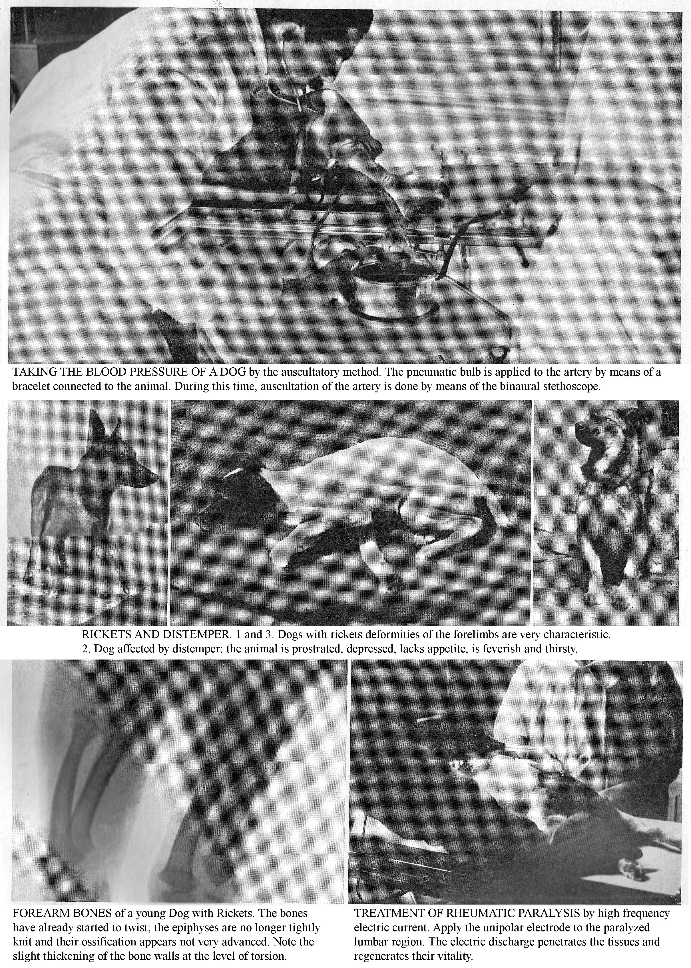
TAKING THE BLOOD PRESSURE OF A DOG by the auscultatory method. The pneumatic bulb is applied to the artery by means of a bracelet connected to the animal. During this time, auscultation of the artery is done by means of the binaural stethoscope.
RICKETS AND DISTEMPER. 1 and 3. Dogs with rickets deformities of the forelimbs are very characteristic. 2. Dog affected by distemper: the animal is prostrated, depressed, lacks appetite, is feverish and thirsty.
FOREARM BONES of a young Dog with Rickets. The bones have already started to twist; the epiphyses are no longer tightly knit and their ossification appears not very advanced. Note the slight thickening of the bone walls at the level of torsion.
TREATMENT OF RHEUMATIC PARALYSIS by high frequency electric current. Apply the unipolar electrode to the paralyzed lumbar region. The electric discharge penetrates the tissues and regenerates their vitality.
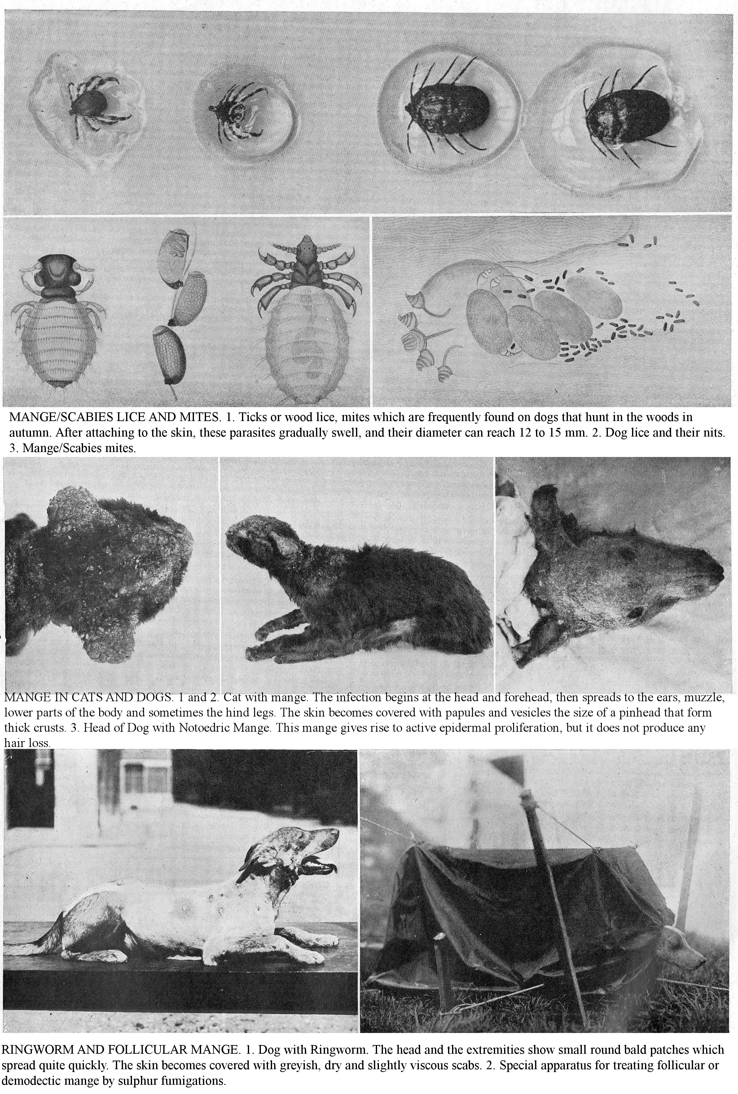
MANGE/SCABIES LICE AND MITES. 1. Ticks or wood lice, mites which are frequently found on dogs that hunt in the woods in autumn. After attaching to the skin, these parasites gradually swell, and their diameter can reach 12 to 15 mm. 2. Dog lice and their nits. 3. Mange/Scabies mites.
MANGE IN CATS AND DOGS. 1 and 2. Cat with mange. The infection begins at the head and forehead, then spreads to the ears, muzzle, lower parts of the body and sometimes the hind legs. The skin becomes covered with papules and vesicles the size of a pinhead that form thick crusts. 3. Head of Dog with Notoedric Mange. This mange gives rise to active epidermal proliferation, but it does not produce any hair loss.
RINGWORM AND FOLLICULAR MANGE. 1. Dog with Ringworm. The head and the extremities show small round bald patches which spread quite quickly. The skin becomes covered with greyish, dry and slightly viscous scabs. 2. Special apparatus for treating follicular or demodectic mange by sulphur fumigations.
THE MOST COMMON INFECTIOUS DISEASES
TAKE INTO ACCOUNT THE MODE OF INFECTION OF CONTAGIOUS CONDITIONS AND, AT THE FIRST SYMPTOMS, APPLY ESSENTIAL ISOLATION AND DISINFECTION MEASURES.
Contagious diseases, by their frequency and severity, constitute the most important chapter of canine pathology. Their study is of considerable interest as a result of the high losses they cause in breeding, whole Kennels can be quickly decimated. The crowding of animals, often the material difficulty of perfectly disinfecting of premises, exercise areas, etc., explain the spread and the reappearance of contagious outbreaks in an endemic state. Apart from poor hygiene and incorrect nutrition, which are predisposing causes, observation shows that contagious diseases are intimately linked to the introduction into the kennel of sick animals or those incubating the illness; to frequent changes (purchase, sale, staying at exhibitions, etc.) are important epidemiological factors.
DISTEMPER
Distemper, also called Canine Typhus, Pox, Strangles, Canine plague, etc., is a general contagious condition, which is mostly seen in young dogs and young cats, and which does not recur as the first occurrence results in immunity. It has a protean character and presents with rather complex symptoms and forms. This condition is specific, transmissible, inoculable, and microbial. Distemper is the deadliest form; when it occurs in dog packs which have been free of it for a long time, it ravages almost all of the animals and causes high mortality.
Aetiology: Because it was sometimes rather difficult to detect the condition, it was often believed to occur spontaneously. To explain its appearance, many causes that are incapable of causing distemper were blamed: age, race, season, food, housing, bad weather, etc. The real cause is infectious illness.
Bacteriology has not yet discovered the specific microbial agent (micrococci, diplococci, streptococci, various bacilli). Bacteriological research has appeared to establish that distemper may be caused by the introduction into the organism of a common germ (Pasteurella), which is very widespread in nature and normally exists on the digestive and respiratory mucous membranes of perfectly healthy Dogs. It has been shown that this specific agent passes through filters and is in the group of currently invisible microorganisms. These considerations predict, due to the pathogen’s obscure nature, the random results of vaccination.
Modes of contagion: Cohabitation is the most frequent and surest means of transmission of this disease. When a batch of young dogs is infected, not a single one escapes the disease. The contagion can remain indefinitely in the Kennels following the frequent transfer of young dogs. The disease is highly contagious, generally endemic and can become epizootic. All products of the illness are virulent: nasal discharge, saliva, eye discharge, tears, contents of skin vesicles, diarrhoea, etc. Young, debilitated, ill-kept, malnourished, unacclimatized subjects are already predisposed. In practice, animals become infected when they are in contact with sick animals, in soiled premises, when they experience artificial inoculation, when they lick sick animals, when they smell, ingest or inhale infectious matter or virulent dust. However, all these modes of viral penetration are caused by cohabitation, deadly mixing within the kennels, and especially by the non-disinfection of the premises, by soiled objects.
Receptivity: The disease mainly affects young animals under a year old; however, it is not specific to young age: Dogs between the ages of 5 and 6 years can also contract it. Remember that a Dog that has previously been unexposed is susceptible to contagion, regardless of its age, and do not assume that a Dog is safe when it is past the usual age for the disease (6 to 15 months). Susceptibility is favoured by mundane influences: cooling, relocation, suckling, incorrect weaning, inappropriate medication (overuse of purgatives, etc.). It varies to a notable extent with breed; animals of pedigree breeds and apartment Dogs are the most affected.
Symptoms: The disease is heralded by symptoms that are more or less marked, depending on the severity of the location of the condition. Animals become depressed, lazy, unheeding, downcast; they have an intense fever, 39 to 40.5 centigrade; the muzzle becomes hot and dry; appetite decreases or disappears; thirst persists or becomes more intense; the mouth is warm; the different functions become confused. Soon more characteristic and more complex signs appear, according to the form taken by the disease. Respiratory, digestive, eye and skin disorders are associated with it, rarely in isolation. Remember that the rash, nasal catarrh and eye catarrh constitute a clinical trinity that has a specific character. The locations of the disease are as follows:
CUTANEOUS FORM. The characteristic vesicles of this form begin as reddish spots or pustules, you then observe a pinkish-grey or yellowish-white epidermal swelling; they acquire the dimensions of a lentil in 24 hours; ruptured spontaneously, they produce a clear liquid which dries up and becomes a crust. You will observe the eruptive form in 3/4 of the cases; it often goes unnoticed in long-haired dogs; do not confuse it with signs of eczema in adult dogs. The normal location of pustules is on the thin areas of skin, stomach, inner side of the thighs, armpits, etc. These rashes can be localized or generalized and reappear several times in the course of the disease. In dogs less than 2 months old the cutaneous form is sometimes the only sign of distemper; it usually develops in 10 to 15 days, without causing itching; it is the simplest, least serious form of the disease. Do not interfere with the development of pustules with empirical treatments (purgations, vomiting); during this period, avoid chilling which could cause serious organic repercussions.
OCULAR FORM. Often, from the onset of the disease, you will see ocular disturbances (swollen eyelids, profuse secretion of tears, bloodshot and suppurating conjunctiva, photophobia). The yellowish, greenish, purulent eye discharge collects on the eyelids, sticks to the eyelashes, accumulates around the eyes and blocks the eyelids. The cornea, continuously bathed in pus, ulcerates, and you see signs of diffuse keratitis, unilateral or bilateral, or purulent internal ophthalmia, followed by evacuation of the contents from the eye and loss of vision. When the ulcerations heal, you will see indelible whitish scarred patches on the cornea that partially impair visual integrity.
RESPIRATORY FORM. In the mild form, there is only Coryza; the bronchi and lungs remain unaffected. The nasal mucosa is the site of vesico-pustules, and the discharge becomes putrid and bloody, Dogs cough, sneeze, breathe painfully, then, after 2 or 3 weeks, you will see these issues gradually disappear. At the same time, you will see the classic trinity of eye, skin and digestive disorders. Coryza and sore throat constitute a frequent clinical duality; more rarely the disease remains localized in the upper respiratory tract is complicated by Bronchopneumonia.
Broncho-pneumonia constitutes the severe form of distemper: the temperature reaches up to 42 centigrade, the discharge is abundant, greyish, yellowish, red-stained, breathing is painful, the labial murmur is pronounced, there is a weak, painful cough; auscultation reveals wheezing and mucous rales. Patients quickly weaken, become exhausted, eyes are sunken, rheumy, labial breathing is constant, coughing nearly impossible, and death is inevitable. Death is the result of asphyxiation, general poisoning, and sometimes epileptiform nervous disorders.
DIGESTIVE FORM. This localization is frequent and is announced by fever, lack of appetite and vomiting. The oral mucosa are red and dry, the mouth is foul. Patients are constipated at first but soon develop profuse, infected, foamy, bloody diarrhoea (dysentery); the abdomen is painful, tucked up, the animals are lethargic, have difficulty moving and emit an unpleasant odour. In a few cases, diarrhoea and dysentery frequently improvements and worsen. The disease persists for more than a month. You often see nervous, skin, and eye problems as complications, and patients usually succumb to secondary gangrenous broncho-pneumonia.
SUPERACUTE SEPTICEMIC FORM. In this form, generally seen in kennels where there is poor hygiene (overcrowding, non-disinfection), death occurs within a few days, before any localization or after having caused nervous disorders due to congestion of the brain. The absence of classic skin symptoms (rashes and eyes) means that this form is often unnoticed.
NERVOUS FORM. Nervous disorders (meningitis, epilepsy, chorea, paralysis, etc.) frequently complicate the course of distemper; these are due to the nerve centres being impregnated with toxins secreted by the pathogen.
Meningitis is characterized by alternating stupor and coma; in a few cases, patients show hyperaesthesia and severe irritability; rabiform attacks, which you should not confuse with the onset of rabies. Epileptiform attacks are frequently observed; sometimes occurring hourly.
The involuntary movements are initially partial and can remain localized, but often progress rapidly, become general, and this usually ends in death; in the best cases, these slowly disappear; involuntary movements are seen in the jaws, head, temples, masseter muscles, eyelids, limbs and sometimes all over the body.
Weakness, motor incoordination, local paralysis, paraplegia, twitching, generally result from the development of infectious foci in the brain, medulla and spinal tissue. You may see vision loss and deafness.
Nervous disorders are more usually secondary and often a long time after the onset of disease. The condition follows its normal course for a few weeks, progressing towards recovery, when suddenly you notice a weakening of the hindquarters or the appearance of involuntary movements. Sometimes these disorders become fixed, the animal totters or is unable to stand, presents with clonic contractions of a limb; sometimes they subside and disappear over time.
Course and termination of the disease: The fever, the initial intensity of the general symptoms, the form of the rash (localized, generalized), do not provide any information on the course, duration or termination of the disease. The course of the condition is filled with unpredictable events and complications. Sometimes it develops in a week: you notice a discreet rash, some digestive disorders (inappetence, vomiting) and slight conjunctivitis. From a prognostic point of view, the form beginning with intense Coryza is the most serious, because this manifestation is usually the prelude to the decisive localization of the virus in the respiratory system. Do not consider the appearance of cutaneous manifestations as a complication, on the contrary it constitutes a beneficial peripheral diversion of disease avoiding, if there is sound health care during this period, serious internal localizations (bronchitis, bronchopneumonia, etc.). Death can result from a systemic septicaemic infection, or be the result of serious internal complications: bronchopneumonia, stroke, epilepsy, jaundice, etc.
Diagnosis. Very often the condition goes unnoticed and is mistaken for infection of the digestive tract mucosa; in fact, the symptoms of distemper are not initially characteristic; clinically the signs of distemper may appear much the same as bronchial catarrh or sporadic gastroenteritis.
Attach great importance to the rash and eye disorders; look for the presence of vesicles when determining the nature of a localised respiratory or digestive issue. The absence of pruritus allows you to differentiate the specific rash from eczema or mange. Do not confuse the irritability, the rabies-like attacks seen at the onset of meningitis with true rabies.
Prognosis: Mortality varies to a large extent with the breed: low in street dogs, but high (60% to 80%) in pedigree dog breeds as these are often weaker; nervous complications (weakness, paralysis, twitching) are frequent in small luxury dogs; these disorders, which darken the prognosis, are frequently observed during recovery or convalescence. The prognosis is especially serious because of its contagiousness and the devastation that the disease causes in the Kennels; it is aggravated by poor sanitary conditions.
Lesions: Lesions are not specific; they affect the skin, the respiratory system, digestive system and nervous system. Skin lesions consist of a vesicular, pustular pose-coloured rash covered with greyish or dark crusts; sometimes abscesses and large areas of peeling skin can be observed. In coryza, the pituitary is congested, oedematous, covered with mucopurulent exudate and presents with bruises and pustules; the sinuses may be filled with muco-pus. The laryngeal, tracheal, and bronchial mucous membranes present similar alterations; the lung tissue presents foci of hepatization and suppuration. The digestive system is inflamed, showing ulceration, severe congestion; the intestine has scattered bruised spots; the liver is often degenerated; the kidneys are congested. The central nervous system presents lesions of meningitis, cerebral congestion, intraventricular effusion.
Treatment: Distemper requires, above all, sanitary therapy intended to support the patient, combat the phenomena of intoxication, and to avoid outside complications, especially chilling. Keep the patient in a warm place; cover it well, feed it with milk, broth, minced raw meat, given often and in small quantities. Give milk, a recommended food, at will, but lack of appetite is almost always the rule, feed the patient with a spoon or use food enemas; coffee and tea, either alone or with added alcohol, are also invaluable tonics.
Medical therapy is based on symptomatology and includes the following indications:
Against fever: acetanilide, antipyrine, quinine (25 cgr. to 50 cgr.).
Rashes: Promote the rash using artificial heat (cotton wraps), sunbathing, administration of hot and stimulating drinks, Tincture of Mars Tartrate (III to VI drops) [ferrous tatrate]. Calm itching with lukewarm washes of weak antiseptic solutions, or sprinkle scabs with talcum powder, dermatol, etc.
Eye disorders: Use washes with boric water, 1% zinc sulphate 50% sublimate; for pruritus, wash with 1% cocaine; repeat washes or eye drops frequently.
Respiratory disorders: Against chest localizations, use cresyl, menthol, or eucalyptus disinfectant fumigations. Practice revulsion using irritant or iodine rubs under the throat and on the sides of the chest. Calm the cough with diacode syrup, ether syrup, codeine, terpine, ipecac (20 to 30 gr.); facilitate expectoration with guaiacol (I to X drops per day in a tea infusion), mineral kermes or sodium benzoate (0.20 to 1 gr.).
Digestive disorders: Put patients on a milk or hydro-lactic diet. Calm vomiting by administering opium extract (0.02 gr.), laudanum (50 cgr. to 1 gr.). Perform intestinal antisepsis with purgatives: calomel (25 to 30 cgr.), bismuth salicylate, benzonaphthol or salol (1 to 3 gr.). In severe cases, fight infection, increase bactericidal power by using injections of collargol or electrargol; use, for a few days, injections of polyvalent antistreptococcal serum (2 to 10 cc); fight weakness, lack of energy, with subcutaneous injections of camphor oil (1 to 5 cc) or caffeine.
Nervous disorders: Use very severe sanitary therapy: no chilling, wide ventilation, local massages, regular walks without causing fatigue; Give one to two 1/4 milligram granules of strychnine arsenate. Calm the convulsive attacks with chloral, sulfonal, or bromides used in syrups. Help early recovery from disease by the extensive use of cod liver oil, a very healthy diet, Fowler's liquor and phosphate preparations.
We do not mention empirical treatments (extirpation of the worm [frenulum] from the tongue or tail, expression of the anal glands to release a viscous liquid, pitch plaster on the skull, stick of sulphur in water, excessive use of purgatives and emetics) except to show their ineffectiveness and their dangers.
Vaccination: Vaccination against distemper would be of great interest to breeders; but currently, despite the amount of scientific research into this subject, its effects are still very uncertain. Attempts by Trasbat, who hoped to vaccinate against the disease, through pox innoculation, have long been abandoned. The use of Lignières' polyvalent anti-pasteurella serum produces uncertain results. Dr Phisalix claimed in 1902 that he had found a vaccine and indicated the operative technique (2 inoculations 12 days apart). These claims caused a stir in the breeding world, and control experiments instituted by the Practical Veterinary Society were established, but the result was negative. In England, where dog breeding is so important, there were numerous trials and almost all with unfavourable results for this method.
Other researchers, instead of attacking a causal agent of the disease, seek to fight secondary infections, in particular staphylococci and streptococci, which follow distemper agent quickly and definitively in an affected animal. Joly advocated the use of the polyvalent antistreptococcal serum; Dassonville and Wissocq modified the anti-strangles serum into a serum against the dog disease. The use of a yeast selected by the chemist de Pury is based on the good effects obtained in human medicine by its use in furunculosis [deep infection of hair follicles leading to abscesses] caused by staphylococcus.
Bissauge and Naudin have studied the therapeutic value of various vaccines on many subjects and have come to the following conclusions: 1, none of them has any prophylactic effect; 2, the antistreptococcal sera from the Pasteur Institute and from Dassonville and Wissocq have given, if applied at the onset of the disease before the appearance of intestinal pulmonary or nerve lesions, satisfactory results by giving 2, 3, or 4 injections of 4 ccs 2 days apart; 3, Pury's Swiss yeast has sometimes given unexpected results, especially in cases of early nerve infection. The uncertain prophylactic and curative results of vaccine therapy gives an indication of the importance of prophylaxis and medical treatment.
Prophylaxis: Avoid contagion by isolating the sick, disinfecting the Kennels and not admitting any Dogs that have not undergone an observation quarantine of at least fifteen days. Apply this last instruction particularly to animals returning from exhibitions and competitions. Disinfect rooms, sleeping boxes, grooming objects (brushes, curry-combs), food and drink containers (wash with boiling water, then with bactericidal solutions: carbolic acid, sulphuric acid, etc.).
CONTAGIOUS BRONCHO-PNEUMONIA.
This disease causes considerable mortality in packs. Nothing distinguishes it from sporadic pneumonia and bronchopneumonia; therefore, isolate individuals who show suspicious signs early on.
Symptoms: The onset is marked by lack of appetite, depression, feverish reaction, frequent coughing with nausea, vomiting and mucopurulent discharge. Auscultation indicates mucous and crackling rales, sometimes tubal murmur; percussion reveals dullness or slight dullness. Subsequently, there is intense dyspnoea, and you observe labial murmur, foreshadowing a disastrous ending. You can observe nervous events, epileptiform seizures or paraplegia.
Prognosis: Very serious due to contagion and high mortality.
Diagnosis: The absence of a rash allows you to differentiate contagious bronchopneumonia from distemper. Lesions: diseased lung tissue forms a compact homogeneous mass, denser than water, very friable and reddish brown in colour; islands of suppuration, gangrene, may be observed.
Treatment: Apply preventive health measures against the spread of contagious diseases: isolate the sick, disinfect the premises. Keep the animals in a well ventilated room, protected from drafts and damp; in cold weather, protect weak dogs and those sensitive to cold with blankets. Build up the sick; give milk, simple broth or broth with added raw minced meat. Practice revulsion; trim the long hairs on the chest; first perform prolonged rubbing using mustard flour mixed with water to a pasty consistency, then apply a large pericostal poultice of flax flour heavily dusted with mustard flour; wrap the chest with a sewn-on strip of canvas, and leave for an hour to an hour and a half. You can also use iodine tincture brushes or stibiate ointment rubs as a revulsant. If the sinapism [mustard dressing] gives insufficient results, have a fixation abscess performed between the armpits (2 cm3 of turpentine).
Calm the cough with diacode syrup, calm fever with quinine (5 to 10 cgr.); fight infection with colioidotherapy (collargol, electrargol, novor, etc.), by hypodermic, intramuscular or intravenous injections; combat heart weakness with injections of ether, camphor oil or caffeine. Give a subcutaneous injection of polyvalent antistreptococcal serum (2 to cm3) for a few days. Convalescence is long and often interrupted by relapses; to avoid these, build up the patient and give them creosoted cod liver oil.
Prophylaxis: As soon as it is noticed, isolate any animals showing suspicious signs: cough, throwing, acceleration of breathing; etc., and disinfect the premises, as phlegm, faeces, urine become vehicles for infection.
HAEMORRHAGIC GASTROENTERITIS.
This disease, also known as Canine Acute Pasteurellosis, is a microbial disease that suddenly appears in Dogs in the spring. The causative agent is said to be a pasteurella.
Symptoms: The disease can take a super-acute form characterized by a marked comatose state, a cyanotic tint of the mucous membranes, convulsions or various paralyses which can lead to death in less than 24 hours. In the subacute form, you observe the following symptoms: deep depression, weakness, tremors, marked fever, anorexia, intense thirst. The predominant symptoms are repeated vomiting of blackish, bloody matter, profuse and foetid diarrhoea preceded by persistent constipation; sometimes there is jaundice. Diagnosis: The absence of conjunctivitis, rash and localization on the lung and bronchi allows you to differentiate Haemorrhagic Gastroenteritis from distemper. Prognosis: Very serious; more than 75% of affected animals succumb.
Treatment: Protect the patient from cold and damp; practice wrapping in cotton wadding. Institute a water or water and milk diet; calm vomiting with tincture of opium, chloroform water, tincture of belladonna. Use intestinal antiseptics against diarrhoea: naphthol, benzonaphthol, bismuth salicylate, salol (2 gr.), cresyl (2 to 3 gr.), calomel (25 to 50 cgr.). Fight weakness with coffee, alcohol or injections of caffeine (10 cgr.), camphor oil, ether (1 to 3 cm3), etc. Prophylaxis: Isolate the patient, disinfect the premises.
ARTICULAR RHEUMATISM [RHEUMATOID ARTHRITIS].
Rare in dogs, rheumatoid arthritis is a generally febrile condition resulting in joint, periarticular, single or multiple muscle inflammation. In all cases, the disease has an moveable character and is above all very prone to recurrence.
Aetiology: Cold seems to play a preponderant role. The disease manifests in animals exposed to cold drafts, sudden changes in temperature; it is more common in damp and rainy weather. Chilling acts as an occasional cause in predisposed individuals. The predisposing causes are hereditary or acquired: young age, fatigue or overwork in hunting or draft dogs, previous involvement of the disease, pneumonia, pleurisy, pericarditis. It is generally accepted that there is a hereditary predisposition to joint rheumatism.
Symptoms: Onset is abrupt and is announced by fever and anorexia. After 24 to 48 hours, signs of localization appear; these tend to be at the knee and the stifle. In Dogs, you notice real painful twinges: limbs not being used for support, sudden stiffness, prolonged recumbency. Diseased joints are the site of painful, hot effusions, which mask the sharpness of normal joint forms. Remember that what characterizes rheumatic disease is the rapidity with which it changes its site, affecting one or more joints. The course of the disease is very irregular; you see periods of remission and aggravation. The duration is very variable. Due to its movable nature, inflammation can reach the pleura, pericardium, endocardium, meninges and cause serious complications. Healing almost always occurs, but is incomplete. Other times the condition becomes chronic.
Chronic rheumatoid arthritis is characterized by swelling and pain of varying intensity; there is usually marked continuous or intermittent lameness; the lesions are located mostly in the femoro-tibial or carpal joints. Over time, the affected joints become deformed, osteophytes develop there, and you see lesions in inflammatory arthritis and sicca syndrome.
Treatment: Carefully avoid chills and drafts; wrap the Dog in woollen blankets with the aching joints wrapped in flannel and keep the patient in a warm room. Give it a light diet: milk, soups and vegetables.
External therapy: Lightly massage the joints with calming ointments: methyl salicylate, camphor, belladonna or chloroform extract, ammoniacal liniment, etc. Alternate with hot water affusions. In the meantime, wrap the joints in cotton wadding.
Internal therapy: Administer sodium salicylate in daily doses of 0.50 to 2 gr., or, when this drug is contraindicated (kidney damage, albuminuria), give salol, salophen. Add milk to alkaline mineral water (Vais, Vichy) or baking soda.
Treat chronic articular rheumatism with arsenio-iodide medication: potassium iodide, 1 to 10 gr.; arsenate of soda, 1 to 5 cgr.; simple syrup, 100 to 200 gr., a teaspoonful or tablespoonful morning and evening. Leave a week of rest after 10 days of treatment. In rebellious cases, apply a hot poker tip to the diseased joints. Prophylaxis: Avoid cold, damp, drafts and sudden changes in temperature.
UMBILICAL SEPTICAEMIA IN NEWBORNS.
Septicaemia in newborns of umbilical origin is a serious disease which is the dominant factor in stillbirth in Kennels. It is necessary to point out some physiological considerations in order for you to understand the mechanism of umbilical infection and to deduce prophylaxis from it.
Aetiology: The young are infected at the umbilicus at birth, especially in poorly maintained kennels where the litter is soiled.
The infectious agents that live in the litter (ovoid bacteria, streptococci, necrosis bacilli, etc.) determine umbilical phlebitis, the starting point of sepsis in newborns.
Symptoms: The course of the disease is characteristic; you notice the disease in the first days after birth, rarely after the first week. Vigorous puppies, in good health, showing no sign of congenital debility, are miserable, depressed, refuse to nurse, show markedly rapid breathing and rather profuse diarrhoea. There are those who die within 10 to 12 hours, without presenting any digestive disorders. Examine the umbilical region and you will see a small suppurating crevice or fistula which is the site of the septicaemic infection.
Diagnosis: The diagnosis is not difficult: the development and progress of the disease in an infected litter where, as a rule, the great majority of puppies die within the first week of birth, examination of the umbilical region leave you in no doubt.
Prophylaxis: There is no curative treatment. Prophylaxis lies entirely in umbilical hygiene; disinfect the umbilicus with boric water; using a dropper apply 1 drop of tincture of iodine for 2-3 days; cover the umbilical wound with iodoform collodion. Place the Puppies on clean litter; in the event of a serious epizootic, disinfect the premises thoroughly.
INFECTIOUS POLYARTHRITIS.
Consecutive to umbilical phlebitis, polyarthritis in newborns most often occurs in the weeks following birth. In poorly maintained kennels with soiled litter, infection of the umbilical cord is very easy. Infectious agents (ovoid bacteria, pyogenic streptococcus, necrosis bacillus, etc.), - their variable virulence explains the diversity of manifestations (infant septicaemia, infectious polyarthritis, etc.), subsequent to umbilical infection.
Symptoms: The illness is sudden, heralded by anorexia and fever, early on, several joints are swollen, hot and painful on pressure; the joints most frequently affected are those of the knee, hock, stifle, elbow or shoulder. Often joint inflammation results in suppuration; the affected joints fluctuate; the periarticular tissues are infiltrated by pus. Determine the nature of the causative condition by examining the condition of the umbilicus; its infection results in suppuration at the level of the umbilical wound.
Prophylaxis: Before she gives birth, give the mother clean litter. Avoid infection of the umbilical wound by disinfecting it with an antiseptic liquid (tincture of iodine, hydrogen peroxide), then cover it with a collodized plaster.
CANINE TUBERCULOSIS.
Dogs which are susceptible, especially after they have reached adulthood, can spontaneously contract tuberculosis either by inhaling virulent dust or, more often, by ingesting tuberculous material from animals or humans. Numerous observations of spontaneous tuberculosis have been recorded in dogs living with tuberculous people or eating their sputum, things they have licked or vomited, the remains of their meals, etc., or ingesting milk or other tuberculous material from animals. In the vast majority of cases, canine tuberculosis is of human origin (Cadiot and Breton) (Canine Medicine and Surgery, 1 vol., By J. Cadiot and Breton.).
Symptoms: After a latent period which usually lasts for a few months, tuberculosis presents with symptoms which, although non-specific, cause suspicion. Depending on the location of infection, you may notice decreased appetite, weight loss, feverish fits, discomfort breathing and panting when walking at a somewhat brisk pace. In subjects with bronchial or pulmonary lesions, you observe a mucopurulent discharge, rich in bacilli. Ascites, pleurisy, pericarditis, and arthritis are common complications.
Localizations of tuberculosis in the skin, the true aetiology of which is often unknown, are seen mainly in the neck area, in the form of ulcers or fistulas with virulent pus. Skin tuberculosis manifests as fistulous wounds, with irregular edges, jagged, peeling, covered with a greyish, viscous coating. From the point of view of social hygiene, Dogs with open tuberculous lesions are dangerous and must be destroyed.
Diagnosis: Clinical diagnosis, especially early on in the absence of specific symptoms, is usually impossible; it gives a presumption and not a certainty. Use bacteriological examination of the discharge, pus or skin or bone lesions, tuberculin injection.
Use of tuberculin: Dogs and cats are very sensitive to tuberculin; the doses vary between 1/4 and 1/2 cc. In the tubercular dog, the reaction is fast; it usually appears around the 5th hour after injection, lasts 4 to 5 hours and may be accompanied by general symptoms: inappetence, dejection, depression, chills; the reaction’s amplitude varies between 1.5 and 2 centigrade. For the Dog to be declared tuberculous, it is not enough to register a temperature rise of 1.5 centigrade or more, it is necessary that the temperature reaches at least 40 centigrade and remain there for 3 or 4 hours. Failure to respond does not necessarily imply the absence of Tuberculosis, as rare failures of Tuberculin are seen in animals with severe tuberculosis where clinical signs are very evident. In cats, the results are less clear than in dogs.
Lesions: Lesions are seen on the abdominal and thoracic organs, sometimes even on the skin and in the joints; they constitute greyish or yellowish-grey granulations, isolated or merging, with or without central curdling. You can find these on the peritoneum, intestine, in the abdominal glands, liver, spleen, kidneys, lungs, etc.
Treatment: Treat the patient with good sanitary conditions; build it up with meat meals. Combat demineralization using mineral phosphates. Address malnutrition with iodized cod liver oil (2 tablespoons per day) and phosphated cod liver oil (Codex) (1 or 2 teaspoons).
With intestinal localization, resort to intestinal antiseptics (salol, benzonaphthol), astringents, opiates. In the pulmonary form, combat the cough with guaiacol, terpine. Stimulate the body with tonics, sodium cacodylate (10 to 50 cgr.).
Prophylaxis: Dogs originating from outside the Kennels must be quarantined; if they show suspicious signs (inappetence, fever, cough, profuse and chronic diarrhoea, skin ulceration, joint fistulas, etc.), have them tuberculin tested.
FELINE TUBERCULOSIS.
Tuberculosis takes many forms in cats; according to Professor Cadiot, 15 of cats are tuberculous. Remember that any ulcerative, fistulous wound resistant to healing is almost always bacillary in nature. Cutaneous tuberculosis is the most common, followed by pulmonary and bone tuberculosis. Feline bacillosis is contagious to humans; because the Cat is an apartment-dweller, we wish to draw your attention to this danger. The cat reacts badly to tuberculin; clinical examination should be carefully directed in questionable cases of Tuberculosis.
Feline Tuberculosis is less frequent than that of dogs; the lung is the organ most often affected followed by, in order of frequency, lesions of the intestine, mesenteric ganglia, spleen, liver, and serous membranes (Cadiot). The two main sources of infection are the digestive mucosa and the respiratory mucosa. In 3/4 of cases Feline Tuberculosis is of human origin. The pulmonary form is characterized by anorexia, cough, tightness, a little nasal discharge, progressive weight loss, dejection, lack of appetite, profuse and persistent diarrhoea; in a few cases, enlargement of the belly (ascites) gives rise to suspicion of Tuberculosis. You can also observe external tuberculous lesions, closed or open (festering wound on the upper part of the neck, face, forehead, ulcerations of the nose, etc.), which constitute a danger from the point of view of social hygiene, especially for children.
LOCAL ANESTHESIA.
To avoid the dangers of general anaesthesia, use local anaesthesia. Subcutaneous injections of 1% cocaine can numb superficial tissues for performing various operations in all areas. By injections in linear or circumferential trails, you can expand the anaesthetic area at will. Fit a long, thin needle to the Pravaz syringe; introduce it into the connective tissue under the skin, or better into the thickness of the dermis, depending on the direction of the incision you are to make; withdraw it gradually while pushing down the plunger in successive strokes, so as to leave behind a trail of the solution.
You can also perform local anaesthesia by sprays of ether chloride and ethyl bromide on the area to be operated on; refrigeration is felt only superficially, therefore, you can only use this method for superficial operations (abscess puncture, tumour removal, etc.). To achieve ocular anaesthesia, spray a few drops of cocaine solution between the two eyelids, and the outer layers of the cornea are numbed within minutes.
RECOGNIZING AND COMBATTING RABIES
WHAT PRESENTATIONS OF THIS DISEASE SHOULD NOT BE CONFUSED WITH PSEUDO-RABIES, AND WHAT MEASURES SHOULD YOU TAKE TO PREVENT OR BLOCK ANY TRANSMISSION TO HUMANS?
RABIES is a very infectious, virulent disease, transmissible by direct inoculation (bite), or indirectly (licking, soiling of skin wounds or mucous membranes, by slime, etc.). Particularly common in predators, this disease is characterized by disorders of cerebral and medullary origin. Though its causative agent is still unknown, its inoculability and transmissibility indisputably prove that it is an organism. Cases of spontaneous rage, attributed to the influence of fear, grief and especially the influence of reproductive arousal, do not exist; this disease has only one aetiology: contagion. Clinical observation and experimentation have fixed the site of the rabies virus; it exists purely in the cerebrospinal system, with maximum virulence in the medulla.
FURIOUS RABIES AND DUMB RABIES. Symptoms: The expression of rabies in dogs varies a lot depending on the case, the breed and the individual; its protean aspect is due to the different parts of the nervous centres and the first to be infected. Canine rabies does not start with fury; it may already have been present for several days before the urge to bite occurs. Besides, in many cases the fury does not show itself at any period of the disease and, under these conditions, it is transmitted by the dog licking. The initial symptoms are invariably abnormal changes in the animal's habits, mood or temperament; unfortunately these go unnoticed in many cases.
Clinically, Canine Rabies has two main forms, essentially identical, but with some peculiarities in their expression. Most often Rabies is accompanied by peculiar barking and rage (Furious Rabies); other times, it does not present with rage, or even barking (Dumb Rabies, Paralytic Rabies). From the start, the Dog becomes dejected, anxious and taciturn; he seeks solitude and darkness; he stops barking as usual; often he is agitated, he comes and goes, lies down the gets up again. At this prodromal stage, he still has no desire to bite; he often becomes more affectionate for his masters; he licks their hands and face and can thus transmit the disease to them, when the licked parts are the site of open wounds suitable for absorbing the virus already present in his saliva. At the next stage, anxiety and agitation become evident; he scratches his bed, turns it over; he frequently changes position, continually moves about, seeks nooks and crannies of the apartment where he can be found. The irritability of patients is very variable; some become a little less docile, others are easily irritated. The Dog's voice soon changes in tone, it makes a particular howl from time to time, a sort of very characteristic cry of distress which is very important for diagnosis; but this particular cry, which resembles barking and howling, does not always appear at the onset of the disease; it is absent in quite a number of cases. When it emits this howl, its jaws do not close completely, instead of closing with each bark as they do in ordinary barking. Skin sensitivity sometimes seems more exaggerated; you see somewhat intense itching on various points of the nose, legs, ears, tail; sometimes so intense that the animals scratch themselves continuously and even bite themselves somewhat deeply. In the following stage, this hypersensitivity gives way to true anaesthesia. The Rabid Dog no longer completely perceives painful sensations. The exaggerated excitability makes Dogs bite; they are greatly affected by the sight of an animal of their own kind and suddenly become aggressive towards it. Genital arousal is more intense, the reproductive instinct more pronounced; the Dog becomes erect, and sometimes this condition persists for long hours. In the female dog, the maternal feeling seems exaggerated; she frequently licks her young, nurses them, then she bites them, killing and sometimes devouring them. In the following stage, you will observe delirium, and hallucinations that the patients translate into unusual gestures, attitudes and movements. At times the animal seems attentive and remains motionless; then he suddenly dashes off and snaps at nothing. At the onset of Rabies, the Dog eats and drinks; it is not hydrophobic; he will eat as long as the difficulty of swallowing (pharyngeal palsy) is not too marked. Very often taste and appetite are depraved, the animal bites and swallows all kinds of foreign bodies in its food, such as litter, straw, wood, wool, hair, linen, plaster, earth, gravel; he laps his urine, eats his excrement. This depravity, although important, is non-specific in nature and is seen in other conditions (stomach neurosis, gastritis, intestinal complaints, pica). The hypersalivation is not constant, but the majority of Dogs drool copiously, sometimes bloody drool, from their mouths; this salivary discharge is also non-specific and can be observed in various diseases (pharyngitis, stomatitis, obstruction of the pharynx by a foreign body, etc.). In a more advanced stage, dysphagia soon manifests, swallowing is impossible, and patients stop eating and drinking.
Usually, the Rabid Dog who begins to be enraged is dominated by an ardent desire to run away from home, to run and wander. Before escaping, it sometimes bites animals, strangers or even its owners. It goes headlong, rushing at any Dog it meets and biting it without warning. Furious Rabies, when the patient does do not die in a seizure, leads to debilitation, exhaustion, paralysis, and death. The average duration of the course of disease is four to five days.
Dumb Rabies is characterized by the silent course of the disease; it is distinguished from Furious Rabies by the following two main characteristics: the animal does not try to bite; it cannot do so because its lower jaw is paralyzed. Sometimes you observe a short premonitory period with signs of restlessness, rabid howl and pica; but these symptoms are fleeting, and the paralysis progresses rapidly.
The physiognomy of a dog affected by Dumb Rabies is characteristic. His eyes are glowing and surprisingly fixed, but there is nothing fierce about the expression. The mouth is open, during the first hours there is a flow of viscous and abundant saliva; the oral mucosa, red at first, darken in colour and become bluish. Contrary to popular belief, the tongue does not always hang.
There are cases where paralysis of the jaw is the only obvious symptom; the Dog is gentle, obedient and harmless as usual; you are led to believe there is some obstruction that prevents it closing its jaws: a bone stuck in the throat, dislocation of the lower jaw. It develops rapidly, paralysis progresses, and death occurs within two to three days. In rare cases, the paralysis, instead of involving the masseters, begins in a limb, or you see paraplegia or hemiplegia. The animal soon dies.
Diagnosis: The diagnosis of rabies presents serious difficulties in many cases as various conditions can simulate its stages. Examine the patient very carefully; take all necessary precautions to avoid any danger; repeat the examination several times and at various times as needed. Meticulously gather information concerning the facts prior to illness, concerning the patient’s health and family, its origin and the cause of its condition. Avoid being misled by the testimony of people who have an interest in hiding the truth. In particular, take into account nervous phenomena, anxiety, hallucinations, irritability, perversion of taste, modification of the voice, dysphagia, paralytic symptoms, the urge to bite or attack, changes in character and habits, symptoms supplied by the digestive system, lesions and in particular the presence of foreign bodies in the stomach, while not losing sight of the fact that this last symptom is not of absolute diagnostic value. Do not base your conviction on, or conclude whether it is Rabies, based on the fact that the presence of another Dog does not cause the suspect animal to become enraged. In somewhat doubtful cases, when it comes to a biting Dog, keep it alive for observation. If it is killed or dies, have it autopsied by your veterinarian. If the post-mortem diagnosis is questionable, inoculation [of a test subject] with a fragment of the medulla of the suspect animal will help clarify the nature of the condition.
PSEUDO-RABIES. Pseudo-rabies cases are common in dogs. Here are symptoms of the different conditions that can cause rabies-like attacks; among these, worm diseases play a dominant role. A stuck foreign body (needle, bone, wood splinter) implanted in the oral or pharyngeal mucosa, or even a dislocated jaw, or violent stomato-pharyngitis, force the animal to spread its jaws and this attitude can resemble the appearance of Dumb Rabies. Usually, when it comes to an occurrence like this, you do not observe the physiognomy of Rabies in the animal. The Dog is agitated, anxious, attempts to reject what bothers him, it vomits and scratches its throat or nose with its front paws; but at no time do you see delirium or fury.
Epilepsy can mimic Rabies, because of the seizures it causes, the drooling during seizures, the distress cries and convulsive movements of the jaws; sometimes the Dog can bite during the attacks. Other ailments - gastritis, enteritis, angina, some poisonings, worms, and especially intestinal helminthiasis - may be accompanied by rabies-like symptoms. But, in all these cases, you do not see hallucinations, alteration of the voice, cutaneous anaesthesia, or rabid physiognomy.
POST-MORTEM DIAGNOSIS. Post-mortem diagnosis is of considerable importance. It involves autopsy, microscopic examination and inoculation. Lesions: The lesions found at autopsy are non-specific; the corpse of the dead Rabid animal is emaciated; dried drool, sometimes mixed with mud, adheres around the mouth and nostrils. Rigor mortis is very prompt; sometimes haemorrhagic spots are seen in the thickness of the neck.
The digestive system often has many changes; mainly in animals that have ingested foreign bodies in their food. The oral mucosa is hyperaemic, dark, bluish, purplish, sometimes excoriated, as is that of the pharynx. Examination of the stomach, sometimes the intestine, reveals the presence of disparate foreign bodies (straw, hay, hair, excrement, cloth, earth, gravel, leather, wood, etc.). Attach great importance to this peculiarity, if the information gathered supports the suspicion of Canine Rabies. The kidneys are congested, the urine is foul, overloaded, sometimes bloody, albuminous or glucosuric. The respiratory organs show all the signs of asphyxiation. It is in the nervous system that you find the most significant changes - to the nerves, meninges, spinal marrow, brain and cerebellum. Under the microscope, congestive and degenerative lesions can be seen in all of these organs.
ANIMAL HEALTH REGULATIONS AND PROPHYLAXIS. Statistics indicate 3,382 cases of canine rabies in 1916, with 1,735 people bitten including 11 deaths (Martel); this rabies epizootic reached its height in 1919. It is through prophylaxis, detection of rabid dogs, the capture of loose dogs, the slaughter of rabid animals, suspected and infected animals that the health services can considerably reduce cases of rabies in the streets, but the application of sanitary measures frequently comes up against inertia, and often hostility, from dog owners.
Article 3 of the Ministerial Circular of June 29, 1916 stipulates that Dogs and Cats bitten or rolled by a rabid animal, or having been in contact with one, will be immediately slaughtered. Article 2 says that Dogs found on the public highway, without carrying the required collar and nameplate, and Stray Dogs, whether or not carrying the collar and nameplate, whose owner is unknown in the locality, will be seized and slaughtered without delay. Although the Dog can be given immunity to rabies after a bite, health measures in all countries require the slaughter of Dogs bitten by rabid animals.
Following a bite or any contamination (licking, scratching, stinging, etc.), prevent absorption of the rabies virus by cauterizing as soon as possible; but, in the meantime, use all means suitable to drive the virus out of the wound, such as washing, compression, ligation, debridement, excision, suction, etc.
Preferably, cauterize with a red-hot or white-hot iron. Use active caustics (antimony butter, Vienna caustic, sulphuric acid, zinc chloride, turpentine, etc.). Before applying this treatment, wash, squeeze and compress the wound; bleed it, debride it, place a ligature over the area, excise any skin flaps, torn or chewed bruised parts; cauterize the wound over its entire extent; apply a hot iron or caustics several times.
Absorption of the virus is usually rapid; deep cauterization or excision of the inoculation wound performed within minutes of the bite does not always prevent the development of rabies, and Pasteurian anti-rabies treatment is required in all cases,
FELINE RABIES. Due to the cat’s means of attack - sharp teeth and claws, agility - Feline Rabies is one of the most formidable forms of animal rabies. It is rare, because the Cat is less exposed to bites than the Dog.
Like the Dog, the disease announces itself by a change in character, habits and physiognomy, by dejection, anxiety, excessive excitability, and restlessness. The physiognomy becomes sombre; the animal takes unusual postures and makes unusual movements, frequently and without apparent cause; he fidgets, comes and goes, prowls, instead of indulging in meals and sleeping as usual. The taste deteriorates, the patient ingests substances foreign to its diet, its appetite decreases and disappears. The voice changes, the enraged cat utters plaintive meows, sometimes similar to those made during the mating season. The eyes become dazzling and threatening; the animal makes confused jumps, extends its claws and tries to attack; it arches its back and its hair bristles; it becomes excessively ferocious, rushes against objects, animals and people, especially when disturbed in its chosen hiding place; attacks are aimed at the face or hands.
Soon weight loss occurs, weakness and then paralysis occur, there is a staggering gait and wild eyes. The fury gives way to coma, the paralysis progresses, and death usually occurs in 2, 3 or 4 days. In summary, rabies in cats is characterized, more or less, by symptoms in the same order as in dogs. However, prodromal symptoms are more often unnoticed, and the propensity to bite is usually the first sign of illness.
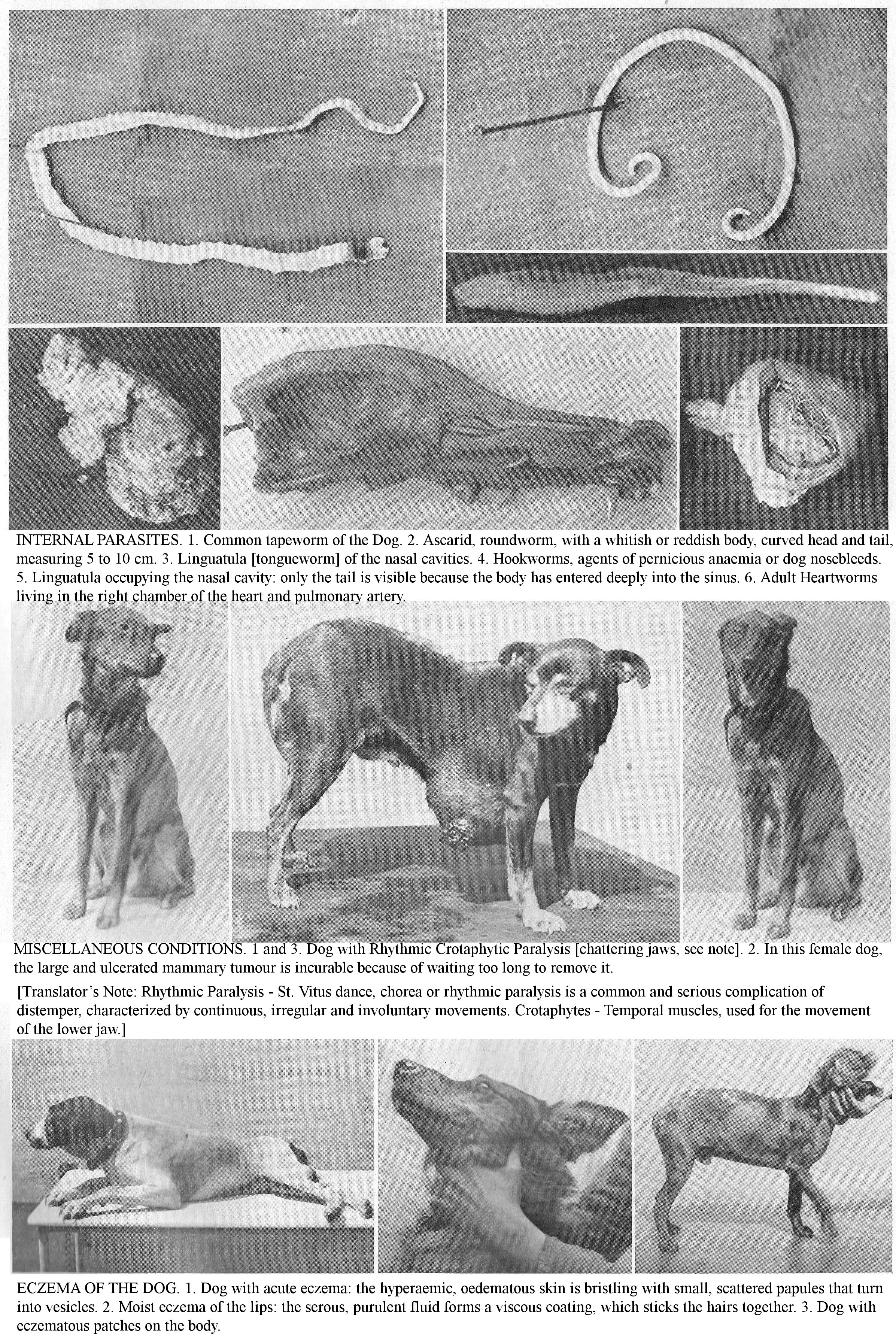
INTERNAL PARASITES. 1. Common tapeworm of the Dog. 2. Ascarid, roundworm, with a whitish or reddish body, curved head and tail, measuring 5 to 10 cm. 3. Linguatula [tongueworm] of the nasal cavities. 4. Hookworms, agents of pernicious anaemia or dog nosebleeds. 5. Linguatula occupying the nasal cavity: only the tail is visible because the body has entered deeply into the sinus. 6. Adult Heartworms living in the right chamber of the heart and pulmonary artery.
MISCELLANEOUS CONDITIONS. 1 and 3. Dog with Rhythmic Crotaphytic Paralysis [chattering jaws, see note]. 2. In this female dog, the large and ulcerated mammary tumour is incurable because of waiting too long to remove it.
[Translator’s Note: Rhythmic Paralysis - St. Vitus dance, chorea or rhythmic paralysis is a common and serious complication of distemper, characterized by continuous, irregular and involuntary movements. Crotaphytes - Temporal muscles, used for the movement of the lower jaw.]
ECZEMA OF THE DOG. 1. Dog with acute eczema: the hyperaemic, oedematous skin is bristling with small, scattered papules that turn into vesicles. 2. Moist eczema of the lips: the serous, purulent fluid forms a viscous coating, which sticks the hairs together. 3. Dog with eczematous patches on the body.
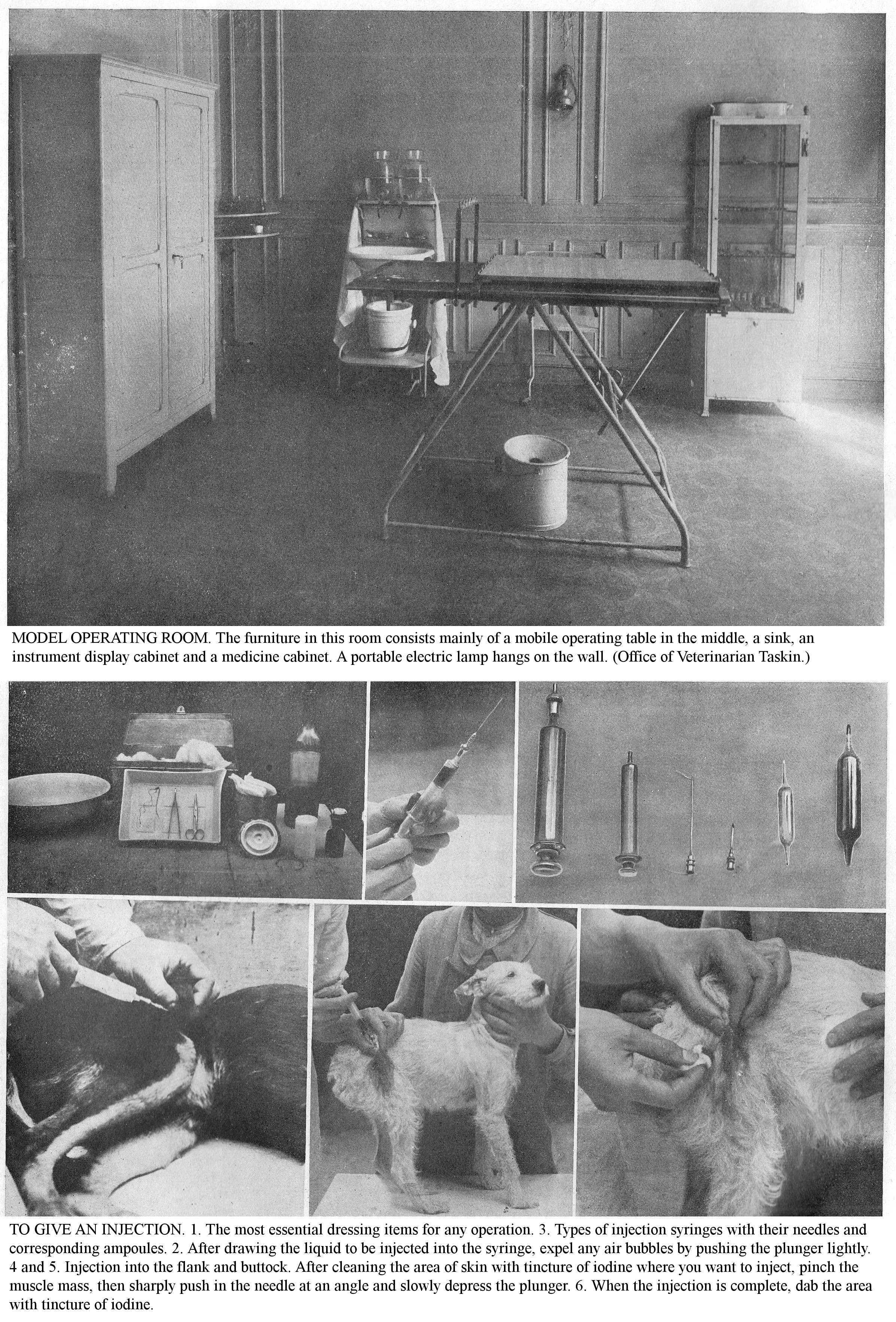
MODEL OPERATING ROOM. The furniture in this room consists mainly of a mobile operating table in the middle, a sink, an instrument display cabinet and a medicine cabinet. A portable electric lamp hangs on the wall. (Office of Veterinarian Taskin.)
TO GIVE AN INJECTION. 1. The most essential dressing items for any operation. 3. Types of injection syringes with their needles and corresponding ampoules. 2. After drawing the liquid to be injected into the syringe, expel any air bubbles by pushing the plunger lightly. 4 and 5. Injection into the flank and buttock. After cleaning the area of skin with tincture of iodine where you want to inject, pinch the muscle mass, then sharply push in the needle at an angle and slowly depress the plunger. 6. When the injection is complete, dab the area with tincture of iodine.
EAR CONDITIONS
HOW TO RECOGNIZE AND TREAT THEM TO AVOID ANY COMPLICATIONs THAT MIGHT REDUCE THE VALUE OF THE ANIMAL BY MAKING IT UNFIT FOR ITS PURPOSE.
EAR CONDITIONS, like eye conditions, should be treated immediately because deafness, an extreme consequence of an untreated condition, renders a Dog unable to perform the services expected of him.
Semiology: In Dogs, examination of the ear is reduced to the exploration of the internal ear. Checking the pinna does not require any special manoeuvring or the use of any instruments. When the ear is sore, the patients defend themselves, so secure, muzzle and lay the Dog on a table, positioning the ear so that natural light illuminates the interior part of the ear. Practice examining the entrance to the ear canal with the ear in a suitable position; this will tell you the condition of its integument, the presence of earwax, abnormal amounts of wax, lumps or foreign bodies. Use artificial light to illuminate the depth of the ear. When the ear canal contains liquid exudate, exert light pressure in order to perceive a characteristic noise at the level of the liquid.
To explore the ear canal more fully, use bivalve speculums; insert them carefully into the depth of the ear then illuminate the ear canal using either natural light or artificial light, focusing the light rays in the speculum cavity using a reflective mirror.
CONTUSIONS.
Contusions of the ear shell present with varying degrees of severity: sometimes there is a simple detachment of the integument producing an effusion of blood into the cavity; sometimes there is a fracture of the cartilage at the same time. Treatment: Immobilize the area with a net; perform repeated lukewarm affusions [water showers].
Ear shell wounds are superficial and not very extensive, or sit on the cartilage and cause rupture. When the pinna has been split on its free edge, if the lips of the wound heal a distance apart, the ear is split. Treat skin wounds by trimming the surrounding hair and disinfecting the wound; if the edges are very wide apart, join them with a few stitches. To prevent healing from being delayed by scratching or constant rubbing of the ears, put a bonnet on the Dog, a sort of cloth or mesh cap, fitted on one side with two or three loops and, on the other with leather straps that allow it to be securely fixed. During a fracture of the base of the ear shell, if the parts consolidate irregularly, the ear canal may be more or less completely blocked.
Haematomas of the pinna, characterized by a fluid effusion between the skin and the cartilage, are common in dogs. Puncture lumps, and after the liquid has drained, give an irritant injection (tincture of iodine), and immobilize the ear.
Ear abscesses may occupy one or both sides of the ear. Avoid otitis externa, necrosis of the cartilage by puncturing abscesses at once. Cartilage necrosis often requires surgery: cauterization or excision.
Ear shell ulcers or ear canker is common in those with long, hanging ears. It often follows a traumatic lesion of the edge of the pinna; it frequently accompanies auricular catarrh and almost always sits at the tip of the ear shell. This lesion has a great tendency to persist, maintained by scratching, rubbing and incessant agitation of the ears.
Treatment: Canker is symptomatic of auricular catarrh so treat the underlying condition. Secure the ears with a bonnet or canvas cap; by covering the canker with a tightly adherent layer of iodoform collodion or salicylic collodion, you can dispense with the application of a cap. Clean the wounds two or three times a day with antiseptic washes; wipe them off and sprinkle with iodoform. If healing is slow, have your veterinarian pass two tow sutures through the ear, near the lesion; in the days following the operation, the edges of the ulcers subside, and the unhealthy secretions soon dry up.
Because this lesion is a frequent localization of arthritic tendency, institute an internal treatment (Fowler's liquor, baking soda).
FOREIGN BODIES AND PARASITES.
The ear may contain clumps of ceruminous material, inert bodies (gravel, small pebbles, fragments of bones or wood, pieces or ears of grasses). In dogs with auricular catarrh, if the ear is not kept very clean during hot weather by frequent washing, you may observe in the external auditory canal larvae, "maggots", from fly eggs deposited at its entrance. Remove foreign bodies with injections of lukewarm water or with fine forceps, a stylet, or blunt curette.
EXTERNAL OTITIS.
Otitis externa or inflammation of the external auditory canal, particularly common in dogs with long hair, with hanging ears (Spaniels, Poodles, Pekingese, Briards), is simple or parasitic, unilateral or bilateral. The main aetiological factors, apart from eczema of which it is often only a localization, are: incorrect diet, insufficient exercise, arthritis, abundance of earwax and foreign bodies in the ear; often it is seen in the course of distemper.
In acute otitis you see intense inflammation of the inner integument; the ear is hot, red, sore and secretes a seropurulent exudate with a foul odour. The oblique head carriage, more or less twisted on the neck, holding the ear in a sloping position, somewhat frequent shaking of the head, continual scratching and rubbing, attract your attention and constitute specific symptoms of the a severe ear condition. Apart from local symptoms of the ear, you may observe reflex disturbances, nausea, vomiting. Perforation of the eardrum as a result of pustular and ulcerative forms of otitis can lead to brain complications.
Treatment: Keep ears raised on the head by applying a bonnet; cut the hairs adorning the ear and clean it with a lukewarm soap. If the pain is intense, use warm emollient, narcotic injections: decoctions of mallow, marshmallow, poppy head, laudanum preparations, 1% chloral solution. If the secretion is profuse, resort to weak antiseptic solutions. In injections, cover the projections in the ear with antiseptic powder, or simply absorbent powder: starch, bismuth sub-nitrate, zinc oxide, iodoform, etc. For Luxury Dogs, use scented powders. Calm the intense pruritus with a boric solution, with the addition of cocaine or laudanum; supplement the action of the injections by depositing a little boric or salol petroleum jelly at the entrance to the external auditory canal.
After reducing the inflammation and pain, cleanse the ear frequently with lukewarm injections (alum, zinc sulphate, tannin and glycerine, potassium permanganate, cresyl, etc.). Ensure the liquid penetrates to the bottom of the duct, rubbing lightly at the base of the ear shell. Alkalis and arsenicals form the basis of internal treatment; avoid recurrence by continuing to administer them for a week or 15 days a month. Give a meat diet to a debilitated patient; give an overfed patient a refreshing diet: soups, lean stews, milk and milk preparations.
Chronic external otitis or auricular catarrh follows acute otitis, or starts from its outset under the influence of still ill-defined causes which are almost always dominated by constitutional issues (arthritic tendencies). You will see localised thickening, oozing from the internal integument of the base of the ear shell and auditory canal, and sometimes papillomatous growths. In a few cases, the inflammation spreads to the middle ear and causes auricular vertigo, deafness or fatal accidents. The prognosis of auricular catarrh, regardless of any complications, is serious due to its tenacity, its tendency to recur and its ambulatory nature.
Treatment: To dry up the secretion, use injections of iodized glycerine, solutions of alum, zinc sulphate (2% to 3%), then dry with cotton wool and place a few drops of iodized glycerine, or balsam of Peru at the entrance of the ear canal. Spray the ears with boric acid, talc and zinc oxide, or a mixture of these. Treat superficial ulcerations of the duct with solutions of silver nitrate or potassium permanganate; if they exist at the entrance to the ear canal, cauterize them lightly; excise growths.
As internal treatment, have recourse to arsenicals (Fowler's liquor); with iodides (potassium iodide); with alkalis (baking soda). Exercise is always a helpful adjunct.
EAR TUMORS.
Ear polyps commonly seen in dogs are usually localized to the lower part of the ear shell and the external ear canal. Very variable in shape, size and consistency, they are usually numerous, pedicled or sessile and arranged in cauliflowers or in clusters. When the condition is bilateral, hearing is more or less lost. Light caustics and ablation form the basis of treatment.
DEAFNESS.
Deafness, common in older Dogs, can be caused by various lesions of the hearing system (accumulation and hardening of earwax, thickening and induration of the integumentary membrane, perforated eardrum, lesions of the middle ear, etc.). The decrease and loss of hearing is unilateral or bilateral. In the latter case, the hearing impairment is usually uneven between the two ears, one is more deaf than the other. Deafness is noticeable by the Dog's inattention to commands or calls, sometimes by his dazed expression.
Treatment for deafness varies depending on its cause. Deafness due to various acute ear diseases usually resolves when the diseases are resolved. When it comes from a general influence or tendency, combat that. With rare exceptions, when it is well established and longstanding, you have little hope of a cure. When deafness is noticed, examine the ear canals, and remove any foreign bodies, earwax or polyps that may obstruct them. Diversions applied near the ear (sutures, blisters) are, along with electricity, the main agents recommended. Internally, use nux vomica and potassium iodide.
Prophylaxis: Use frequent soaping to prevent the accumulation and hardening of earwax; combat arthritic tendencies through diet and a healthy lifestyle (meat-and-vegetable diet, exercise) which is the dominant aetiological factor in Otitis, and you will thus avoid deafness which particularly hampers watchdogs and hunting dogs.
CONDITIONS OF THE EYE AND ITS APPENDAGES.
DO NOT NEGLECT ANY CARE TO MAINTAIN THE VISUAL INTEGRITY ESSENTIAL FOR SERVICE DOGS.
THE EYE is one of the most precious organs, and one of the most fragile, because it is exposed to all kinds of mild or serious ailments that you must prevent or treat without delay.
EYE EXAMINATION.
The examination of the ocular apparatus includes a series of methodical explorations: internal examination of the eye in direct light and without an instrument, exploration of the conjunctival fornices, examination of the internal parts by lateral illumination, examination of the deep parts with an ophthalmoscope.
Due to the difficulty of diagnosis and the methodical application of treatment, the veterinarian’s intervention is necessary for various eye conditions and its appendages. Remember that the judicious use of the treatment depends on the visual integrity of the diseased eye and that of its congener (sympathetic ophthalmia). Visual integrity, especially in Guard Dogs and Hunting Dogs, plays an important role in their use. Also, in this chapter, we insist on ocular prophylaxis, pathology and therapy.
CONTUSIONS.
Eye contusions are common in dogs and are usually produced by bumps or tree branches. Symptoms: In the simplest cases, you will see subconjunctival bruising, closed eyelids and tearing. If the contusion is severe, you may notice traumatic ophthalmia, corneal erosions, iris rupture, lens dislocation, etc. Sometimes the interior vessels of the eye are torn, blood rushes into the organ and mixes with the humours (hypohaema [collection of blood in the anterior chamber of the eye]) and forever compromises visual integrity. Sometimes you can see the opening of the eye.
TRAUMATIC INJURIES.
Wounds: Eye wounds, usually caused by sharp or prickly objects (cat's claw) or bites, may be seen by the explosion of powder or the action of a projectile. Eye wounds caused by lead grains are frequent in hunting dogs.
Superficial lesions are limited to the eyelids, conjunctiva and superficial layers of the cornea. Deep lesions affect the whole of it, damage the internal parts of the eye (iris, choroid, lens) when infected and most often lead to the loss of the eye. If it is punctured, the eyeball is noticeably reduced in volume and its window is pleated.
Eyelid wounds are frequently observed, are divided in variable directions, are partial or full thickness; the removal of a skin flap is not uncommon.
Conjunctivitis wounds are usually mild, but extensive loss of substance heals only slowly.
Superficial punctures are rarely followed by a serious accident; they are only dangerous when they are deep. When the point has penetrated deeply, the injury is almost always followed by intense inflammation. Sometimes the vitreous humour, or even the lens, drains through the wound. Inflammation from a simple wound causes atrophy of the eye and even loss of vision.
Burns to the eye are usually produced by flames or caustics. Superficial burns, especially if they are narrow, may allow the eye to recover completely after the scabs fall away. Deeper burns are usually followed by a more or less extensive opacity. Finally, burns can cause irremediable dislocation of the eye.
The foreign bodies usually found in the eye tissues are grass pieces, thorns, splinters, grains of lead, etc. They can adhere to the eyelid, conjunctiva, or internal surface of the cornea, or occupy the descending part of the anterior chamber, or even be stuck in the iris or lens capsule. The prognosis varies greatly depending on the site of the lesion. Do not confuse these foreign bodies, which cause inflammatory disorders, with those adhered to the outer surface of the globe and simply stuck between it and the walls of the orbit.
The most resistant part of the internal eye is the anterior chamber. In other areas, foreign bodies most often cause inflammatory and nervous events and, when infected, almost always cause more or less complete loss of the eye; sometimes the retina is paralyzed by the pressure of these bodies, especially when they are heavy, such as lead shot. Foreign bodies in the anterior chamber may be eliminated spontaneously, either emerging whole by a corneal ulcer, or else they may be subject to the dissolving action of the aqueous humour and disappear slowly; in other areas elimination is extremely rare.
Treatment: The treatment of ocular wounds lies entirely in their antisepsis; the treatment applied in the hours following an eye injury most often affects the preservation of the damaged eye and its congener. It is, in fact, proven today that accidents that occur to the neighbouring eye (sympathetic ophthalmia) are almost always due to the penetration of dangerous germs, to infection. Achieve asepsis of the eye by abundant washing with a 1% sublimate solution; irrigate the conjunctival fornices, the internal corner of the eye, the ciliary border, and lacrimal passages with this liquid. Protect the eye with an antiseptic dressing; this is a very important part of the treatment.
CONJUNCTIVITIS.
Conjunctivitis, the acute inflammation of the conjunctiva, is a common disease. Aetiology: Its ordinary causes are: trauma, repeated rubbing, foreign bodies, dust introduced into the conjunctival fornices, entropion, ectropion, eyelid eczema, follicular mange. Conjunctivitis is one of the first manifestations of distemper.
Symptoms: You will notice photophobia and tearing, the conjunctiva is red and tender, and often the inflammation has spread to the cornea. Usually after a few days the condition goes away. If the underlying cause remains, conjunctivitis persists in a chronic state and is characterized by a purulent secretion with a little tearing.
Treatment: Treat the underlying condition; extract foreign bodies from the eye and fornix, address entropion, ectropion, eyelid inflammation, etc. Wash the conjunctiva repeatedly with a lukewarm antiseptic solution (1% boric water, 1 part per 4,000 sublimate), or an astringent solution (zinc sulphate, methylene blue, tannin, white water [lead acetate], etc.); combat the purulent secretion by touching the conjunctiva with a swab soaked in 1% or 2% silver nitrate solution and immediately afterwards wash the eye with a 1% boric solution; if the pain is severe, instil cocaine or atropine. Destroy the granulation by cauterizing with silver nitrate and lotion with a mild antiseptic solution.
CORNEAL CONDITIONS.
Keratitis, inflammation of the cornea, can be superficial, parenchymal [affecting functional tissue] or deep. The most common causes are: irritation, burns, contusions, foreign bodies (dust, grass husks), conditions of the conjunctiva or eyelids; it often accompanies the distemper. Symptoms: At first, the painful eye, covered by the eyelids, weeps constantly, the cornea is infiltrated, becomes opalescent; sometimes an abscess can form.
Treatment: Frequently apply a lotion of weak, warm antiseptic solution (borated or boric acid solution) or 1 part per 3000 sublimate; calm the pain with instillations of atropine or cocaine; As soon as the inflammation subsides, give insufflations of calomel to hasten absorption.
Corneal ulcers. Corneal Ulcers are a complication of Keratitis; they affect the cornea to a variable thickness and extent. When the middle layer is punctured, the inner membrane herniates through the ulceration (keratocele).
Treatment: Use hot antiseptic lotions: boric water 3% to 4%, sublimate at 1 part per 3000. Treat the cloudy patch with insufflations of calomel or daily anointings of yellow mercury oxide ointment.
EYE ABSCESS.
EYE ABSCESSES are suppurative inflammations of the membranes and media [middle coat] of the eye. An eye abscess is characterized by swelling and infiltration of the eyelids and conjunctiva; the cornea is inflamed and oedematous. Suppuration of the eye is accompanied by general symptoms: depression, anorexia, fever. The healthy eye may be affected secondarily (sympathetic ophthalmia).
Treatment: The only treatment is enucleation. To remedy the deformity resulting from this operation, Cadiot and Breton recommend suturing the eyelids. In unilateral blindness, the head is tilted, oblique, the healthy eye always carried higher than the other.
AMAUROSIS.
This condition is characterized by the weakening or loss of sight, without loss of transparency of the media and without obvious damage. Single or double, transient or permanent, it is either due to lesions of the nervous centres of the optic nerve or lesions of the retina. The causes of Amaurosis are violent bruising of the eye, some types of brain damage, infectious diseases, especially distemper. The immobility of the iris and the fixed pupil diameter characterize complete amaurosis.
Treatment: Treat the causative condition if possible; give iodine medications; if unsuccessful, use subcutaneous injections of strychnine.
CATARACT.
Cataract, cloudiness of the lens, is a relatively common condition; it is caused by bruises and sores of the eye, sometimes it is congenital; in general, it is an acquired condition resulting from heredity, old age (senile cataract), arthritis, diabetes, etc.
Symptoms: On examination of the eye after atropine instillation, the lens appears opalescent or marked with white, greyish, bluish, or yellowish spots, of varying sizes, covering part or all of the lens. Cataracts decrease vision in the eye when recent and peripheral, or completely eliminate vision.
Treatment: Medical treatment with vesicants around the eye, setons, purgatives, etc., does not work. Essentially surgical, treatment consists of moving, modifying or extracting the lens, so as to allow the light rays to reach the retina.
INFLAMMATION OF THE IRIS.
Inflammations of the iris (iridocyclitis, irido-choroiditis) usually occur as complications of eye trauma or in the course of various infectious diseases (distempers). They are characterized by marked acute symptoms (inflamed, sore eye, bloodshot conjunctiva, hyperaemic iris, red, brown or yellowish in colour, presence of serous, purulent exudate in the anterior chamber, etc.). These recurrent conditions end up causing blindness.
Treatment: Apply moist, warm compresses to the eye (1% boric acid solution, 1 part per 4000 sublimate); instil atropine sulphate eye drops in the eye.
LUXATION OF THE EYE.
Dislocation of the eye means its complete ejection from the orbital cavity. This accident is caused by violent traumas, particularly in Dogs and Cats, and scratches.
Treatment: The treatment varies according to the lesions: if the eyeball is intact, has retained vascular and nervous supplies sufficient to maintain it, perform repositioning, which includes the following stages of operation: application of boiled cocaine compresses, slight enlargement of the eyelid opening, placement of the eyeball, application of stitches to the eyelids to keep the eye in position. If the lesions are too severe, remove the eyeball.
Eyelid conditions: Adhesion of the edges of the eyelids is sometimes congenital, sometimes follows wounds or inflammation of the eyelids. Surgical intervention constitutes the basis of treatment (separation of the eyelids by incision, suture of the mucous membrane and skin). Combat the adhesion of the eyelids to the eyeball, which creates an obstacle to vision, with daily instillations of sterilized oil. The deviation of the eyelashes inwards towards the eyeball most often results from chronic inflammation of the eyelids; vision is disturbed, it can result from Conjunctivitis, simple or ulcerative keratitis. Use tweezers to pluck the eyelashes that irritate the eye or, in the event of recurrence, excise the eyelid flap that supports the deviated eyelashes.
Entropion is characterized by the inward turning of the eyelid. It is noticed as a result of ophthalmia with photophobia, by lumps developed on the eyelids, etc. Entropion is a cause of conjunctivitis and keratitis as a result of continued irritation by the eyelashes. Only surgical treatment (excision of a skin flap of varying width and extent in the region of the inverted skin) is effective.
Ectropion, the turning outwards of the free edge of the eyelids, is most often seen in the lower eyelid; it is usually due to inflammation accompanied by swelling of the conjunctiva or to a wound, with loss of substance that sits on the eyelid or periocular region; surgery is the only treatment indicated.
BLEPHARITIS.
Blepharitis is characterized by local or generalized inflammation of the eyelids. Apart from bruises, the causes are scratching, excoriations resulting from eczema, follicular mange.
Symptoms: The swollen, hot, tight, painful eyelids more or less completely cover the eyeball. Sometimes only the free edge of the eyelids are inflamed. You will mainly encounter this form in old and poorly kept Dogs and Cats: the free edge of the eyelids is thickened, bloodshot and produce a mucopurulent exudate that accumulates towards the inner corner of the eye and clumps the eyelashes. Sometimes the skin near the free edge of the eyelid is the site of a small abscess the size and consistency of a barley grain.
Treatment: Do frequent applications of lukewarm 2% boric solution, or with very light lead acetate. After these washes apply boric petroleum jelly. You can also place a wet bandage over the eye; sew an absorbent cotton ball on a sizable square of ordinary canvas with two openings corresponding to the ears, fitted with cords to secure it by tying them, two by two, under the throat and under the lower jaw; sprinkle every two hours with a cold or lukewarm solution of mild lead acetate. In the event of a stye, facilitate the opening of the abscess using hot wet compresses, or have a puncture performed. Treat ciliary blepharitis by cleansing; wash the eye and eyelids with a lukewarm boric or borate solution; change the diet and give Fowler's liquor or cod-liver oil.
Prophylaxis: Avoid ciliary blepharitis by daily washing of the eyes.
LACRYMAL SYSTEM CONDITIONS.
Conditions of the lacrimal system (inflammation, dropsy, occlusion of the lacrimal sac) are manifested by tearing and depilation below the inner corner of the eye.
Treatment: Empty the tear sac by finger pressure, several times a day, and instil a lukewarm solution of boric acid or 2% zinc sulphate in the inner corner of the eye; cover the eyelids with a thin layer of petroleum jelly.
PURULENT OPHTHALMIA IN NEWBORNS.
This condition is observed in puppies or kittens whose mother is affected by vaginitis. Purulent ophthalmia, caused by intravaginal infection, develops from day 1 to day 20 after birth. As young subjects do not open their eyes until around the 9th day, the disease is recognized late. Often they die of exhaustion or the eyes empty completely due to suppuration.
Prophylaxis: In female dogs and cats with metritis or vaginitis, practice before, during and after the birth, antiseptic vaginal irrigations. Immediately after the birth, wipe the eyes of the young with an absorbent cotton ball; then wash them with 4% boric water, then instil a few drops of 2% silver nitrate. After the onset of the disease, protect the healthy eye by applying compresses soaked in boric water to the diseased eye, because the disease is easily transmitted; wash your hands thoroughly with 1% sublimate solution after touching diseased eyes.
MISCELLANEOUS CONDITIONS OF DOGS AND CATS
HOW TO TREAT: BRUISE, BURNS, BOILS, ABCESSES, LUMPS, TUMOURS, MISCELLANEOUS HERNIAS AND RUPTURES.
OUTSIDE of the diseases which fall within the main categories discussed so far, there is a whole series of very varied conditions which constitute special events of greatly varying severity, but which generally require immediate treatment.
CONTUSIONS.
Contusions – bruises - are lesions without loss or apparent break in continuity of tissue, produced in living tissue. The most common causes involve kicks, sticks, falls, collisions, being crushed by vehicles, etc.
Symptoms: Contusions are manifested by signs of more or less intense local inflammation: hot, tender, painful swelling; when on one limb, you observe a limp of varying intensity. We distinguish three degrees of Contusion. The first degree is manifested by bruising of the skin, with slight peripheral oedema. In the second degree, the capillaries are ruptured, and the effused blood forms a haematoma. In the third degree, the lesions are sometimes considerable; muscle tissue is reduced to mush, vessels and nerves are ruptured, tendons are severed, bones re fractured.
First and second degree contusions frequently end in resolution; sometimes the haematoma persists as a cyst, or suppuration occurs and an abscess forms.
Gangrene is a frequent termination of third degree contusions. Various complications can occur with severe or extensive bruises; the most frequent are: thrombosis and venous embolism; hernia or peritonitis when the contusion sits on the abdomen; pleurisy, when it exists on the chest walls; exostosis [bony spur] in the case of contusion of the bones; paraplegia, when the spinal tissue is compressed in the lumbar region, etc.
Treatment: For first degree contusions, use cold showers, water compresses or cold or lukewarm lead acetate; if the pain is severe, apply laudanum poultices or cocaine petroleum jelly. Puncture the haematoma, drain its contents, and inject tincture of diluted iodine. To begin with, treat third-degree contusions with showers, cold or hot compresses; if the tissues are crushed and leaking blood, open it widely and wash the cavity frequently with a strong antiseptic solution. If the dead skin falls off, treat it as an ordinary wound and encourage healing with iodoform or salol dressings.
BURNS.
Burns are injuries produced by solids, liquids or gases.
Symptoms: The symptoms depend mainly on the degree of burn. A mild burn manifests as varying degrees of inflammation: redness of the skin, increased sensitivity; if the burn is more severe, there is inflammation proper, the skin swells, the epidermis dies and is separated from the dermis by a limpid serous secretion which constitutes the blisters; this secretion is accompanied by greatly increased sensitivity and an infiltration of the surrounding and underlying tissues.
Prolonged action of burning to the same part of the body leads to its death; the Burn can spread from the skin to nearby muscles, vessels, nerves, tendons, bones. The general symptoms consist of fever, dejection, inappetence; when the Burns are extensive, they may be followed by visceral congestion or by asphyxiation because the skin cannot breathe.
Treatment: For first degree burns, use cold water and astringents: lead acetate, 1% solution of iron or zinc sulphate; then sprinkle the burnt area with starch powder. Treat second degree burns by draining the blister, then apply antiseptic solution followed by iodoform ointment, sterilized petroleum jelly, or absorbent powder (starch, boric acid, tannin, and iodoform) or 1% picric acid compresses. If the Burns are located at the extremities, use prolonged lukewarm antiseptic baths then cover the wounds with an insulating powder and a cotton bandage. Prevent animals from biting the wounds, which are often very itchy; apply a lotion of 12 parts per 1000 picric acid solution to calm the pruritus. For third degree burns, encourage the shedding of charred or dead tissue; prevent complications by washing wounds with antiseptics and applying absorbent powders or boric, iodoform or salol salty ointment; if the pain is severe, use cocaine preparations.
The treatment of choice is the use of specific, polyvalent serum from Leclainche et Vallée; wash the wound with a warm boiled solution of 9 parts per thousand sodium chloride; brush it with the serum, then apply a few layers of wadding or sterilized gauze to isolate the dressing; repeat the treatment until healing is complete.
Combat various visceral complications and septic accidents by administering diffusible stimulants (wine, alcohol, coffee, ammonia acetate) internally, by subcutaneous injections of caffeine or salt water.
BOILSS, ABSCESSES, ANTHRAX.
AN ABSCESS is a collection of purulent fluid that develops in the tissues of the body. Depending on the mode of development, Abscesses are divided into hot, cold, superficial and deep. Hot abscesses are caused by contusions, trauma, and penetration of foreign bodies; but, whatever the origin of the abscess, it is due to the penetration of pyogenic microbes (white and golden staphylococci, streptococci, bacilli, colibacilli, etc.) into body tissue.
Symptoms: An Abscess manifests as swelling, heat, and pain preceding the fluid collection. The inflammation is accompanied by general symptoms: fever, inappetence, functional problems, the severity of which varies with the pyogenic disposition and site of the abscess.
Diagnosis: Examination of general and local signs allows fairly easy diagnosis; in doubtful cases, confirm it by performing an aseptic capillary puncture. Prognosis: Depends on a superficial or deep site, anatomical region, and functional reaction.
Treatment: Calm the pain with emollient poultices; as soon as the fluid collection is noticeable, open the Abscess; do not act too prematurely, only open the purulent cavity when the fluid occupies two-thirds of the lump; cleanse the cavity by irrigation with an antiseptic liquid: carbolic acid, 3% to 5%; sublimate, 1 part per 1000; cresyl, 2% to 4%; tincture of a third iodine; quarter hydrogen peroxide.
If suppuration persists beyond the normal time or the puncture wound fistulizes, there is a foreign body or a fragment of dead tissue in the walls of the abscess; surgical intervention is then indicated (debridement, counter-opening, drainage, etc.). For cold abscesses, wipe daily with tincture of iodine.
GROWTHS, TUMOURS.
Common in Dogs and Cats, Growths (Epitheliomas, Fibroids, Papillomas, Sarcomas, Lipomas, Cysts) are particularly frequent in elderly animals. Defective diet, arthritis, heredity, traumatic irritation, slight and repeated bruising, intervention of infectious agents (bacteria, sporozoa or fungi) are the most common environmental factors in tumours.
Symptoms: The most common epitheliomas are those of the skin, mammary glands and anus; then come those of the kidney, liver, testis, vulva and vagina. Epitheliomas of the skin appear as hard, bumpy tumours that ulcerate and grow more or less quickly.
Sarcomas, many of which are soft in consistency, grow rapidly, eventually becoming enormous in size, and become generalized or recur after removal.
Fibroids, usually small in size and hard in consistency, grow slowly, remain sessile or pedicle, always remain clearly demarcated, are easily operable and do not recur.
Papillomas, especially found in young animals, develop on the skin and mucous membranes; they are most often multiple and sometimes very numerous; their normal sites are the lips, cheeks, ears, dorsal region, mucous membranes of the mouth, penis, vulva, vagina. Those of the skin, warts, are generally consistent, small in size and well delimited; quite often merging, they are arranged in a mushroom or cauliflower shape.
Lipomas, found mainly in overfed, elderly or obese dogs, are primarily located in the subcutaneous connective tissue. Soft in consistency, well defined, and covered by intact skin, they grow slowly and often remain static for long periods. Cysts are characterized by fluid contents and the absence of any inflammatory phenomena.
Prognosis: Considered from the point of view of their development and gravity, Growths are distinguished as either benign or malignant. The former (Fibroids, Papillomas, Lipomas, Cysts) usually grow slowly or soon stop growing and do not recur after removal. Malignant growths, Cancers (Epitheliomas, Sarcomas) develop rapidly, have a marked tendency to spread, generate secondary tumours and, very generally, they recur.
Treatment: For benign growths which do not interfere with any function, refrain from any intervention; if treatment is decided (e.g. due to rapid increase), resort to ligation (strangulation); the application of a very tight band at the base of the tumour causes it to die; also use caustic or hot iron cauterization or excision. For warts, use a caustic liquid that you apply with a small mounted swab dipped in the caustic, then drained. For malignant neoplasms, early and complete ablation should be performed.
Goitre is a growth of the thyroid bodies located on the anterior face of the neck. Acute thyroiditis results in enlargement, tenderness, difficulty swallowing, and difficulty breathing. Chronic thyroiditis is characterized by sluggish parenchymal [functional tissue] hypertrophy and often ends in cystic or cancerous degeneration. Besides local disorders, Goitres produce weight loss due to insufficient thyroid secretions. In most cases, the enlarged thyroid is bilateral, varies in consistency, soft, firm, with fluid fluctuations in places, or uniformly hard.
Treatment: Treat the patient with iodine medication; continue this treatment for a long time with varying lengths of rest, depending on the development of the tumour and the general condition of the patients. Neoplasmic goitre is amenable to surgery.
HERNIAS.
Hernias are characterized by the emergence of a viscera from the abdominal cavity through natural (inguinal passages, umbilicus) or accidental openings (tearing of the ventral walls or the diaphragm). Congenital hernias that exist at the time of birth are due to defective closing of a provisional opening intended to close soon after birth (umbilicus). Acquired hernias are consecutive to trauma or stenuous effort.
Aetiology: Heredity, young age and old age, the relaxation or abnormal dilation of the inguinal, crural, or umbilical rings, the too slow organization of the tissue that must form the umbilical opening are all predisposing causes; violent contractions of the abdominal muscles, jumps, expulsive efforts, abdominal contusions are determining causes.
Symptoms: The most important symptom of Hernias is the existence of a growth of varying shape and size in some part of the abdomen. Palpation detects an elastic consistency; pressure exerted on it reduces its volume or even reduces it completely. Uniform consistency of the growth, its sounds and rumbling, indicate the presence of the intestine in the Hernia. The functional symptoms of uncomplicated hernias are nil or little marked; you see only various digestive disturbances, and sometimes slight colic. The engorgement and strangulation of Hernias are marked by signs of severe pain, colic and vomiting; the enlarged growth is strained, painful under pressure, usually irreducible.
Do not confuse Hernias with soft growths, abscesses or cysts; in doubtful cases, confirm the diagnosis by an exploratory puncture. Treatment of Hernias is complex; it includes the use of bandages, vesicants, caustics, destruction of the sac, and surgical treatment.
Using bandages, the contents of the sac are forced back into the abdomen by a ball applied at the level of the hernial growth; this lacks fixity and is not recommended. Irritant (sinapism), vesicant (revulsion) or caustic (nitric acid, sulphuric acid) applications on the skin covering the hernial sac are intended to cause exudation in the subcutaneous connective tissue which pushes the herniated organs back into the abdomen, and the formation of a fibrous support capable of blocking the hernial path. These applications are very difficult to do.
Injections into the periumbilical connective tissue of irritating fluids (alcohol, saturated sea salt solution, zinc chloride 10%) work in the same way. The destruction of the hernial sac, applicable to simple easily reducible hernias, is done by ligation or suture. Surgical treatment is a more modern process; performed under antiseptic conditions its results are excellent.
Regardless of the site and size of the hernia, fight the choking and strangulation with hot packs applied to the growth and manipulation. The latter is most advantageously performed with the animal lying down and anesthetized: grasp the pedicle of the hernia with your left hand, compress the growth with the fingers with your right hand, and attempt to gradually reduce the displaced organs; start by reducing the parts closest to the hernial opening. The collapsed growth tells you that shrinking is taking place. Manipulation should not be too lengthy or too violent, it requires the intervention of the veterinarian.
Inguinal hernias in puppies which show little tendency to heal spontaneously are treated by castration*; the different operating stages include: incision of the scrotum and dartos [thin layer of muscle lying immediately under the skin]; isolation, reduction and amputation of the vaginal sheath; suture the lower vaginal ring. Old or large hernias require surgery; this is urgent in cases where strangulation occurs.
The inguinal hernia, frequent in bitches, can acquire rather large dimensions; the sac most often contains part of the omentum or one of the horns of the uterus, sometimes the intestine or bladder. It is particularly serious when the female is pregnant and one or more foetuses develop in the herniated horn.
[* Translator’s note: castration in French refers to both male castration and female spaying.]
EVENTRATION [EVISCERATION].
This occurrence is extremely grave; it consists of the emergence of the intestines through an opening covering the entire thickness of the abdominal walls; in Dogs, it can be caused by a deep bite or be the consequence of the wounds made by the tusks of wild boars.
This condition sometimes accompanies certain operations (castration of the male and the female, untimely cauterization of an umbilical hernia, etc.). Eventrations are usually formed by the small intestine or the floating colon, rarely by the large intestine.
Symptoms: When the hole which gives passage to the intestine is narrow, the viscera appear in the form of a small growth which lengthens from minute to minute so that, after a few moments, an intestinal loop emerges from the abdominal wound. The pain felt by the injured individual then becomes sharp; it is gripped by extremely violent colic, kicks, lies down and rolls about, crushing the intestines. Prognosis: Very serious; death is the usual outcome.
Treatment: Return the viscera to their normal position, perform manipulation; however, only perform mitigation if the herniated viscera is not the site of extensive tears, such as those made by the animal with its teeth or feet. When the gut is intact, give yourself the chance of mitigation, however disturbing the appearances of the leaked abdominal organs may be. The treatment is surgical.
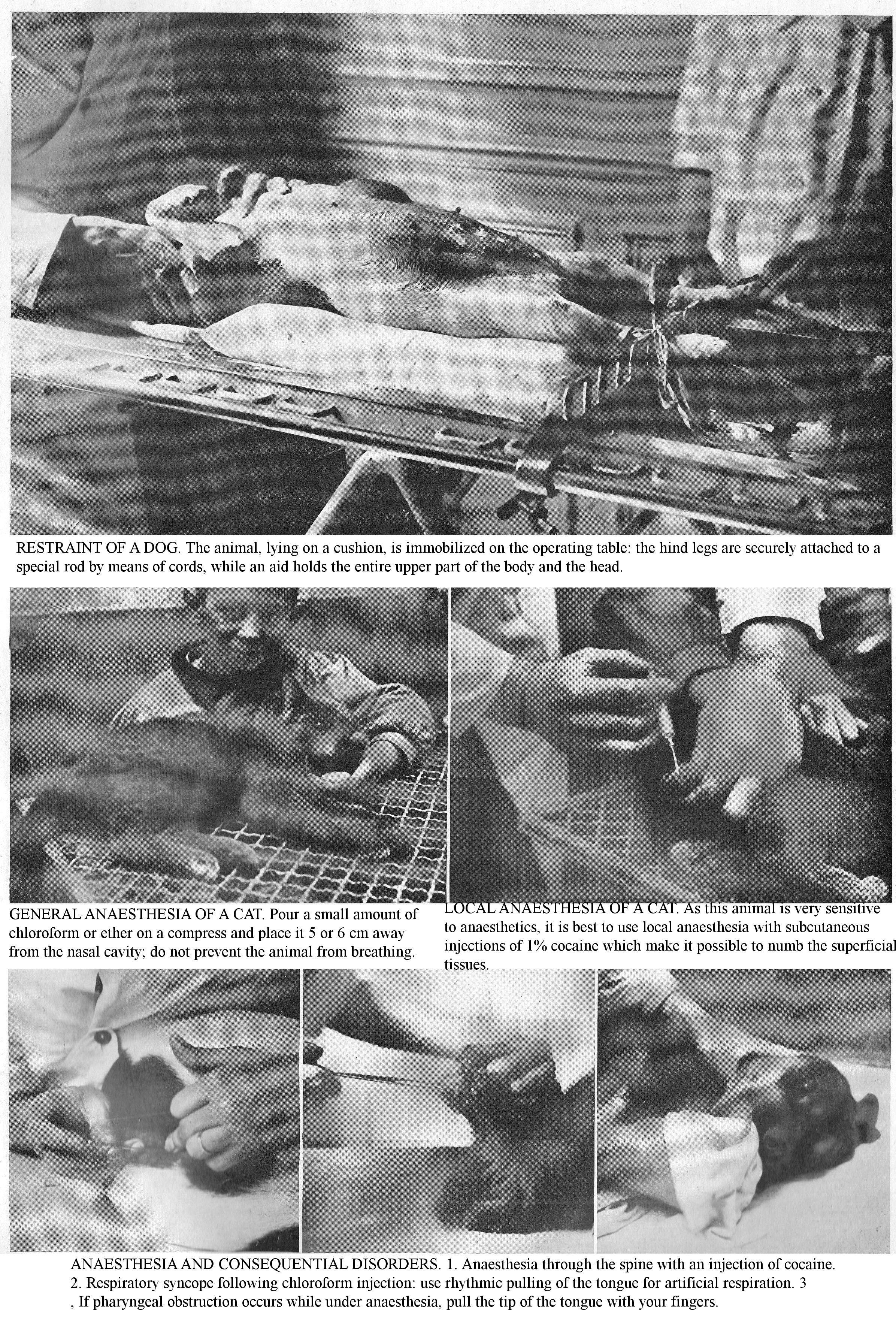
RESTRAINT OF A DOG. The animal, lying on a cushion, is immobilized on the operating table: the hind legs are securely attached to a special rod by means of cords, while an aid holds the entire upper part of the body and the head.
GENERAL ANAESTHESIA OF A CAT. Pour a small amount of chloroform or ether on a compress and place it 5 or 6 cm away from the nasal cavity; do not prevent the animal from breathing.
LOCAL ANAESTHESIA OF A CAT. As this animal is very sensitive to anaesthetics, it is best to use local anaesthesia with subcutaneous injections of 1% cocaine which make it possible to numb the superficial tissues.
ANAESTHESIA AND CONSEQUENTIAL DISORDERS. 1. Anaesthesia through the spine with an injection of cocaine. 2. Respiratory syncope following chloroform injection: use rhythmic pulling of the tongue for artificial respiration. 3, If pharyngeal obstruction occurs while under anaesthesia, pull the tip of the tongue with your fingers.
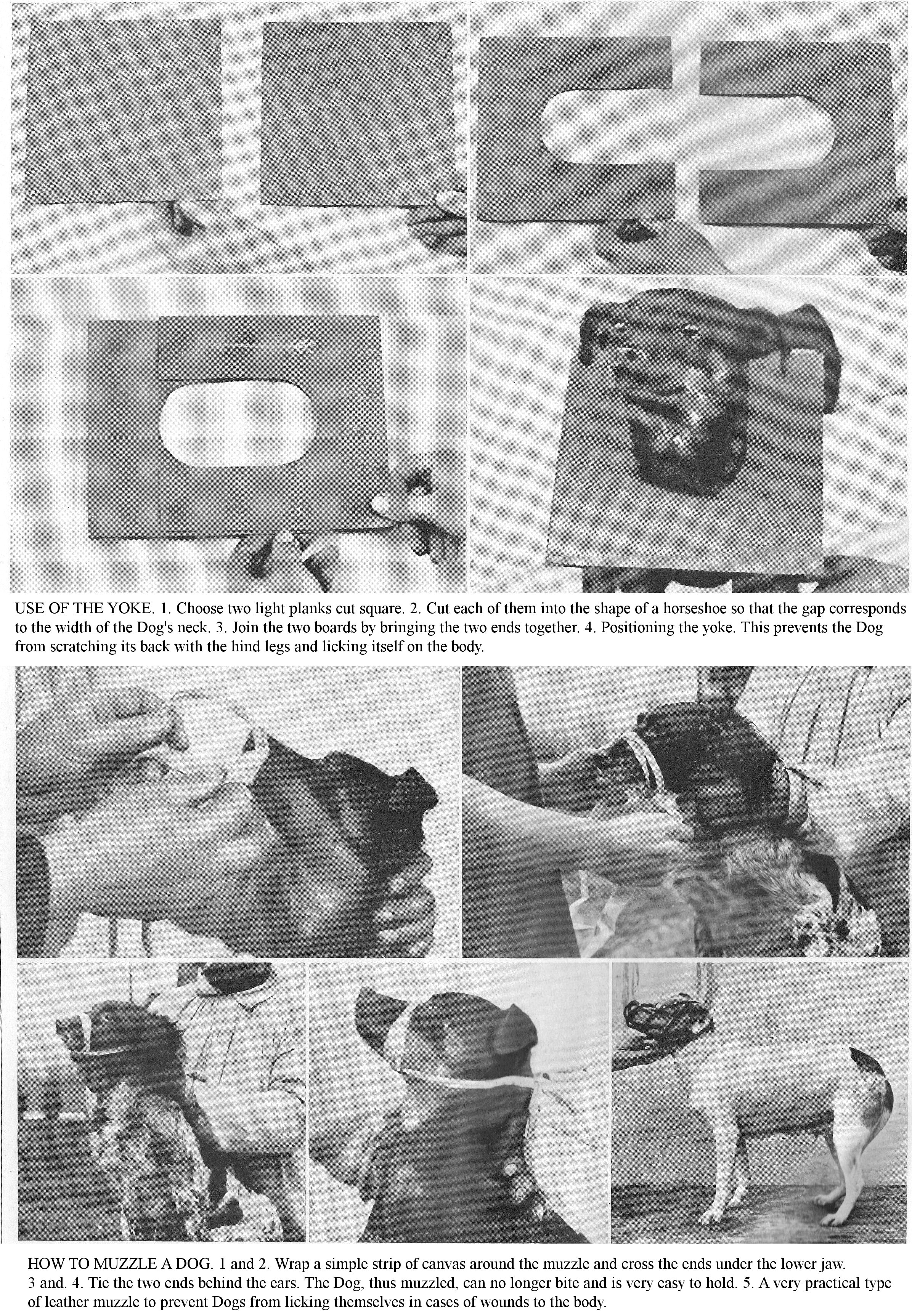
USE OF THE YOKE. 1. Choose two light planks cut square. 2. Cut each of them into the shape of a horseshoe so that the gap corresponds to the width of the Dog's neck. 3. Join the two boards by bringing the two ends together. 4. Positioning the yoke. This prevents the Dog from scratching its back with the hind legs and licking itself on the body.
HOW TO MUZZLE A DOG. 1 and 2. Wrap a simple strip of canvas around the muzzle and cross the ends under the lower jaw. 3 and. 4. Tie the two ends behind the ears. The Dog, thus muzzled, can no longer bite and is very easy to hold. 5. A very practical type of leather muzzle to prevent Dogs from licking themselves in cases of wounds to the body.
DISEASES OF THE MUSCULOSKELETAL SYSTEM
THESE COMMON CONDITIONS, WHICH OFTEN DO NOT APPEAR TO BE SEVERE, MAY, IF YOU DO NOT PAY ATTENTION TO THEM, MAKE YOUR DOGS UNFIT FOR ANY SERVICE.
DISEASES of the musculoskeletal system (joint injuries, tendons, sprains, dislocations, etc.) constitute, by their frequency and their relative severity, an important chapter of canine pathology, particularly in racing Dogs (Greyhounds) and draught dogs (sled dogs); because they can, in fact, cause significant discomfort when the part is used. Lameness is very variable, not only from the point of view of its location and its nature, but also its intensity.
Objective symptoms allow the lesion to be located: changes in shape, volume, heat, observation of oedema growths, joint movements, etc., of the limbs examined. Examine the whole limb methodically and successively, from bottom to top; do not neglect any region. In doubtful cases, where you find diffuse tenderness, it is convenient to examine the opposite limb. Use your memory to identify accidents, bruises, slips, falls, etc., and do not neglect the information provided by the attitude of the lame limb.
AGGRAVEE [CONTACT PODODERMATITIS].
Aggravee refers to the inflammation of the elastic tubercles that line the underside of the digits.
Aetiology: This condition is seen in Dogs which have raced on hard and stony ground or which have hunted for a long time in ploughed fields or on ground bristling with dry stubble. Dogs are most exposed to it during the summer, when the sun is fierce and the ground has become hot.
Symptoms: At first, the patient is hesitant to put weight on the paw due to pain; while the animal is hunting it does not appear too ill-at-ease, but as soon as it stops it shows signs of pain; it often lies down and, afraid to put weight on the paw, remains stretched on the ground. On examination of the paw, you find the plantar tubercles hot and swollen; palpation and pressure provoke complaints or threats.
Treatment: Treat the mild form with rest, lukewarm baths and sprinkle the plantar area with starch; for the severe form, use wet dressings with compresses soaked in an astringent or antiseptic solution, alum, iron sulphate, carbolic acid; calm sharp pains with cocaine petroleum jelly. Prophylaxis: Progressively train Hunting Dogs, in the days before the hunting season give them daily alum foot baths.
INFLAMMATION OF THE NAIL MATRIX.
Frequent in dogs, inflammation of the nail matrix is caused by traumas, bruises or wounds, and especially interdigital eczema. The bulge is swollen, hot, red, sensitive to pressure, sometimes oozing or ulcerated. Lameness is pronounced, often the limb shies away from supporting weight.
Treatment: At first, use warm antiseptic baths and wet swaddling. If an abscess develops, have it punctured; on ulcers, apply dressings made from tincture of iodine and iodoform, protected by a sheepskin shoe. When the lesions are old or the nail deviated, removal is indicated.
DEW CLAWS.
Dew claws may become abnormally long. Due to their curvature, they eventually reach the skin, pass through it and enter the pad that covers the matrix: the injured part becomes inflamed, ulcerated and suppurates.
Treatment: Cut off the claw in its middle part with scissors; in a few cases where the injuries are old, the tissues indurated, the removal of the digit is the only treatment.
Violent detachment of the claw, partial or total, is a very painful issue, resulting in severe lameness; if the detachment is extensive, have it removed.
PHLEGMON OF THE DIGIT.
Panaris Phlegmon of the digit or Interdigital abscess is especially common in hunting dogs and occurs as a complication of sores in the digital area. The condition is initially located first in the integument, spreading to the subcutaneous tissues, tendons, synovial membranes, phalanges (synovitis, arthritis, decay, necrosis) which, in the majority of cases, require surgery. Prevent these complications by using antiseptic baths and dressings early on, and having abscesses opened.
FOREIGN BODIES OF THE FOOT.
Foreign bodies (broom spikelet, tip of a prickly body, thorn, splinter, piece of wire, etc.) can become implanted in the tissues of the extremities; these lesions are mostly seen in Hunting Dogs and Pulling Dogs. Apart from noticeable lameness, these various infected foreign bodies cause an inflammatory reaction, fistular suppuration of the wound. In other cases, compression accidents (an elastic band applied around the paw by a child, a snare that the Dog has become caught in while hunting) can cause gangrene of the limb extremity.
Treatment: Extract foreign bodies using forceps; if necessary, debride the wound or fistula, address inflammation with antiseptic baths and dressings. For accidents caused by a ligature, cut the ligature and remove it; then treat the wound as a simple wound; in cases where the ligature has caused gangrene of the foot, have the injured part amputated.
TENDINOUS LESIONS.
In dogs, it is quite common to find that the lower limbs are bowed as a result of a congenital defect. This condition is further characterized by permanent flexion of the metacarpals on the forearm, produced by the contraction of the flexor muscles of the foot or the retraction of their tendons. Tightening may exist in one or both limbs: the forelegs are deformed, the knee is more or less deviated inwards; the flexor muscles and tendons are hard, tense, and the Dog’s general condition is otherwise unaffected.
The prognosis is not serious, the condition disappears spontaneously by the sole influence of rest; encourage healing by massaging the limb. In rebellious cases, have a tenotomy performed (section of the flexors of the metatarsals and the perforator).
RUPTURED HAMSTRING.
Rupture of the hamstring is most often caused by a violent contusion, a cut, or crushing of the leg. Sometimes it is the result of malice. You will see locally, on the postero-inferior part of the leg, a largely open wound or, when the skin is intact, a simple depression which corresponds to the separation of the tendon tips. When the injured individual tries to support itself on the injured limb, the limb flexes under its bodyweight, and its croup collapses; it walks on three legs; the entire metatarsal region of the affected limb touches the ground.
Spontaneous healing is the rule, and the limb regains function within a few months; but, especially with large breeds, if the distance separating the ends is considerable, the tendon segments heal at a distance and the animal remains crippled.
Treatment: Immobilize the hock joint in a forced extended position; Apply a bandage of potash silicate, plaster or gutta-percha, bandage with a window for open healing if the wound requires it.
SPRAINS.
Sprains are caused by the incomplete displacement of the articular extremities. The main causes of this lesion are external violence, falls, jumps, vigorous muscle contractions. Youth is an important predisposing cause.
Symptoms: The symptoms are local and functional: a few hours after the event, the injured joint is the site of an oedematous swelling, hot, painful, very sensitive to pressure. These objective signs can be difficult to recognize if the lesion is located in a deep joint (hip).
There is also a functional symptom: the injured joint is unable to perform its physiological role. For a vertebral joint, the animal is stiff, but moves easily; for a single limb joint you see a limp of varying intensity: in general, it is very intense, the Dog can hardly put the limb to the ground, but unlike dislocations and fractures, its general conformation and length are unchanged. Usually the sprain heals in 8 to 15 days. Sometimes lameness persists: it occurs with periarticular induration and bone growth.
Treatment: At first, reduce the inflammation with damp dressings: thick tow, placed on the joint, held in place by a band and sprinkled every 2 hours with lukewarm lead acetate. This dressing has the immense advantage of immobilizing the diseased joint. The encourage the absorption of the effused exudates by the massaging with a hand coated with petroleum jelly; massage in the direction of the venous flow; make a series of light passes over the area first, then progressively harder as its sensitivity dulls.
For chronic sprains, use hot baths, massage, and tincture of iodine applications. If you notice periarticular lesions (bony growths), apply cauterization; shield the cauterized surface from being chewed by covering it with cotton wadding or by applying a muzzle or a cardboard collar to the animal.
DISLOCATIONS.
Dislocations are characterized by an abnormal and permanent displacement of the articulating joint. Apart from trauma, these lesions are particularly seen in Hunting Dogs following falls, jumps, vigorous muscle contractions. Dislocations may be complete or incomplete (connection still existing to a variable extent); they can be simple or complicated; in the latter case, the dislocation is accompanied by bone fractures, joint wounds, vascular ruptures, etc. Dislocations can be congenital, developmental abnormalities, due to intrauterine trauma.
Symptoms: The articular deformation is more or less marked, the pain is always very intense on palpation; the dislocated joint is the site of abnormal mobility, limited in direction and amplitude, quite different from that of fractures. Lesions: In dislocations there is almost always a partial or complete tear of ligaments, joint capsule, sometimes tendons, vessels and nerves; joint cartilages can be crushed or torn off; sometimes the skin is torn and the joint open.
Diagnosis: It is easy to differentiate between Dislocations and Sprains (possible and normal movements of the injured joints), joint contusion, fracture (crepitation, normal mobility of the bone ends). In doubtful cases, resort to radiography.
Treatment: The treatment of recent dislocations involves reduction and restraint of the injured extremities. If manipulation is very painful, the contracture of the muscles is strong and the reactions violent, have general anaesthesia performed. While awaiting the arrival of the veterinarian try, by continuous traction, exerted in opposing directions on the two displaced bones, to restore the normal relationship of the articular surfaces and to reduce the dislocation; if necessary, vary the manoeuvres and continue them until the correlation is perfect. Apply an irremovable bandage, as in the case of a fracture; remove it after three weeks; prevent muscle atrophy with exercise and massage.
JOINT WOUNDS.
Periarticular wounds, frequent in the limbs, are generally serious because they can be complicated by damage to the tendons and the tendon and joint synovial membranes. These produce a viscous, yellowish liquid, the synovia.
Treatment: Treat periarticular wounds with antisepsis and apply cotton wadding: cut the hairs around the wound, irrigate it with a warm solution of 1 part per 1000 sublimate; sprinkle with iodoform; apply a layer of collodion and cover with a bandage.
Closed arthritis: This lesion resulting from wounds and periarticular contusions is marked by a hot, painful, oedematous swelling; movements of the limb joint is hampered or suppressed; lameness is intense, sometimes the limb provides no support.
Purulent arthritis is characterized by swelling, heat, tenderness, and fluid build-up in the joint at different places. The pus shows up outside, making one or more fistulas. Treat Simple Acute Arthritis with warm baths, a damp wrap, and immobilization of the joint.
Arthritis sicca, common in Great Danes, is a localization of rheumatic origin; it normally occurs in the knee and stifle joints. It is announced by swelling of the diseased joints, articular crepitation and pronounced lameness. In addition to internal treatment (iodine medication), make topical applications of tincture of iodine.
TO TREAT FROSTBITE.
Frostbite consists of an alteration of tissues caused by cold; it can occur on the Dog’s limbs and ears. The determining cause of Frostbitten limbs is a prolonged stay in cold mud, sleet, icy water or irritating mixtures used to melt snow in the streets.
Symptoms: In the first degree, the skin is congested; in the second degree, you notice an exudation in the altered areas which lifts and removes the epidermis; in the third degree, you observe death of skin and underlying tissues to a varying thickness.
Treatment: Rub the affected areas with dry cotton pads; carefully avoid contact with hot liquids, and cover affected limbs with a thick, loose layer of ordinary cotton wadding. If there is oozing, use absorbent powders: talc combined with zinc oxide, boric acid, starch. Then, with hot antiseptic baths and wet swaddling, encourage the shedding of necrotic skin; finally activate wound healing.
ENSURING NORMAL REPRODUCTION
THE SCIENTIFIC PRACTICE OF BREEDING, PREVENTIVE AND CURATIVE TREATMENT OF STERILITY, NURSING AND WELL-UNDERSTOOD SEPARATION REQUIRES YOU TO HAVE VALUABLE KNOWLEDGE.
Despite contradictory opinions on this subject, Dog breeding, prudently and wisely conducted using elite breeding stock, based on healthy and practical data, can give interesting results from a speculative point of view. For this to be so, it requires, in addition to the technical knowledge essential to any breeder, active surveillance and great activity on your part. The envisaged profits (income from matings carried out by pedigree studs, sale of puppies bred from animals who have appeared in competitions, etc.) may be reduced to nothing if the methods of reproduction, hygiene, diet, housing are incorrect.
Abandon traditional practices and base your breeding on rational methods whose basics and the essential directives we provide here. Applied judiciously, they will allow you to reduce, to a notable extent, general morbidity and mortality, to suppress the contagious foci which often decimate a Kennels and to obtain the precocity and the hardiness essential to working breeds.
COUPLING PRACTICE. The timing of reproductive instincts varies with breed and also with the individual, the climate, healthy maintenance and diet. Males are lustful animals able to mate in any season; their fertility period varies from 2 to 8 years; but, from the 6th year onwards, you often observe a decline in the vitality (lack of hardiness) of their offspring.
Fertilization is only possible in the Bitch when she is in heat; during this period, she is restless, runs, gambols, likes to play, and engages in a host of unusual acts which have given heat the characteristic name of "follies." Locally, you will see an abundant discharge, quite often bloody, spreading a strong odour that attracts males. The Bitch then frequently leaves her master's home, leading an entire pack in her wake, and does not return until her ardour has died down. The duration of heat varies between 10 to 15 days; during this period, the female dog can mate several times. The manifestation of heat may suffer disturbances (absence or frigidity, excessive intensity or nymphomania), these abnormalities playing an important role in maternal sterility.
Not only is fertilization only possible during heat, but the period during heat (beginning, middle and end) also has a leading effect on fertility. In fact, practice shows that maximum fertility is observed during the moment when her reproductive instincts are the least marked, that is to say towards the end of heat, a period coinciding with maturity, dehiscence and shedding of ova.
For these physiological reasons, consecrated by practice, therefore breed your bitches towards the end of heat, from the 10th to the 12th day. In the practice of mating, present the dogs in heat to the stud, twice at 24 hour intervals; in this way all the Puppies will be born on the same day; do not overuse repeated coverings; often they can cause genital over-sensitivity, a common cause of sterility.
In primiparas, especially in miniature breeds, avoid large litters, by only mating them once. Although this rule is debatable from a physiological point of view, it is adopted by breeders. Mate the females in the middle of January, so that the Puppies will have Spring and Summer in front of them, favourable times for rearing; attach great importance to the time of birth, as it plays a major role in the morbidity and mortality of the young. After mating, the two sexes remain united for a fairly long time, the separation being delayed by the enormous swelling of the two erectile bulges of the penis, which hold this organ in front of the lips of the vulva; ejaculation is also very slow. Afterwards, to encourage fertilization, isolate the bitch far from other Dogs to remove her from the excitement of the Kennel; if she has to travel, give her a few days before departure.
Cats can mate in the first year, but it is not until the second that they are fertile. The female goes into heat 2-3 times a year, usually in late autumn and early spring. She is more ardent than the male; she calls out, chases after him with plaintive meows. However, copulation causes her pain, both because the male clings to her with his claws and teeth and because his organ is bristling with horny papillae. She screams furiously, she defends herself, and mating resembles a fight.
GESTATION PERIOD. Gestation, true internal incubation during which the foetuses are formed and develop at the expense of the mother, is of great interest to breeders, both in terms of its diagnosis, duration and anomalies. The gestation period in female dogs is 63 days in large breeds and about 59 to 63 days in small breeds. The number of offspring in each litter naturally varies with breed and individuality; under-fertility is the prerogative of miniature breeds (1 to 2 young); hyperfertility (4,10,12 and above) is normally seen in working breeds, especially in those showing strong signs of hardiness.
The duration of gestation, in the cat is about 50 to 56 days; ordinary litters contain 5 to 6 young, born with their eyes closed and not opening them until around the 8th day.
The signs of gestation are rational or certain. Among the rational signs, let us quote the development of the belly and mammary glands. Belly growth is very slight in first-time mothers, especially when the number of foetuses is small. Take into account not only the enlargement, but also the special shape of the belly; it increases in all directions, especially in its lower third; it widens at this point, so that its projection appreciably exceeds the plane of the crural region. In cases of ascites, dropsy, which could simulate pseudo-pregnancy, you can clearly perceive the effused fluid by palpation; moreover, the fluid level shifts with the position imposed on the patient.
Confirm the diagnosis of pregnancy by feeling the abdomen. In Dogs and Cats, the thinness of the abdominal walls and the ease you experience in depressing them would seem to allow an easy diagnosis of pregnancy. However, owing to the small size of the foetuses and the tension of the uterine walls, it is not always easy to observe their active movements and to get a clear idea of their movement, at least during the first half of pregnancy. position. It is only later, when the amount of fluid has decreased and the foetuses have developed, that you can feel them quite distinctly. Methodically feeling the abdomen then allows you to detect a solid body as it floats in the uterine fluid; its characteristics and position prevent it from being confused with any of the intra-abdominal organs.
In doubtful cases, perform vaginal exploration. Introduce the index finger, previously disinfected and oiled, into the vagina; you will thus recognize the volume, consistency, and degree of dilation of the uterine cervix. Complete the examination by placing the animal on her back and depressing the abdominal wall between the umbilicus and the pubis with your free hand; you can thus appraise, between your two hands, the volume, consistency and sensitivity of the uterus. The functions of the mammary glands are closely linked to reproduction. Around the 40th day, you will notice them swelling; they become elastic; in the days preceding parturition, a simple squeeze gives an abundant and good quality milk flow.
Pseudo-gestation: Some abnormalities in pregnancy include false pregnancy, super-fertilization, and ectopic pregnancy. In bitches which have not been impregnated, you sometimes observe extremely interesting phenomena at the time when the birth should take place. In the bitch, from the 40th to the 50th day, a very noticeable increase in volume of the mammary glands occurs. On the 60th day, that is to say the time when she was due to give birth had she been impregnated, you will see local signs: the edges of the vulva enlarge; its opening dilates, the vaginal mucosa is red and secretes a viscous liquid. She becomes agitated, anxious; she looks for a place where she could deposit the offspring she thinks will arrive soon. If she is free, she prepares a bed with the greatest care, for the babies whose arrival she expects; her firm and elastic teats secrete abundant and good quality milk, which you can easily extract by a simple squeeze. If you give her a newborn puppy, she will adopt it without difficulty.
Super-fertilization means two ova from the same ovulation period are fertilized by different males. Super-fertilization is relatively frequent in bitches, which can give birth to Dogs of very unequal size, and belonging to very different breeds in the same litter.
In ectopic pregnancy, the fertilized ovum, instead of descending into the womb, may stop at any point on the course it must travel, or even escape the grip of the fallopian body and fall into the abdominal cavity; it can adhere in the normal way wherever it stops and undergo the various phases of its development outside the womb.
FAILURES DUE TO STERILITY.
Many improvements are delayed and remunerative enterprises suspended, because the breeding on which they depend is temporarily or permanently halted due to sterility; furthermore, the high rate of infertility, hindering hereditary transmission, prevents the improvement of dog breeds. The framework of this work is only to examine the subject in general, but I have already dealt with this elsewhere (1).
(1. Ed. CUROT, "Fertility and Sterility", 2nd edition.
Cause of sterility in males: In males, fertility only requires the relative integrity of two functions, the secretion and excretion of sperm. The spermatozoa, essential elements which give the sperm its activity, arise exclusively from the seminiferous cells; consequently, any sterility through failed secretion necessarily presupposes a morbid state of spermatic secretion. But to cause sterility, the problem must affect both testicles and each of them must be almost entirely affected. Partial or unilateral lesions of the testes are, by virtue of the possible functional replacement, powerless to cause sterility.
MALE STERILITY.
1. Sterility by secretion disorders: Anomalies (testicular atrophy). Inflammation of the testis (hydrocele, sarcocele, orchitis). 2. Sterility by obstacles to the excretion of sperm: Neoplasms (fibroids, cysts, sarcomas, etc.). Lesions (epididymis, vas deferens, seminal vesicles, urethra). 3. Impregnation disorders: Chemical cause (hyperacidity, blennorrhoea, polyps of the penis). 4. Infertility due to impotence: Age (youth, old age), general condition (obesity, reproductive overwork, malformation, deformation of the penis).
Causes of sterility in females: The causes of sterility in females are very numerous; disorders of ovarian function may exist in very varying degrees, from complete absence of ovulation to imperfect maturation of the ova; the result is the same, and sterility is inevitable. All conditions (oophoritis, degeneration, tumours), all stoppages in the development of the ovary can, by this mechanism, cause sterility. But, as with the testicles, the lesion must be bilateral and extend to almost all of the gland. The normal development of the external genitalia and mammary glands is a favourable indicator for ovulation. In a female that previously been impregnated, the question is simpler than in first-time mothers, since the ability to ovulate is not in any doubt. Irregularity in the manifestation of heat (duration, intensity, periodicity) should raise suspicions of sterility due to disturbance of ovarian function.
Infertility due to ovular impregnation disorders includes all the physical or chemical obstacles that sperm encounter on their way from the vulva to the ovary. Physical causes include acquired or congenital lesions: vaginal scarring, cervical lesions (stiffness, induration, complete obliteration). Utero-vaginal growths play an important role, depending on their location, size and method of implantation; in maternal infertility, these growths can indeed cause displacement of the uterus, tubes and ovaries; they can obliterate surface cavity, the uterine body or cause its deviation, lesions that can constitute mechanical obstacles to the fall of the ovum into the uterine cavity, and to the penetration of sperm. Vaginal mucus, in some pathological cases (vulvitis, vaginitis, metritis, etc.), has a harmful effect on the vigour of spermatozoa and is a frequent cause of sterility.
FEMALE STERILITY.
1. Ovarian function disorders: Imperfect development of the ovary (atrophy, degeneration, growths, cysts, fibroids, sarcomas). 2. Ovular impregnation disorders: Physical causes (vaginal atresia, cervical stenosis, stiffness, induration, complete obliteration). Chemical causes (vaginal hyperacidity). 3. Ovular enlargement disorders: Tubal obstruction, change in ratio of the tubes and ovaries (uterine growths, peri-uterine growths).
Relative sterility: In some cases you may observe sterility without any disease of the reproductive organs being able to explain it. There is no doubt that, all special circumstances being ruled out, there are females who, placed under the same conditions as others, prove to be much more fertile. In daily practice, you can observe cases of sterility which are attributable to neither the male or female involved, but to their pairing and where each one shows fertility when to another animal. Physiologically, it is impossible to explain these paradoxical facts which constitute a variation in fertility.
The power of proliferation is largely influenced by climate; acclimatization has a depressive effect on fertility. The first year after a foreign hunting dog has been introduced to the Kennels may be one of relative sterility. The age of procreators, which directly depends their vigour, is an important factor in fertility; extreme states, such as youth or senescence, can cause relative sterility; the rare exceptions cited does not invalidate this physiological rule. Temperament influences fertility in males; sexual ardour is generally combined with a nervous temperament, while a lymphatic constitution [sluggish temperament] is accompanied by a kind of frigidity which makes studs poorly fertile. In bitches, on the contrary, the lymphatic [sluggish] temperament would be the most favourable to fertilization. Frigidity in bitches is characterized by a kind of sexual atony, causing the total or partial abolition of heat.
Poor diet, youth, cold temperature, obesity, and inbreeding all have a depressive effect on the manifestation of heat. Observation shows that frigid females often have such poor development of generative organs (atrophy of the vulva, mammary glands) that they are often unsuitable for reproduction.
Genital over-sensitivity, characterized by exaggerated nervousness, may be considered the first degree of nymphomania, manifested by a continuous urge to mate. Nymphomania, an important factor in canine sterility, is often the consequence of pathological lesions of the ovary (oophoritis, fibroids, sarcomas, cysts, etc.); it can cause disturbances of general sensitivity, sometimes weakness or paralysis of the hind limbs, as a result of spinal cord injuries. Cats, especially She-Cats, of a very nervous and impressionable nature, often manifest hysterical attacks: the patient suffers from nervous crises, falls to the ground, clenches its teeth, drools saliva through the corners of the lips, and then everything returns to normal. At other times the She-Cat often makes the signs of mating; she twists, lifts her tail, tries to rub against neighbouring bodies, an intense itching manifests itself in various regions. The prognosis becomes serious when the attacks are numerous. Calm this neurosis with dosimetric granules of camphor bromide (at 1 cgr.); give one granule morning and evening for 5 to 6 days.
The influence of sexual characteristics on fertility is not in doubt: the breeding male or female must not only have intact genitalia but, in all respects, must be an excellent specimen of their gender. Observation shows that studs that show signs of feminism and bitches that show masculine traits (angular and abrupt lines, strong skeleton, highly developed muscular system, large head, broad chest, etc.) have poorly developed fertility. Therefore, attach importance to sexual characteristics when choosing animals for breeding.
The intimate relationship between nutrition and procreation suggests the influence of the diet, both quantitative and qualitative, on fertility. In abundantly nourished individuals, reproductive power is maximized because there is always a close relationship between it and vital activity. Carefully avoid overfeeding, which causes obesity, a common cause of infertility. The sperm’s richness in phosphorus products (nuclein, lecithin, etc.) shows the importance of mineral nutrition in the breeding male: physiologically "the most mineralized male is the most fertile".
Obesity produces sterility by a double mechanism: it impairs general nutrition and it causes atrophy of the ovaries. Associated with inbreeding - which is common in pedigree breeds - obesity produces a kind of neutral gender, often leading to impotence or infertility. Combat the effects of obesity through diet and especially through exercise, the latter being an essential factor in animals intended for breeding.
In the male, repeated matings are a cause of temporary sterility; physiologically, the prostate fluid precedes the semen in ejaculation, and when you overexert the studs they are more prostatic than spermatic and therefore sterile. Inbreeding is accused of having a depressive effect on fertility and on the vigour of the offspring. To those authors who accuse inbreeding of causing sterility by the accumulation of hereditary factors, we will say that the experience of breeders has shown that when the mated animals are free from any pathological defect, inbreeding does not diminish either hardiness or fertility. On the other hand, do not mate two animals possessing the same imperfection because the offspring would inherit their predisposition to disease in double dose, and the vital resistance would be decreased. So, avoid the excess inbreeding that produces size reduction, spread of white in the coat, etc., by refreshing the blood with the help of other breeding stock.
Diagnosis of sterility: In this diagnosis, two important points must be resolved: whether sterility is male or female in origin, and identifying the cause of this sterility. The first point, in most cases, is fairly straightforward to elucidate; the second is tricker and requires careful diagnosis. The following method, based on clinical and micrographic data, which we have been using for many years, provides a scientific solution to these various questions.
Male sterility: This diagnosis is based on verification of the male’s fertility based on microscopic examination of the sperm and sexual organs, which alone constitutes a reliable criterion. Use a magnification of 300 to 500 diameters; if the spermatozoa are found to be normal in terms of number and vitality, rule out male sterility; if, on the contrary, semen examination shows you the absence of sperm or their deterioration, it incriminates the stud. Note that the sperm in average semen should occupy about a quarter of the surface being examined. Complete the diagnosis of male infertility by clinical examination of the sexual system; check if it meets the conditions for normal mating.
Female sterility: If the male is found to be unharmed, attribute the sterility to the female. To clarify the diagnosis, perform clinical examination of the sex organs, investigate and determine the acidity of the vagino-uterine mucus. Perform genital examination by inspection, intravaginal and rectal explorations; the inspection will tell you about vulvar lesions; vaginal exploration, performed using your finger, will help you understand the condition of the walls of the vagina and cervix.
Investigating and measuring the acidity of vaginal mucus is a serious part of maternal infertility. Collect vaginal mucus with a special probe, the end of which is covered with a cotton swab. Determine its acidity using special litmus paper which, when brought into contact with the vaginal mucus, takes on various shades which are intimately linked to variations in acidity (normal acidity, hypoacidity, hyperacidity). Fertilization ability peaks with normal acidity and decreases markedly with vaginal hyperacidity.
TREATING STERILITY.
Since infertility is not a disease, but the consequence of a multitude of morbid conditions, there is no single, invariable treatment. Our experience has shown us, eight times out of ten, that female sterility is due to local causes and must be treated locally. In the case of acquired morbid lesions, the indications relate to two important factors: inflammation and mechanical obstacles. The therapeutic indications to be fulfilled are to moderate the first and eliminate the second. The methods used to combat infertility both relate to health care - therapeutics and surgery.
Combat vaginal hyperacidity, a common cause of infertility, by using alkaline solutions (baking soda, 5 parts per 1,000). Calm general overexcitement with a debilitating diet (half-diet, meatless diet); give the patient serious exercise; cause anaemia with weekly purgations; for specific drugs, use sedatives: chloral, camphor bromide or potassium or sodium bromide, 5 gr.; distilled water, 200 gr.; ten days out of fifteen, give a teaspoonful or tablespoonful depending on the size of the patient.
Apart from a tonic, highly digestible, fortifying diet, institute the following treatment for frigidity: tincture of cantharides, V to IX drops in the mash. Preferably use the effect existing in yohimbine [bark of Pausinystalia yohimbe, an aphrodisiac] on the genital tract (1 to 2 cgr. several times a day). Often, putting frigid female dogs in contact with female dogs in heat provokes reproductive awakening in the former. Use this hygienic process before resorting to drug treatment; in a few cases, stimulants (coffee, alcohol, ammonia acetate) effectively combat frigidity.
Treatment for impotence varies with the cause; if it is due to reproductive overstrain, decrease or temporarily stop the matings; if it is linked to a state of weakness, give a highly digestible and tonic diet. Medical treatment involves the use of aphrodisiac substances (tincture of cantharides, yohimbine, etc.).
CANINE OBSTETRICS. When the time for parturition arrives, from the 58th to the 65th day, nature alone is generally enough for this task; during the preparatory period for parturition (preparation and dilation of the cervix), do not intervene and do not disturb the mother’s tranquillity; leave her in a darkened, secluded place. Bitches always go to their bed to give birth; they lie on one side, their body bent in an arch, the head facing back and close to the genital opening; in this way, each puppy, as it emerges from the vulva, is within reach of the mother who licks it, turns it with its muzzle to face her teats, after severing the umbilical cord with a bite.
Post-parturition care: After a difficult labour, to avoid any subsequent infection (septic metritis), irrigate the sexual organs with antiseptics; a warm solution of 4% boric acid, 1 p part per 2000 potassium permanganate, hydrogen peroxide cut to 1/3, etc. Give the mother to a half-diet and a refreshing diet: milk, milk formulas, broth, soups and very little meat. Combat weakness or lack or energy with stimulants (coffee, alcohol, injection of caffeine, ether, camphor oil, etc.). Calm fever with quinine sulphate, etc. Carefully avoid colds that are harmful to the mother and her offspring.
NATURAL NURSING. Natural nursing is the most advantageous for the mother and young. In the former it eliminates mammary gland issues. Breast milk provides newborns with the standard physiological food which exerts a decisive influence on their rate of development. Use mother’s milk even if only temporarily for a few days so that the young can absorb the colostrum which allows them to cope better with artificial feeding.
Usually, maternal affection is well-developed in the Bitch and is less pronounced in the She-Cat, especially in first-time mothers. Watch the female dog while nursing in order to know how she is disposed towards her young; if she has pain or injury to the teats caused by the ever-voracious young, she may trample them under her feet and be tempted to devour them. After parturition, make sure that all the teats, as many as possible, are giving milk; sometimes one or more of the teats are barren and in this case the stronger puppies get hold of the ones that are productive while the others suck the atrophied teats in vain, tiring the mother unnecessarily and naturally not prospering.
Litters vary among breeds from 1 to 12 pups, most often 4 to 7, generally the larger they are the fewer in number. As they are born, puppies instinctively steer themselves to the nipples, grab one and suck the colostrum. If the number of young is excessive or if the milk yield is in deficit, remove some of them; base your selection on gender, specifics of their coat, and signs of health and vigour.
Many Bitches, except first-time mothers, can breed up to 8 pups provided they are put on an intensive diet, but usually only leave about 4; it is always better to have a smaller number of beautiful puppies than a higher number of sickly, puny ones who, having suffered in their youth, will never regain their normal development. Do not disturb or worry the mother by visiting too frequently; during the first week, or at least the first days, she does not leave the Puppies; she always takes care of them, licks them, cleans them or feeds them. Towards the end of the 2nd week, the little ones can see and hear, and start to move; soon they taste the milk you offer them and the food intended for their mother. Trim the puppies' claws to prevent them from scratching the nipples during compressions while sucking. As long as nursing continues, the Bitch eats her puppies’ excrement; that's why you find their sleeping kennel clean.
In small litters, it may happen that only some of the teats are used; to avoid congestive problems, milk the others regularly. If you notice signs of starvation with normal milk yield, you can attribute this to poor milk quality and modify the nursing Bitch's diet, both qualitatively and quantitatively. If poor growth persists, resort to the use of a foster mother, artificial feeding or combination of natural and artificial feeding.
Excreta examination during the nursing period is the best criterion for feeding; if there are no digestive disorders, the dominant symptom of which is diarrhoea, and the weight curve is normal, you can confirm that the diet is correct. If, on the contrary, you observe cases of digestive intolerance, temporarily stop nursing; put the puppy on a half-diet and look for the origin of these disorders, which, in the majority of cases, are related to a faulty diet.
Nursing rarely lasts more than two months; the mother regulates the duration herself, by limiting the number of feedings, according to her ability to produce milk.
The rules for the healthy state of natural nursing are much simpler than for artificial nursing. Ensure the puppies absorb the colostrum, and give the mother highly digestible food.
ARTIFICIAL NURSING. Artificial nursing involves the use of milk from another species; it becomes necessary due to the death of the mother, the insufficiency or absence of mother’s milk, a malformation of the teats. Apart from the difference in chemical composition of the milk used (cow, goat) compared to Bitch’s milk, one of the reasons artificial nursing is inferior to natural nursing lies, in the absence of specific ferments which play an important biological role.
The health concerns of artificial feeding involves the following guidelines: use the mother's colostrum if possible, and use sterilized milk. Use a baby bottle when nursing; Puppies may initially refuse this mode of feeding; get them used to it by running a little lukewarm milk into their mouths and then they will eagerly take the teat. To make the young lap milk, do not put their noses in the dish, which will cause coughing; but dip a finger into the liquid and give it to them to lick; this way they quickly get used to drinking on their own. Observe great regularity in the number and interval of meals and rigorous cleanliness of the containers used. With artificial feeding, it is not a question of knowing how to prepare milk (dilution, sterilization); it is, above all, necessary to determine the amount of milk to be given daily, as insufficiency and overfeeding are both detrimental to the development of puppies. The absence of digestive disorders (diarrhoea) and the regular growth observed with the help of regular weighing, constitute, in the absence of precise data, the best criterion of the physiological milk ration.
Avoid giving meals too close together to prevent serious digestive disorders (milky indigestion, gastroenteritis). At first, schedule feedings every two hours; later, space them out more, let the infants drink what they want; it is only important that the opening of the nipple is not too large, and the flow of milk is not too abundant. Keep the containers very clean; the smallest amount of milk that remains will ferment and become sour and spoils the milk that settles there; disinfect the bottles morning and evening by immersion in boiling water; in this way, you will avoid the many disorders of the digestive system, which lead to a high infant death rate.
The dilution of cow's milk carried out by adding 1/4 to 1/5 of boiled water per litre, as recommended by many authors, has the serious drawback that, by reducing its nutritional value, it further increases the nutrient deficit. Also, far from advising this practice, we advise, on the contrary, that you compensate for the shortage of protein matter by adding dried casein or eggs; in this way, by achieving the nutrient balance you avoid the stunted growth observed during the period of artificial feeding. For this same reason, the habit of sweetening cow's milk is irrational since this milk already has double the sugar content compared to that of Bitch’s milk.
Because of the problems encountered with artificial feeding, however well-managed, and the ease with which you can find a foster mother in dog breeding, you should give preference to the latter. The prophylaxis of artificial feeding lies entirely in healthy feeding; in addition, avoid breeding from Bitches that are poor nurses.
FOSTER MOTHER. Feeding using a foster mother involves giving the Puppies to another Bitch that is lactating, a condition often found in dog breeding. If you are giving the Puppies to a foster mother, have them absorb their own mother’s colostrum before giving them to their wet nurse. You can also have a Cat nurse Puppies, but this is seldom necessary, since female dogs in the same Kennels go into heat at around the same time, making it easy for you to find nursing foster mothers. To achieve adoptive feeding, use a wet nurse who gave birth 2-3 days earlier; leave her 2 or 3 babies and give her the infants without showing them to her, preferably making this substitution in the dark. Using a foster mother is only a simple variation of nursing and has all the health advantages of the latter.
If there is no replacement, use mixed nursing, a combination of natural and artificial feeding; this diet gives good results when the proportion of breast milk is the dominant component. Done well, it fills the deficit in nursing and ensures normal growth.
WEANING OF PUPPIES. Weaning consists of stopping nursing and giving the puppies more substantial food than milk, gradually approaching the characteristics of ordinary foods. Weaning is a dangerous phase and has an indelible influence on growth, especially in pure breeds.
The physiological time of weaning varies between 45 days and 2 months; from the age of 6 to 7 weeks, the mother, who is starting to be bruised by the small teeth of her "pupils," pulls away from them and does not let them take the teats. Never use sudden and premature weaning, because it causes nutritional disorders which alters the conformation and harmony of form; the subjects become thin and puny, and despite strict health care they never manage to recover from their stunted growth. Do not wean straight away, do not remove all the puppies at once, but one after the other; start with those who appear most precocious; let the weakest ones feed longer from the mother; these will later catch up with the first in terms of bone structure and musculature.
Perform weaning gradually: reduce the number of feedings; allow the Puppies to be with their mother only a few hours a day, 2 or 3 times; feed them corrected milk, simple or sweetened milk preparations, broths, light mash, a little meat. Give these foods in small amounts between feedings. If there is nothing to oppose it, don't wean completely until around 3 months of age. Wean cats around the age of 3 to 4 weeks; perform this in the same way as weaning puppies.
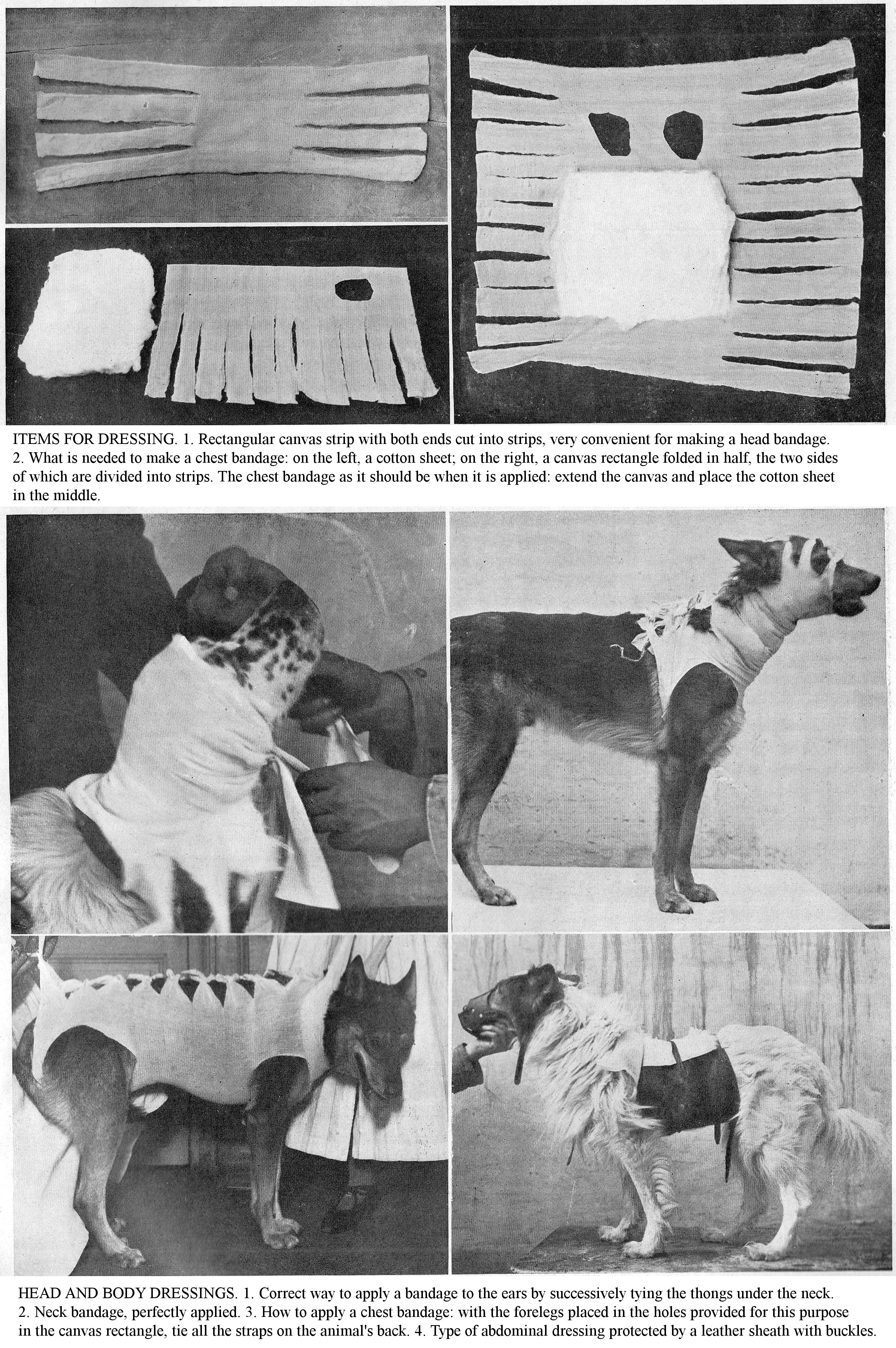
ITEMS FOR DRESSING. 1. Rectangular canvas strip with both ends cut into strips, very convenient for making a head bandage. 2. What is needed to make a chest bandage: on the left, a cotton sheet; on the right, a canvas rectangle folded in half, the two sides of which are divided into strips. The chest bandage as it should be when it is applied: extend the canvas and place the cotton sheet in the middle.
HEAD AND BODY DRESSINGS. 1. Correct way to apply a bandage to the ears by successively tying the thongs under the neck. 2. Neck bandage, perfectly applied. 3. How to apply a chest bandage: with the forelegs placed in the holes provided for this purpose in the canvas rectangle, tie all the straps on the animal's back. 4. Type of abdominal dressing protected by a leather sheath with buckles.
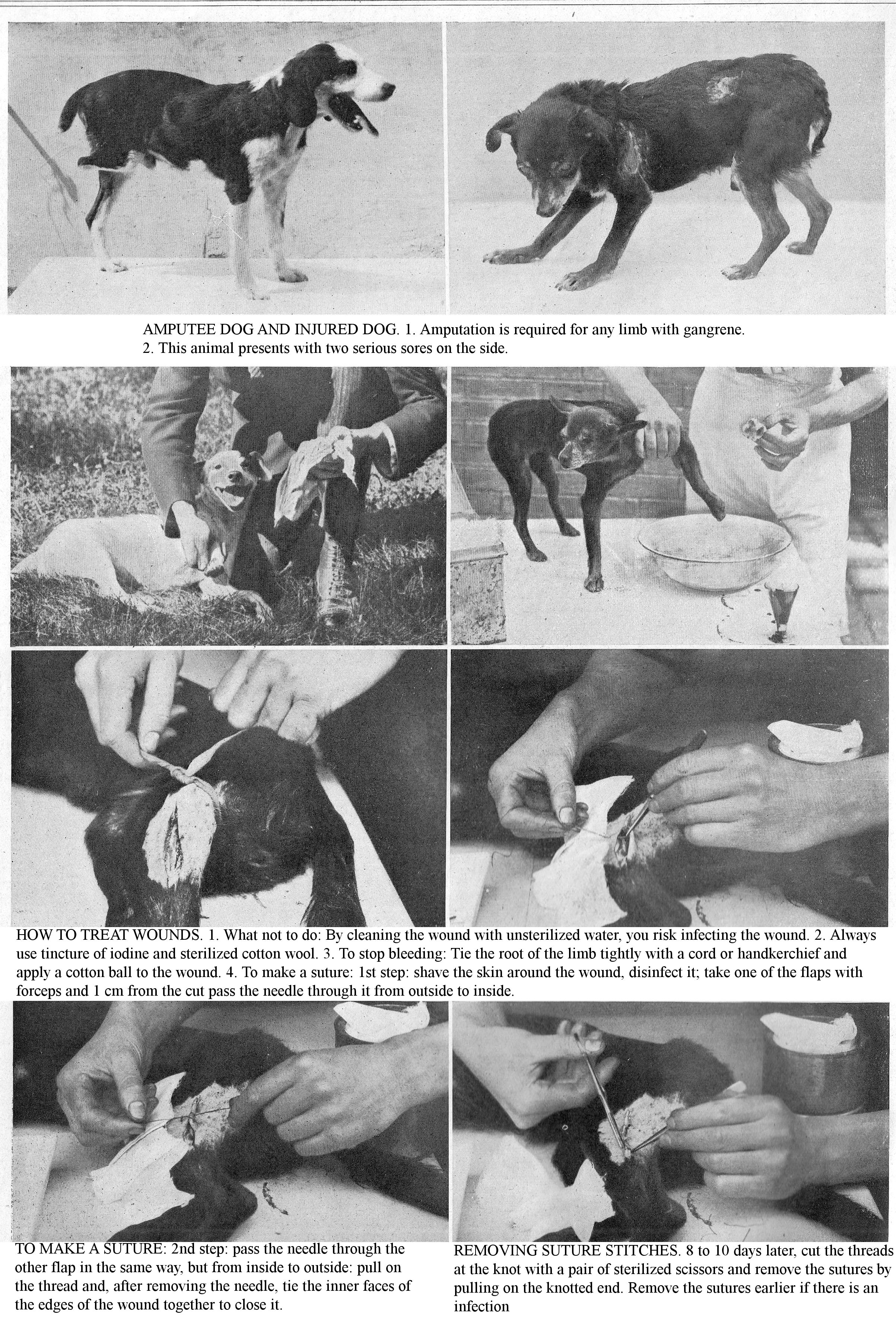
AMPUTEE DOG AND INJURED DOG. 1. Amputation is required for any limb with gangrene. 2. This animal presents with two serious sores on the side.
HOW TO TREAT WOUNDS. 1. What not to do: By cleaning the wound with unsterilized water, you risk infecting the wound. 2. Always use tincture of iodine and sterilized cotton wool. 3. To stop bleeding: Tie the root of the limb tightly with a cord or handkerchief and apply a cotton ball to the wound. 4. To make a suture: 1st step: shave the skin around the wound, disinfect it; take one of the flaps with forceps and 1 cm from the cut pass the needle through it from outside to inside.
TO MAKE A SUTURE: 2nd step: pass the needle through the other flap in the same way, but from inside to outside: pull on the thread and, after removing the needle, tie the inner faces of the edges of the wound together to close it.
REMOVING SUTURE STITCHES. 8 to 10 days later, cut the threads at the knot with a pair of sterilized scissors and remove the sutures by pulling on the knotted end. Remove the sutures earlier if there is an infection.
MINOR CANINE AND FELINE SURGERY.
HOW TO PERFORM VARIOUS OPERATIONS WITH OR WITHOUT THE INTERVENTION OF THE VETERINARIAN TO ENSURE THE BEST HEALING OF TREATED ANIMALS.
RESTRAINT, anaesthesia, asepsis, and post-operative care are relatively easy to perform in dogs. The more complete clinical investigation methods (external and internal palpation, catheterisation, etc.), specifying the diagnosis of lesions, explain the importance of canine surgery and justify the documented study and the advice given to you. We particularly emphasise facts which you will have to apply frequently in the practice of minor surgery, concerning anaesthesia, asepsis, dressings, haemostasis, sutures, etc.
RESTRAINING THE PATIENT.
To perform an operation safely, carefully secure the patient to be operated on. The pain resulting from surgery arouses, so to speak, the self-preservation instinct and, if not immobilized, the worried, frightened patient tries to escape the hand of the surgeon, defending itself with teeth or claws. To prevent this and to prevent any accident, use simple or gentle methods (the influence of caressing, voice, gaze and gesture), anaesthesia or mechanical means of restraint.
To prevent the Dog from biting, muzzle him. If there is no muzzle, use a string or a ribbon of thread that you first tie around the lower jaw, then fold both ends over the upper jaw, where you secure them with a straight rosette knot. By this means the jaws are kept closed. Take care that the Dog does not get rid of these ties with its forelegs by having a helper hold them together, or else secure the forelegs by tying them together.
Sometimes you have to deal with rebellious, snarling Dogs that are difficult and dangerous (Rabid Dogs) to get hold of. In this case, use long iron tongs, a sort of pincer with curved jaws, and form a collar in which you firmly hold the animal’s neck without tightening too much. If you do not have collar tongs, use two long sticks with a noose on each to do the same thing. With these knots placed firmly around the animal's neck, the sticks serve to keep it at a distance between the two helpers.
The Cat is one of the most difficult and dangerous animals to restrain. If the operation is short-lived, such as castration of males, have the animal grabbed by a helper, with one hand on the scruff of the neck and the other by the loins; place the patient on a table, pressing down on them so that they cannot scratch or bite. If the operation requires complete immobilization, use anaesthesia.
ANTISEPSIS AND ASEPSIS.
Antisepsis and asepsis are the basis of modern surgery, by informing you of their use, their microbicidal value, the instructions for using the main antiseptic agents utilised in canine surgery, we give you the facts of these two major Pasteurian methods.
Use carbolic acid 1% to 2% and 3% solution to disinfect instruments, the surgical area, to clean up abscesses, suppurating wounds; avoid it for surgical wounds and recent traumas because it strongly irritates the tissues
Sublimate (mercury bichloride) is one of the most powerful chemical antiseptics; due to its toxicity, use weak solutions 1 part per 2000; use the 1 part per 3000 and 1 part per 5000 for the asepsis of most mucous membranes; do not use it for disinfecting of metal instruments because it blackens them, and alters the polish and cutting edges.
Potassium permanganate is non-toxic and owes its microbial properties to the oxygen it gives off. As it has no irritating action on tissues, use it on all wounds, in particular those of the mucous membranes; it is soluble in all proportions in water and convenient to use. Use the 1 part per 1000 weak solution for the disinfection of mucous membranes and serous cavities, the stronger one 10 parts per 1000 for infected wounds.
Avoid the use of large amounts of iodoform on recent wounds because, after dissolution by bloody secretions, it could lead to poisoning. Iodoform is fixed on the gauze and cotton wool in the proportions of 10%, 20% to 30%.
Cresyl (creolin) is an antiseptic widely used in canine surgery. Moderately priced, miscible in all proportions in water, possessing only very low toxicity, attacking neither hands nor instruments, it is easy and advantageous to use. Use the strong 3% solution, for disinfection of the surgical site and in the treatment of infected wounds; use the weak 1% solution for irrigation of recent wounds and for mucous membranes.
Pure iodine tincture can replace strong antiseptic solutions for disinfection of suppurating wounds.
The different stages of asepsis, an essential complement to antisepsis, include asepsis of the hands, disinfection of the surgical site, instruments and dressing material. Asepsis of the hands is of utmost importance; achieve this by soaping the hands with boiled water and prolonged washing in a 1 part per 1000 sublimate solution, cresyl at 3%; during the operation, avoid putting your hands on the skin of the surgical site, on the table, or on non-disinfected objects; if you touch a suspicious body, perform another disinfection.
Perform disinfection of instruments by alcohol flaming; arrange the instruments on the bottom of a metal dish, douse them with a little alcohol and light it, or use direct flaming through the flame of an alcohol lamp. All surgical equipment (sponges, cotton balls, compresses, threads, etc.) must be aseptic; sterilize the silk threads, the wound drains by immersing them in boiling water for half an hour, and store them in a 5% carbolic solution or sublimate at 1 part per 1000.
The surgical site and its vicinity should be carefully purified; cut the hairs, brush, wash and soap the skin with boiled water, or with carbolic or sublimate solution.
These are the main measures of asepsis and antisepsis to protect those operated on from infection.
PERFORMING HAEMOSTASIS.
In surgery, preventive haemostasis, used mainly in operations on the limbs, is achieved by circular compression by means of a rubber tie applied above the knee or hock, or Esmarck's elastic band.
Capillary bleeding usually stops spontaneously; if capillaries continue to ooze, dry them up with an affusion of cold boiled water or by the use of local haemostats: antipyrine, hydrogen peroxide, adrenaline, rye ergot, ergotine, Rabel water, iron perchloride, etc. When larger volume arterioles, or venules, or ducts are divided, use compression, tamponade, cauterization, ligation, twisting, and forcipressure. Ligate the vessels with silk or catgut threads and twist them using haemostats. Forcipressure is the best method; it consists of applying, to the ends of the severed vessels, stopper forceps that you leave there permanently.
SUTURING.
Sutures are common surgical operations that bring the edges of a wound together using needle and thread. Perform them with silk or simple hemp thread, previously disinfected. Wash and brighten the edges of the wound if they are dried or blackish; bring the edges together so that they are in contact.
Hold the needle in your right hand like a regular sewing needle; cross the edges of the wound more or less at right angles; avoid pricking any nerves, vessels, membranes or tendons; calculate the distance between the stitches, so that the wound does not gape between the sutures; space the stitches regularly. Usually start the sutures in the middle part of the wound, which allows you to line up the edges more precisely and more evenly. Only moderately tighten the threads, to the degree necessary to hold the edges of the wound together; do not pull the threads violently, to avoid cutting the skin over a wide area; tighten the threads so that they keep the edges of the wound in contact, without bringing them too close, to avoid strangulation of the flaps that occur during inflammatory swelling.
The time to remove the stitches varies depending on the nature and thickness of the tissue sutured. In general, remove them from the 2nd to the 4th day; if you observe abnormal swelling, profuse suppuration, constriction of the wound edges by the threads, immediately remove the suture. When removing sutures, take some precautions so as not to destroy the adhesions that have established. As a general rule, remove only one stitch at a time and start with the less essential stitches; if the adhesion of wound edges is weak or non-existent at some stitches, leave these in for a day or two more.
Let us know your Ideas, your Opinions, the results of your Experiences and even your Criticisms.
MAKING EFFECTIVE DRESSINGS.
Protect the surgical wound with a bandage, when its position allows it; cover it with layers of wadding arranged in various ways according to the case, and fixed by means of a bandage. Applying a bandage protects the wound from contact with the air and protects it from external bodies; in this way, you alleviate pain, decrease inflammatory irritation and speed up healing.
Wound dressing materials include cotton wool, compresses, gauze, silk, Florence horsehair, sponges, tape, tarlatan [stiffened cotton gauze]. Cotton wool and gauze are excellent dressing substances, not very irritating to the tissues; they have remarkable absorbent properties. Most sutures are made with silk or Brittany thread. Dry the wounds with small compresses, swabs, preferably sponge. Before applying a bandage, clear the wound of blood clots, scabs, foreign bodies, mud, manure, etc., which may be on its surface or in its immediate vicinity. Act gently and promptly so that the wound is quickly removed from contact with the air to avoid excessive irritation which would delay healing. When applying a bandage, be careful not to interfere with functions, not to deform parts and especially not to hinder circulation by excessive compression. To apply a strip regularly, hold the rolled strip with your right hand; unroll it to wrap it around the parts by its external face; return a little obliquely over the first turns by unrolling the strip as it is applied, and partially or completely cover the turns. If you bandage a conically shaped part, such as a limb, one of its edges presses more than the other and thus forms pleats. Avoid this inconvenience by practicing turnovers; for this, obliquely bend the outer face of the band on itself, from the widest part of the limb towards the narrowest, while you support this fold or turnover; by these means, you apply the strip successively on both sides. Secure the terminal end with a safety pin; split the end of the strip lengthways to have two ends that you tie securely.
The application of the first dressing is very important; it determines the outcome of the wound: if the general condition of the surgical patient is good, and the traumatic fever is moderate, do not repeat the dressing prematurely. Otherwise, apply another; avoid mechanical actions or tugging at the edges; if the wound is festering, cut the sutures and give abundant antiseptic irrigation.
Complications of traumatic injuries: Surgical septicaemia, a formidable complication of wounds, is an infectious, microbial disease determined by the septic vibrio (Bacillus septicus ganqrenae). More or less rapid in its course, gas gangrene is marked by intense inflammation around the wound, spreading rapidly in all directions, by the oedematous circle which borders it, progressive tissue death, putrefaction and the foetid odour which emanates from the seat of sepsis, finally by pronounced fever, dejection, and complete loss of appetite. Surgical wounds made under conditions of asepsis, not contaminated by hands, instruments or dressings, seldom become complicated by sepsis. Place the surgical patient in well ventilated and clean premises; prevent blood clots and secreted fluids from accumulating within the wound in the shallows.
Treatment: When the wound is surrounded by a taut, crackling swelling, have numerous deep scarifications made at the centre of the invaded area and a little beyond its limits. Then direct strong antiseptic irrigations (carbolic acid, lysol, cresyl at 4% to 5%) into the infected tissues, combat debility using diffusible stimulants (wine, coffee, alcohol) or internal antiseptics (cresyl, carbolic acid); fight weakness with subcutaneous injections of ether, caffeine, etc.
Prophylaxis: Prophylaxis includes asepsis of surgical wounds and antisepsis of accidental wounds.
ABLATION OF GROWTHS.
From the point of view of the need for surgical intervention, growths should be distinguished into benign (fibroids, papillomas, lipomas, cysts) and malignant (cancers, epitheliomas, sarcomas). For the former, ablation is indicated when they are large or when they cause functional problems; for the others, given their rapid development and marked tendency to become generalized, early excision is indicated.
Technique: Disinfect the surgical area, use general or local anaesthesia; make curved skin incisions delineating the skin flap that is to be removed along with the growth; enucleation the growth after dissecting the skin, and then the underlying tissues, on each side; apply haemostasis and clan the wound, suturing with or without drainage depending on the extent and depth of the trauma; apply a wadded bandage. For malignant tumours, the ablation should be large, radical, and include the removal of the area of neoplasmic infiltration that involves adjacent tissues.
CAUTERIZATION AND BLEEDING.
Cauterization by a hot iron causes a sharp pain which the animal shows by sudden movements and plaintive barks; muzzle it and have it held in place by several helpers. The animal must have an empty stomach; if the hairs are long and bushy, trim them. Depending on the nature of the lesions, have your veterinarian apply a hot iron in superficial points or in thin and penetrating points.
Indications: Combat, by the application of heat, with thermocautery, lameness due to the presence of exostoses, at the level of the humero-radial, femoro-tibial, and coxo-femoral joints; paralyses justify the use of this treatment. Prevent licking and chewing of the cauterized area by using cotton wadding or a muzzle.
Bleeding: In Dogs, bleed with the jugular, cephalic and saphenous veins using a lancet.
Securely subdue the patient; to this end, lay it on a table, and have it supported by several helpers, muzzle it appropriately. To stop the blood flow, stop the compression; the quantity of blood to be drawn varies with the size and weight of the subjects; in large breeds, do not remove more than 100 to 150 g. of blood.
POSITIONING SETONS.
[Translator’s note: revulsion was an old belief that deliberately causing a suppurating wound in one area draws out disease from another area. A seton suture acted like a wick, it kept the deliberate wound open and drained the fluid.]
Suppuration is easy to induce in dogs; apply this outlet mainly on the back of the neck, exceptionally on the region of the ribs. Setons determine and maintain suppuration in the parts of the body where they are applied. Their application produces pain and blood flow. Use them as revulsants and derivatives in the treatment of various diseases; do not use them in weak and debilitated animals or in septic diseases.
Use a small needle, called the Dog's seton needle; muzzle the animal to be operated on, lay it on a table or hold it upright. Lift the skin of the nape of the neck and pull it strongly so as to form a high fold that you will cross right through in one stroke with the seton needle.
After securing the wick with a knot at one end, insert it into the eye in the heel of the needle. Remove the instrument and release the skin; the fold collapses and the wick is thus placed in the path travelled by the needle. Secure it in place by tying a stopper knot at its free end. This seton is indicated for auricular catarrh and external ophthalmia; use these bindings to combat bronchitis, pneumonia and in cases of distemper. To prevent animals from pulling out the seton, muzzle them. Help the pus drain by squeezing the path of the wick and cleaning the seton openings.
To put a seton through the ear, use a small, ordinary needle, the eye of which is threaded a canker wick. Cross the ear shell near its free edge, at 1 to 2 cm from the base of the ulcer and pull on the needle; engage the canker wick in the perforation; cut it to 1 cm. outside and inside the ear shell, then spread the two ends of the seton into a rosette; leave the setons in place until the ulcers heal.
Complications that sometimes appear after application of setons are haemorrhage, gangrenous engorgement, abscesses, and fungus or indurations. Bleeding results from injury to the vessels that travel in the connective tissue under the skin; combat it with cold lotions and rest; if this is insufficient, remove the wick from the seton, dab the openings with cotton wool soaked in haemostatic fluids (Rabel water, iron perchloride). The gangrenous engorgement which shows itself after the use of setons, in particular during the hot seasons, in weak, debilitated animals, kept in unhealthy conditions, is noticed by its rapidly and invasive progress, so that after a few hours the region where the wound drain was located is heavily swollen. The animal is prostrate, the pulse is small and rapid; if you compress the course of the seton, very foetid blood-tinged pus escapes. Finally, death is not uncommon due to some kind of poisoning resulting from the absorption of the septic material. To prevent the progress of gangrene, remove the wick from the seton, extract the blood clots contained along the path of the wick, cauterize this path with a red-hot iron rod, apply a few points of fire in the swelling. Abscesses sometimes occur 5 days after application of setons; they result from injuries to the muscles when the outlet is applied. Fungous growths [polyps] appear at the orifices of the setons when they have been left in place for a long time; to remedy this, preferably perform simple excision, cauterize with a hot iron or caustic chemicals. The induration of the seton is also a consequence of the duration of the seton; combat this with applications of melting ointments based on mercury or iodine. These post-operative complications show that setons are wrongly considered to be minor surgery; they should be applied by a veterinarian.
TREATMENT OF INJURIES.
The injuries frequently observed in canine medicine are open traumatic lesions whose prognosis varies with their characteristics: superficial injuries, deep wounds, simple wounds, penetrating wounds, etc., and their severity. Aseptic and superficial wounds heal quickly; extensive and deep wounds are usually complicated by general phenomena, almost always of an infectious nature (traumatic fever, thrombosis, embolism, lymphangitis, sepsis, pyaemia).
Cuts are continuous linear wounds, generally with clean edges. The amount of bleeding depends on the vascularity of the area and the depth of the injury.
The punctures caused by penetration into the tissues of bodies with a tapered end (thorn, nail, needle) are narrow, conical wounds of varying depth; sometimes the narrow tip breaks and remains implanted in the tissues where it becomes encysted and is the starting point of a violent inflammatory reaction, resulting in suppuration.
Contusions are sometimes caused in pack dogs by the antlers of red deer and fallow deer, and in livestock guarding dogs by ruminant horns. They are always accompanied by crushing or tearing of tissue and leakage of varying amounts of blood from the blood vessels. But these lesions vary in their severity and the consequences they entail, depending on the force of the blunt body.
There are four degrees of contusions, from the lightest to the fatal one. The first degree is characterized by a slight wrinkling of the tissues and bruising; the second, by a blood clot due to the rupture of large vessels and an alteration of the tissue structure; the third, by the mortification and dislocation of the affected parts; finally, in the fourth degree, the tissues are crushed, mangled and form a sort of livid mush.
Firearm wounds, seen mainly in Hunting Dogs, have a severity related to the size and force of the penetrating agent that causes them. The explosion of the powder produces a simple burn. Lead shots can cause a bruising wound or mutilation, depending on the distance from which the shot is fired: fired from afar, they penetrate the tissue in isolation and, except in the case of an important organ, they usually do not cause any noticeable damage. Bullets of any calibre make a regular path and exit at a point opposite to their entry wound, or remain in tissue. The wounds thus produced are usually narrow, rounded or irregular. The prognosis varies with the complications: internal bleeding, damage to the lung, brain, heart, etc.
Bite wounds: These are common in Dogs due to fights between them or with the animals they hunt: foxes, badgers, etc. Bites share both the nature and the characteristics of stings, bruises and tears. Carnivores with sharp, conical teeth produce real holes; if the animal, after sinking its teeth into the tissues, pulls violently on them, tears occur and flesh is shredded; a broken bone is also a possible accident. A wolf’s teeth and a boar’s tusks produce very serious wounds; they lacerate tissue, crush bones, tear the abdominal wall, penetrate the thorax and often damage the viscera. Hernias are common occurrence. Large birds with strong beaks (barnyard poultry, aviary birds or pet birds) can cause deep wounds, puncture the eyes, or even puncture the skulls of young puppies.
Superficial and benign in appearance, bite wounds are most often very serious on their own. Grazed, bruised, torn, or crushed tissues become the starting point for slight bleeding, intense pain and very marked inflammatory phenomena. The severity of bites, apart from the immediate lesions, lies in the possible transmission of rabies by inoculation.
Treatment of cuts and punctures: For small areas, trim the hair around the wound; irrigate it with well boiled water and disinfect it with an antiseptic liquid or tincture of iodine. If the colour is bright and the site of the lesion allows it, use lotions, antiseptic baths and wet dressings. If the old wound sits on one of the plantar tubercles and is callused, thin the horny covering, bandage it and apply a permanent bandage. In the event of complications (abscess formation, presence of foreign bodies), call your veterinarian, because surgery is necessary (puncture, debridement). In cases of deep cuts, stop the bleeding with an affusion of hot boiled water and practice ligation, twisting of important vessels. In traumatic anaemia, following a significant haemorrhage, have a subcutaneous injection of physiological serum performed (50 to 100 gr.).
Penetrating Injuries: Penetrating injuries, especially those of the abdomen and chest, require the intervention of your veterinarian; while waiting for his arrival, apply the following treatments: trim the hair around the wound; disinfect the area with tincture of iodine and apply a cotton wool dressing, held in place by a bandage. The surgical treatment of "disembowelled dogs" includes debridement of the wound, examination of the abdominal viscera, resection of the omentum flaps, replacement of the abdominal viscera, stopping the bleeding, disinfection of the abdominal cavity, application of sutures, etc.
CRUSHES, TEARS.
In crushes, tissue is torn, deeply bruised, and bones are fractured. Clean up, excise the flaps liable to gangrene, extract the bone splinters, cut the hair around the wound, immerse the extremity for 20 minutes to half an hour in a light antiseptic solution at 40 - 50 proof, cover it with a cotton pad, renewed every 24 hours. You will sometimes encounter fistulous sores on the Dog’s foot due to necrosis of a tendon or a bone. Have the debridement and counter-openings necessary for drainage performed by your veterinarian. Only in the event that a section of the limb is completely dead will the practitioner perform amputation.
Firearm wounds: Only extract projectiles that cause discomfort or those which are fixed in a delicate organ (eyelid, cornea) where they can cause serious disorders (inflammatory reaction, cerebral compression, spinal cord). Treat bullet wounds the same way as contusions.
Bite wounds: Apply the same care as for contusions; if the skin is widely torn, sew it up with a few stitches.
Venomous wounds: In Dogs, these wounds are made by the Asp Viper and the Common European Viper. The first is the most dangerous; its venom causes serious problems, often fatal. The therapeutic instructions are as follows: stop circulation in the injured parts to hinder or prevent the venom entering the body; remove any venom that is still on the wound or in the injured tissue; combat local and general issues. If the area is suitable, apply a fairly tight ligature over the wound to interrupt the flow of blood; in the absence of a rubber tie improvise a tourniquet with a piece of rope or a twisted handkerchief, arranged in a loop and a stick acting as a tourniquet. Thoroughly cleanse the wound by washing with plenty of cold water; debride it, compress the edges to expel any venom that may have seeped into the tissues; if the venom fang is stuck there, extract it using the tip of a pin or the tip of a knife. Complete the local treatment by cauterizing with a strong caustic (chromic acid, sulfuric acid, zinc chloride, etc.) or red-hot iron. Light caustics (ammonia, silver nitrate) are insufficient. If the edges of the wound are inflamed, make small incisions in the swollen area, and inject antiseptic (carbolic water 3%, potassium permanganate, chromic acid).
As a general treatment, use diffusible stimulants (wine, alcohol, ether, acetate, ammonia) and mainly use antivenom serotherapy. The Pasteur Institute is currently preparing several antivenoms: these sera, supplied by immunized horses treated by repeated injections of venom, are delivered as liquid in 10 cc vials. They are devoid of any toxicity and have both preventive and curative properties. Their effectiveness is much greater the earlier you use them, that is, a shorter time after the accident. At 24 hours (and even less) after the bite, they are ineffective. Inject 10 cc under the skin when the bite is very recent, 20 cc for late intervention.
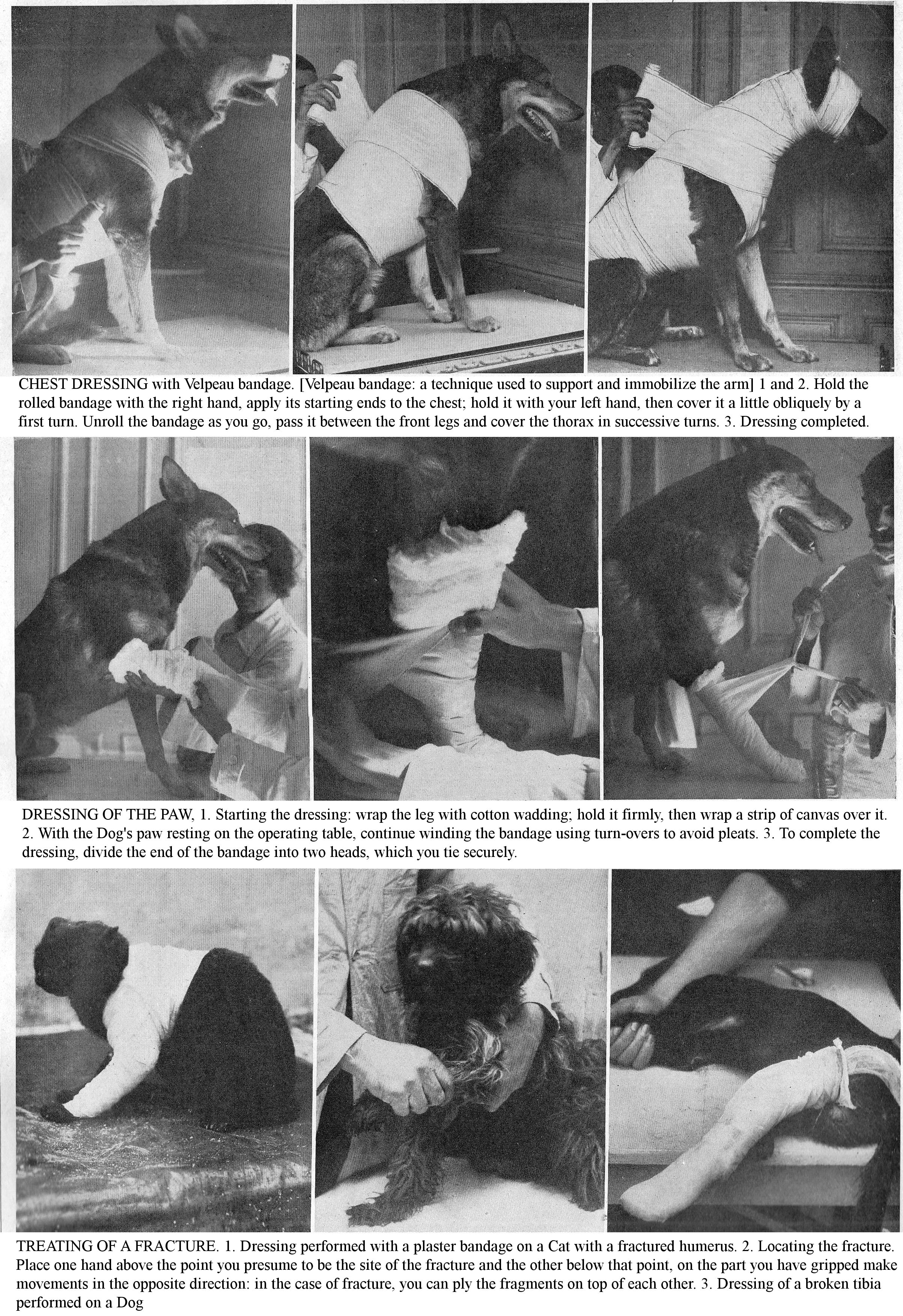
CHEST DRESSING with Velpeau bandage. [Velpeau bandage: a technique used to support and immobilize the arm] 1 and 2. Hold the rolled bandage with the right hand, apply its starting ends to the chest; hold it with your left hand, then cover it a little obliquely by a first turn. Unroll the bandage as you go, pass it between the front legs and cover the thorax in successive turns. 3. Dressing completed.
DRESSING OF THE PAW, 1. Starting the dressing: wrap the leg with cotton wadding; hold it firmly, then wrap a strip of canvas over it. 2. With the Dog's paw resting on the operating table, continue winding the bandage using turn-overs to avoid pleats. 3. To complete the dressing, divide the end of the bandage into two heads, which you tie securely.
TREATING OF A FRACTURE. 1. Dressing performed with a plaster bandage on a Cat with a fractured humerus. 2. Locating the fracture. Place one hand above the point you presume to be the site of the fracture and the other below that point, on the part you have gripped make movements in the opposite direction: in the case of fracture, you can ply the fragments on top of each other. 3. Dressing of a broken tibia performed on a Dog
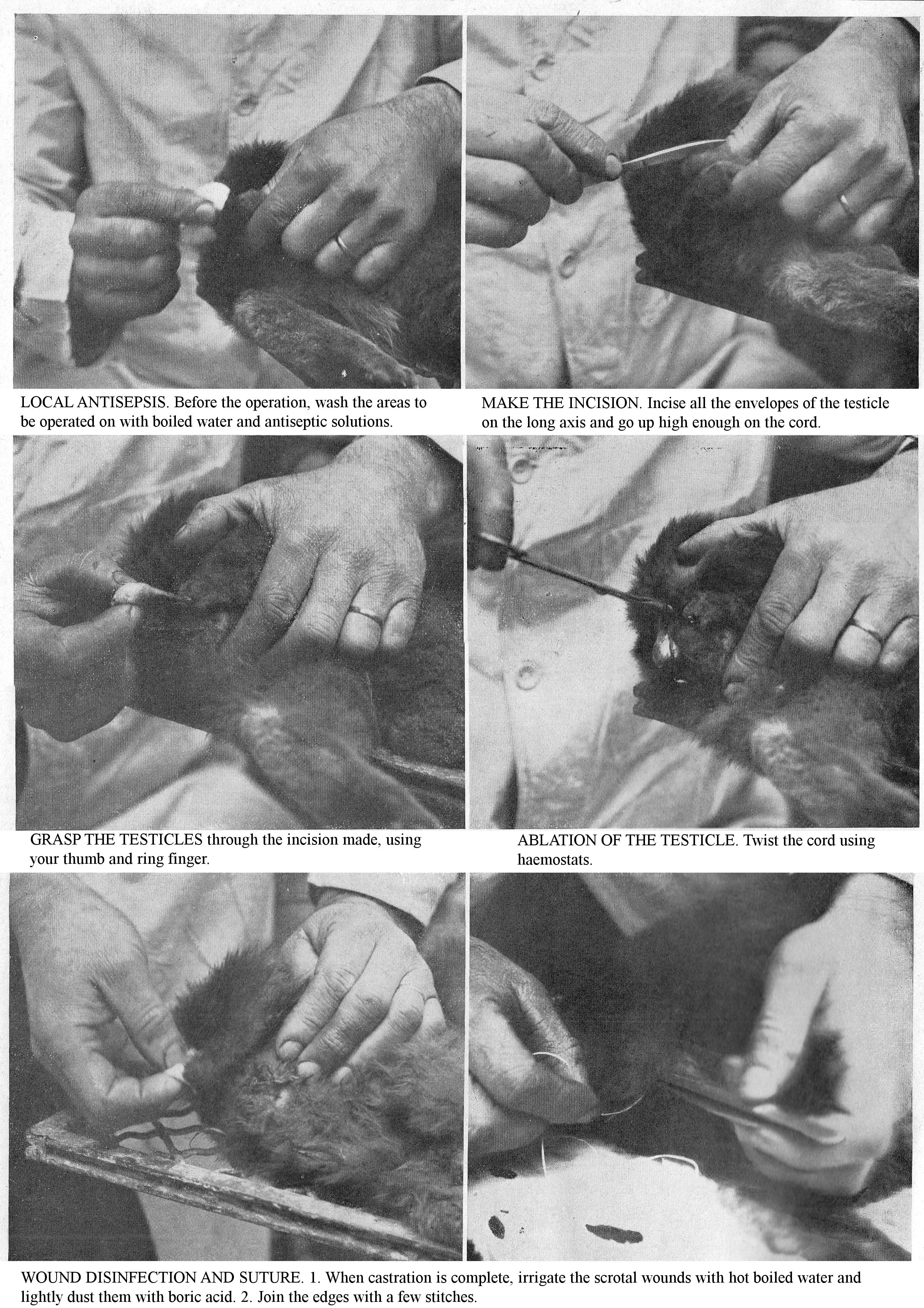
LOCAL ANTISEPSIS. Before the operation, wash the areas to be operated on with boiled water and antiseptic solutions.
MAKE THE INCISION. Incise all the envelopes of the testicle on the long axis and go up high enough on the cord.
GRASP THE TESTICLES through the incision made, using your thumb and ring finger.
ABLATION OF THE TESTICLE. Twist the cord using haemostats.
WOUND DISINFECTION AND SUTURE. 1. When castration is complete, irrigate the scrotal wounds with hot boiled water and lightly dust them with boric acid. 2. Join the edges with a few stitches.
SPECIAL OPERATIONS.
Resection of canine teeth and incisors. In Dogs, the teeth very early become covered in a yellowish, tartar. Clean them by scraping them from the crown to the end using a blunt instrument. When a quantity of tartar has accumulated at the base of the teeth, wash it off with a small swab dipped in water sharpened with hydrochloric acid. If you do not use these principles of dental hygiene, the gums ulcerate, the saliva drains in long strands from the corners of the mouth, and the mouth exudes a foul odour. When dental disease is generalised, as frequently observed in very old animals, remedy it by having the teeth extracted; do not evict all the diseased teeth in one session, for fear of subsequent fatal bleeding. The teeth are extracted using needle nose pliers or special forceps.
You only observe dental caries in old subjects; most often it follows a crack in the enamel. This condition causes, at varying intervals, sharp pains marked by disorders of chewing and depression: in addition, the mouth produces a very foetid odour. A decayed tooth appears cracked or hollowed out of a small cavity with blackish walls. Temporarily give Dogs who have undergone dental operations to a special diet based on liquid foods and easily chewed foods (soups, breads, milk, etc.).
Prophylaxis: Maintain oral hygiene by removing tartar deposits and monitor the condition of teeth and gums.
Cataract operation: Lens opacity is common in Dogs; old-age-related and bilateral cataracts are the most common; these cause loss of sight. This most delicate operation requires local anaesthesia (cocaine), but chloroform anaesthesia is preferred. The main surgical steps are excision and extraction.
Excision of the skin of the eyelids or conjunctiva: Excision of the skin of the eyelids is indicated in cases of entropion characterized by the inversion, inwards, of the free edge of the eyelid. The different operating stages include: excision of a skin flap of a width proportionate to the degree of Entropion and suturing of the edges of the wound. The retractility of scar tissue determines the straightening of the eyelid. Excision of the conjunctiva is indicated in the case of Ectropion, characterized by the outward turning of the free edge of the eyelid. When it is due to the swelling of the conjunctiva, scarify the conjunctiva and apply a cold compress to the eye; preferably excise a strip of the mucous ridge.
Extirpation of the eye: This operation is indicated in cases of eye growths and exophthalmos common in Dogs, following the scratching they get from cats. Enucleation of the eye requires anaesthesia.
Ear amputation: Ear amputation is performed either for therapeutic purposes (persistent auricular canker), or to remedy defective ear carriage, or, most often, by snobbery. Since dog shows in England and a few in France no longer admit Dogs with tails or cropped ears, the removal of these appendages is less common.
Do not cut the ears prematurely, wait until 3 months of age; at this point they are sufficiently grown that you can judge how much you need to shorten them. If you do it sooner, you risk having to start over in the event that they grow more than you expect. Have the amputation carried out by your veterinarian or by persons duly authorized by their knowledge of the trade and their experience, ear carriage contributes enormously to the beauty of the Dog.
If you find it useful to operate yourself, use the following technique: bring the skin of the ear towards the head, in order to avoid the denudation of the cartilage which would otherwise occur immediately after the operation, as a result of the retractability of the skin. Use curved or straight scissors; invert the ear, so as to expose the inside of the ear shell, then incise the skin and the cartilage at the same time, starting from the tubercle which is at the base of the part to be cut off, so as to keep a pointed flap, more or less long; but, in this ablation, always observe great uniformity. Using scissors, you are often required to trim the cartilage several times and, as a result, the ears are cut unevenly. This operation, simple in appearance, requires, in order to be carried out properly, special instruments to prevent haphazard amputations which make the subject ugly, spoiled and diminishes its value. So use ad hoc limiting pliers; the use of these allows you to judge the shape of the ear after ablation; cut overflowing flap with a scalpel or scissors; do not leave an excess of ear shell in the ear, otherwise it will appear heavy, wide and unsightly; after the operation, immobilize the two ends of the ears for 3 or 4 days by fixing them with a double cord tied behind the head. Post-operative care is zero. The straightening of the ears occurs naturally in a variable time depending on their attachment to the side of the head.
OPERATIONS ON THE DIGESTIVE SYSTEM.
OESOPHAGEAL CATHETERISM. Secure the Dog on a table, standing or lying down, head extended, jaws spread using one or two ligatures pulled in the opposite direction. Use catheters, similar to those used for exploring the urethra. Catheterize the oesophagus gently; violent manoeuvres expose you to taking the wrong path, introducing the catheter into the pharynx and tearing the oesophageal membranes.
LAPAROTOMY. Laparotomy consists of the surgical opening of the abdominal cavity, in whatever region it is performed; it is usually only the preliminary to surgery on the abdominal organs. Foreign bodies, wounds, abdominal tumours, intussusception, volvulus, adherent ventral and umbilical hernias, caesarean section, removal of reproductive organs, castration of females, etc. are all conditions which require laparotomy. Thanks to the progress of modern surgery, this operation can be successfully attempted in canine surgery.
PARACENTESIS. Paracentesis is an operation that punctures the abdominal walls, using a trocar, to release fluid that has escaped into the peritoneum.
Indications. Paracentesis of the abdomen is just a stopgap. It is performed in dogs for ascites or abdominal dropsy. However, this lesion is rarely essential; it frequently results from an organic disease (chronic diseases of the pericardium, endocardium, lung, pleura, liver, or tuberculosis).
Surgical technique. Prepare and disinfect the surgical area, puncture the abdomen on the white line with the trocar, a little behind the umbilicus, slowly evacuate the liquid, apply collodion to the wound and a cotton dressing. Often the operation is completed by an iodine injection into the peritoneal cavity.
HERNIA OPERATIONS.
Kelotomies, Instructions: Surgical intervention is a necessity for Hernias that are chronic, large, or causing serious functional disorders (strangulation).
Umbilical hernia: Umbilical hernia, common in puppies, gradually decreases after weaning and eventually disappears. Only operate if the lump has been static for several months or if it is growing in size.
Perineal hernias, located at the top of the perineum, between a deviated anus, the base of the tail and the tip of the buttock, require surgery, perineal kelotomy, or attachment to the abdominal wall of the herniated organ.
OPERATIONS ON THE CHEST.
Surgical interventions on the chest are limited to Thoracentesis; wisely practiced, it plays a major role in the healing process of thoracic effusions.
Thoracentesis: Thoracentesis is an operation that involves entering the sac of the pleura, across the walls of the chest, in order to release fluid accidentally accumulated therein. Indications: acute or chronic pleurisy, when the effusion is abundant and the dyspnoea is strong; hydrothorax, pneumothorax. Surgical technique: Disinfect the surgical site, perform a thoracic puncture using the trocar at the declining part of the thorax and at the level of the 6th and 7th intercostal spaces; evacuate of the exudate, apply a layer of collodion on the surgical wound and a wadded dressing held by a bandage.
GENITOURINARY OPERATIONS.
Apart from castration which is the main operation, surgery of the genitourinary organs involves catheterization of the urethra, bladder, and amputation of the penis: these operations require the intervention of the veterinarian; however, for documentary purposes, we will tell you the main operating stages.
CATHETERISM OF THE URETHRA. Catheterization or sounding of the urethra is an operation most often performed to release the urine accumulated in the bladder, or to clarify the diagnosis in cases of calculus or injury. Technique. Use of a rubber probe 30 to 35 cm. long and 2 to 3 millimetres wide; secure animal on its back; extend the penis; introduce the sterilized and oiled probe into the urethra.
CATHETERISM OF THE BLADDER. Use a rubber catheter, used in the male, or a shorter sound. It is sometimes difficult to introduce it into the meatus, due to the narrowness of the vulva and the folds of the mucosa. Having reached the bladder and after producing urine flow, the catheter makes it possible to detect the morbid state of the bladder wall. When there is a stone and you use a metal catheter, the contact of these two bodies produces a specific metallic noise.
PENIS AMPUTATION. This operation, which requires anaesthesia, is indicated in cases of gangrene, cancer of the penis or when the penis is lined with fleshy growths [polyps] which make urination more and more difficult, at the same time as they secrete a purulent substance that ferments, giving off an foul odour. Surgical technique. The main operating steps are as follows: anaesthesia, incise the sheath on the median line and over its entire length, excise of each of its parts, suture. Remove the anterior part of the penis and penile bone; suture, apply a wadded cotton dressing.
CASTRATION. This operation, from a medical point of view, is indicated in conditions of the testis (tumours), the cord and the prostate; sometimes it is performed as a convenience operation. An emasculated Dog becomes nonchalant, obese and no longer has the same hunting skills.
Technique. Prepare and disinfect the region, incise all the envelopes of the testicle on the long axis; section the posterior part of the cord, ligate the vascular part with a silk thread; remove the testis by sectioning the pedicle or by twisting. Disinfect scrotal wounds, suture, apply a wadded cotton dressing.
The cat is castrated by simple excision. This operation makes this animal more sedentary; it grows fat and its fur becomes bushier and silkier; besides, his excrements, especially his urine, loses that persistent and repulsive odour which makes the entire Cat's neighbourhood so unpleasant.
Castration of the female dog. In towns this operation is sometimes required to avoid the inconveniences of heat (apartment dog) and pregnancy. Castrated female dogs can be used for guarding and even for hunting. Castration can be, carried out by the flank or the white line; the operation should be performed under conditions of the most rigorous asepsis to avoid postoperative complications.
Flank technique: Disinfect the surgical area; make a skin incision of 4 to 5 cm. parallel to the last rib; divide the connective-adipose tissue; perforate the muscle layer and peritoneum. Locate the ovaries. Remove the ovary by twisting or excision, after ligation of the pedicle. Close the incision by a skin suture. Apply a wadded cotton dressing.
By the white line: Perform laparotomy on the white line behind the umbilicus. Introduce the index finger into the peritoneum, which easily detects the body of the uterus; grasp the ovary and remove by twisting or by sectioning the pedicle. Join the edges of the abdominal wound using a double suture; apply a wadded cotton dressing.
LOCOMOTOR SYSTEM OPERATIONS
FRACTURES
Fractures which make up the dominant aspect of canine surgery are breaks in the continuity of bones or cartilages; they may be incomplete or complete. The former include cracks, hairline cracks, incomplete fractures, and curvatures. Cracks may affect only the superficial parts of the bone or extend to the medullary canal. Incomplete fractures are characterized by depression of part of a bone as a result of external violence. Curvatures are most evident on the ribs and are seen as lesions specific to rickets. Complete fractures present the following varieties: simple fracture, when the bone is only broken in one place; multiple fracture, characterized by several breaks in continuity of the same piece of bone; comminuted and splintery fractures, accompanied by crushing of the bone or its division into several small fragments called splinters; complicated fracture, when the lesion is open to the air. Considered from the point of view of their site, fractures can be distinguished as diaphyseal, epiphyseal or intra-articular, with the break in continuity occupying the diaphysis [shaft], or epiphysis [end] of a long bone, or the articulating surface.
Aetiology: Among the predisposing causes of fractures, will mention the situation of the bones, their functions, diet, diseases of the bone system, and the age of the animals. Bones situated immediately under the skin, such as the radius, tibia, etc., are much more prone to fractures than the scapula, humerus and femur, which are situated deeper and are protected by muscles. The bones of the limbs are more often affected than those which form the abdominal cavities, and this is due to the active role they play as the main organs of the supportive and locomotor systems.
The extreme ages of life predispose animals to fractures. Nothing is as common as seeing fractures in young dogs, attributable on the one hand to the delicacy of the bones during this period, and on the other hand, to the frequent falls they make and the knocks the receive. Older dogs are prone to fractures because, over time, the bones undergo changes that make them less strong. The determining causes are external violence, such as bumps, bruises, collisions with the wheel of a car, bites, etc. The indirect causes act at a distance, by a sort of counter-reaction. Thus, a jolt to the incisors can produce a fracture of one or other branch of the jawbones; a very high jump can cause a fracture of the humerus or radius as the Dog touches the ground. Finally, muscle contraction can play a big role in causing fractures.
Symptoms: Symptoms vary depending on whether the fracture is complete or incomplete. The symptoms which characterize complete fractions are: deformation, abnormal mobility and crepitation. The deformation of the fractured region is determined by the mechanical displacements of the bone ends, by the blood effusions and by the congestion and inflammation which rapidly occurs at the site of the fracture.
Abnormal mobility is a very noticeable and characteristic sign in the Dog when a bone such as the radius, tibia, etc., is fractured; when the injured bone is surrounded by thick muscle masses, such as the femur, this symptom is more difficult to notice. In this case, use the following technique: apply one hand above the point you presume to be the site of the fracture and the other, below, apply movements in the opposite direction to the parts you have grasped, and if the continuity of the bone is really broken you can play the fragments on top of each other.
Crepitation or sounds of crepitation, a major diagnostic sign, is a kind of clicking or cracking sound, caused by the friction of the bone ends against each other. Sometimes this noise is clearly perceptible, as in the case of a superficial fracture; sometimes the hand feels it more than the ear hears it. To highlight this sign, hold the fragment closest to the trunk with one hand so as to immobilize it, and with the other hand apply various movements to the opposite fragment. Carry out this manoeuvre cautiously, taking all necessary precautions to not aggravate the fracture.
Scientific signs of fractures include the colour and functional irregularity of the fractured area. The pain is exaggerated by movement, by oscillations of the fractured ends and by muscle contraction. The functional irregularity of the apparatus, of which the fractured bone is a part, is usually very evident. Thus, the fracture of the radius of one limb results in that limb’s extreme lameness or inability to support weight; fractures of the lower jaw make chewing difficult, depending on whether the neck of the bone, one or both branches of the jaw are fractured.
Prognosis: The prognosis for fractures varies according to the site, nature, and complexity of the fracture, and the age and size of the patient. Determine the severity of a fracture based primarily on its site, relative to the joints, the degree of displacement of the bone ends, the presence or absence of splinters, and breaks in continuity of surrounding tissue. If the fracture is intra-articular, splintery, the task of healing the bones will be accompanied by serious complications (arthritis, suppurative synovitis, ankylosis); in such a case do not hesitate to destroy the patient. Healing fractures is generally more successful in small, lightweight dogs than in large, heavy dogs.
Treatment: There three main instructions for treatment: 1, realignment; 2, support; 3, combat inflammation. When a fracture occurs, it is quite rare that you can apply the device needed at the scene of the accident itself, the animal having to be transported following limb fractures. Take some precautions to prevent complications that decrease the chances of fractures healing. For the Dog, improvise a bandage using a handkerchief and temporary splints held in place with a ligature; you thus prevent the tears which result from the movement of sharp bone fragments in the flesh.
Fracture reduction includes: extension, counter-extension and coaptation. These various manoeuvres, which require the intervention of the veterinarian, are intended to restore the fractured limb to its natural length, direction and shape. In his absence, practice extension and counter-extension using ties fixed that you fix at a distance from the fractured ends, and on which you exert forceful traction. Anaesthesia is indicated to reduce reactions, muscle contractions and to facilitate realignment manoeuvres.
Coaptation consists in replacing the ends fractured ends in their proper physiological situation; it is important that the realignment be as perfect as possible, because the wrong direction given to a limb will causes part to protrude, thus producing an incurable deformity, sometimes accompanied by persistent lameness.
The support of fractures, in canine surgery, is one of the most difficult to achieve, not in terms of support itself, but in terms of the final result because it is impossible to completely immobilize the fractured ends. This requires you to use easily removable support appliances which allow the fractured parts to be inspected. Non-removable supports remain applied to the injured region for the time necessary for the fracture to heal. Use removable appliances for a Dog who presents crush fractures, with skin wounds and crushed bones, all complications which imperatively oblige the surgeon to frequently inspect the fractured parts. Apart from these cases, give preference to non-removable devices, because of the indocility of the animals and the need to wrap the entire fractured part in a sufficiently resistant sheath, made rigid by two splints to counterbalance the muscular contraction which constantly tends to make the fractured ends overlap each other and thus cause shortening of varying importance. Splints, placed one outside and the other in, should be strong enough not to flex under the weight of the body or move around. Make them out of cardboard, wood, or wire mesh, and form them into the shape and length of the fractured limb.
There are some general peculiarities of support device application that you should be aware of. Place wadding and cotton wool over the fractured area so that it exactly fills the voids and protects these parts from the pressure of the splints. Secure the splints with a plastered or starched bandage, moistened with plain water, either with an ordinary bandage (cloth or tarlatan) and solution of potassium silicate, or one of the following preparations: gutta-percha, 50 gr.; pitch-resin, 150 gr., or pitch-resin, 150 gr.; wax, 75 gr.
Preferably use plaster bandages as these allow rapid implementation of the dressing and ensure its strength. For fractures of the lower regions, use the bandage to cover the entire free part of the limb or at least the fractured radius and the regions below: wrap the bandage from the foot towards the root of the limb, and tighten it to the proper degree.
Unless there are complications, only remove the dressing when the following phenomena indicate normal healing of the fracture, that is to say after a delay of 3 weeks to 1 month; bolster it if necessary. Signs of severe pain, a strong febrile reaction, or loss of appetite, indicate some complication and require the appliance to be removed. Remove it carefully in order to avoid any movement which would destroy the adhesions of the fractured ends. Cut the bandage with scissors or secateurs, soak it with water at 40 – 50 centigrade; break the plaster or divide the gutta with the hot iron.
Left to themselves, the fractures can heal, but the end result is not always favourable as it is accompanied by a deformation of the region, which, by its voluminous callus, produces discomfort or irregularity in function. Abstention is not advisable except for fractures where there is little abnormal mobility of the ends (metacarpus or metatarsus). With exercise and massage, the bulky calluses gradually decrease, and the muscle atrophy following immobilization gradually disappears. In cases of intra-articular fracturs resulting in ankylosis, functional impairment; when the immobilization has failed, the fragments do not join together, and pseudo-arthrosis develops. Help heal fractures in young patients with rickets by administering bone powder or phosphate drugs.
Exposed fractures: In these fractures, the most serious from a prognostic point of view, the site of the trauma is exposed to the outside through an external wound. Treatment of exposed fractures includes the following instructions: disinfect the wound with hydrogen peroxide, a 3% carbolic solution, or tincture of iodine; apply to the wound, first powdered with iodoform or salol, cotton wadding that you change daily. In cases of gangrene, amputation is a last resort.
Here we provide, for information purposes, the particulars concerning the mitigation of the more common fractures seen in dogs, because the various manoeuvres (reduction, realignment, application of a brace) require the intervention of your veterinarian; but you can always apply first aid while waiting for his intervention to avoid sacrificing the patient.
Scapulum Fracture: Lay the subject on its opposite side and hold the limb extended with a helper. Arrange the bandages, dipped in pitch, under the arm, and cross them under the shoulder; with others, encircle the entire upper part of the forearm; set the appliance by wrapping the thorax in a belt and attach other bands to the base of the neckline. Remove the appliance after 20 to 30 days.
Fractured humerus: Fractures of the humerus heal well in dogs; sometimes the muscles of the arm and shoulder are sufficient to keep the fragments in contact. Muzzle the injured person and lay him on a table, on the side opposite the fractured limb, and have him held him securely by helpers. Apply tow around the fractured radius first, then to the rest of the limb, so as to remove any unevenness on the surface, then apply tow soaked in pitch or dextrin solution. Apply dextrin splints to one side of the area; methodically wrap the strip from bottom to top, with each turn covering at least half or third of the one before it.
Fracture of the radius: Well detached from the trunk and little protected over much of its internal surface, the radius is often affected by fractures usually produced by trauma, falls, sometimes by muscle contraction. Use a dextrin bandage with cardboard splints as described for fractures of the humerus.
Ulna fractures: In Dogs, the ulna is a bone distinct from the radius and can fracture in isolation. Splinting is provided by the latter. Healing occurs in all cases, even without a dressing; nevertheless apply a plaster, dextrin or starched bandage.
Fractures of the femur: In Dogs, set this fracture using a pitch plaster. Apply a pitch bandage to the croup, the outer face of the thigh and the leg; you see healing after a month.
Broken tibia and fibula: A fracture of the tibia is one of the most common in dogs. It heals very easily: apply two pieces of cardboard, shaped like the limb, and going from its lower end to above the stifle; wrap tow around the affected area; hold it in place with turns of tape coated with dextrin, potassium silicate or pitch; leave this bandage on for 3 weeks to 1 month.
Tarsal fracture: Tarsal fractures are very rare and are caused by external violence; jumps, slides, falls, etc. Apply a non-removable fenestrated bandage. Leave the dressing in place for 15 days to 3 weeks.
Fractured vertebrae: These fractures are usually the result of falls from a height, violent bruises, crushing; they lead to serious complications, almost always immediately fatal.
Rib fractures: These fractures can be complete or incomplete, single or multiple; they heal on their own; let animals rest and combat inflammation with appropriate treatment. Sometimes splintered fractures with sharp edges perforate the blood vessels and cause fatal haemorrhages.
In cats, fractures are less frequent; apply the same mitigation manoeuvres; use lightweight cardboard splints and silicate or dextrin bands as dressing materials.
TAIL AMPUTATION.
Tail amputation is often performed in dogs for therapeutic purposes (crushing, gangrene or growths); for a utilitarian purpose (tail is too long or poorly carried); for a fanciful purpose. In young or small dogs, cut the hair on the amputation line, cut off the tail with scissors, as far as possible at an inter-coccygeal joint; when bleeding stops, brush the wound with iodine tincture. Before cutting off the tail, pull the skin towards the rump; this way, as soon as it is released it goes back and covers the bone stump, ensuring rapid healing of the wound. For subjects with a large tail, use a tail-cutter and stop the bleeding by cauterization with a small annular cautery; in case of subsequent bleeding, perform haemostasis by applying a rubber band ligature; remove this after 5-6 hours to avoid gangrenous events.
The consequences of this operation are always the simplest when it is carried out on healthy dogs, purely by whimsy, to put animals in the fashion of the day; on the contrary, when performed to remove a localization of chronic eczema or for "caudal" canker, the stump may be the site of recurrence.
TENOTOMY.
Young dogs sometimes show a retraction of either of the forelegs; this defect of conformation can be to such a degree that, in some animals, they support themselves on the anterior aspect of the toes, and walking takes place by a sort of crawling. Only surgical tenotomy (sectioning the tendons of the metatarsal flexors) can restore normal function.
ABLATION OF DEWCLAWS.
The dewclaws are growths located on the lower and outer part of the hind legs; these are absolutely useless and can hamper walking; their presence, in a few selected breeds, constitutes a defect; so ablation is often practised. Amputation is very simple; do it with scissors, being careful of the living parts of the tissues; touch it with iodine tincture.