PERSISTENT PUPILLARY MEMBRANE
Persistent pupillary membranes (PPMs) are remnants of the embryonic pupil. Shortly after birth these membranes usually break down. Those that don’t are termed Persistent pupillary membranes and the remnants of these membraned sometimes stick to the back of the cornea, producing localised, or more generalised, opacity where they are attached. These often look like filaments or starbursts and usually don’t affect vision. In humans, atropine is sometimes used to dilate the pupil in order to break up PPMs that affect vision. Where they don’t affect vision, they are best left alone.
In dogs, PPM is known to be inherited in the Basenji. In cats, PPM is usually due to uveal disease and there is no known breed dispositions to PPM. However, because of the possibility of a genetic link, it’s recommended that animals with PPM shouldn’t be bred. I’ve seen a few reports of PPM in Bengals – one to a brown Bengal and two to silver Bengals. There are also reports of PPM in a European Shorthair and in a Persian.
CASES IN MEDICAL LITERATURE
The first account I have found is “Adhesion of a Persistent Pupillary Membrane to the Cornea in the Eye of a Cat” by By Edward Treacher Collins, published by the Ophthalmological Society of the United Kingdom in 1907. These are the most relevant excerpts:
“A cat about a year old was found to have on the posterior surface of the cornea in its right eye, at about the centre of the line of junction of its upper and middle thirds, a small, irregularly circular, pigmented patch. Proceeding backwards from this pigmented patch, across the anterior chamber to the anterior surface of the iris, were a number (fifteen to twenty) of delicate thread-like attachments. They varied considerably in thickness ; some were so delicate that they could hardly be seen without magnification, others were much stouter and were pigmented like the iris, showing also in places a reddish hue, suggesting that they contained blood-vessels. The position where they joined the iris corresponded to its small circle, a little distance external to the pupillary border. Some of them seemed to be prolongations forwards of the teeth-like processes forming the notched margin of the small circle. Many of the fibres started from the iris by several roots which united to form one. Some just previous to joining the cornea split up into several finer filaments. In all other respects the eye appeared normal.
The cat was killed with chloroform ; the right eyeball was removed and hardened in formol. Afterwards it was frozen and divided into two by an antero-posterior vertical section. The section passed through the pigmented patch behind the cornea, some of the filaments uniting it to the iris being in one half and some in the other. The front part of one half was embedded in celloidin and cut into microscopical sections; the other half was embedded in glycerine jelly and preserved as a macroscopical specimen .”
“It is conceivable that an adhesion of the pupillary membrane to the back of the cornea might occur in one of two ways : (a) The result of inflammation ; (b) the result of faulty development. “
“Von Hippel has described the microscopical appearances of an adhesion between the pupillary membrane and the cornea in a three day old, not prematurely born, child. It had also double microphthalmos [under-sized eyes], coloboma [incomplete closure of the retinal fissure], partial irideremia [where the iris is so rudimentary it appears to be absent], cataract, hare lip, cleft palate, polydactylism, and a congenital defect of the skull. “
Although that child evidently had multiple serious issues, Von Hippel’s comments may be relevant because more recent cases have occurred in a Bengal breeding line that has also produced micropthalmia, anopthalmia and corneal abnormalities.
According to SJ Aldavood and Sh Montakhabi, in “Persistant Pupillary Membrane in a Cat” presented at the World Small Animal Veterinary Association World Congress, 2004, “Pupillary membrane is a foetal structure, which covers the pupil prior to Birth. Shortly after birth it resolves. Sometimes part or all of it persists, therefore, it is called persistent pupillary membrane (PPM). This disorder is hereditary but the way of inherence is unknown. Most cases of PPM are not with visual deficits although sometimes some of those attending to the lens and cornea may cause blindness due to opacity. In November 2002, A 3 month old European Short hair kitten referred for inspection and preliminary Vaccination to small animal hospital, faculty of Tehran University. During clinical examination, The PPM diagnosed in this case. Some of pupillary membrane strands were free and did not induce cataract and keratitis. There were no adhesions. PPM has been reported in dogs and cats. Animals with PPM shouldn't be bred.”
Another account comes from R Khalkhal, SJ Aldavood and M J Ahmadi in “A Persian cat with Persistent Pupillary Membrane (PPM)” in the Proceedings of the 3rd ISVS & 9th ISVSAR, 2011, “Persistent pupillary membrane (PPM) is a congenital condition. It presents by remaining foetal membrane that persist as strands of tissue crossing the pupil. The pupillary membrane in mammals exists in the foetus as a source of blood supply for the lens. It normally atrophies from the time of birth to the age of four to eight weeks. PPM occurs when this atrophy is incomplete. It generally does not cause any symptoms. The strands can connect to the cornea or lens, but most commonly to other parts of the iris. Attachment to the cornea can cause small corneal opacities, while attachment to the lens can cause small cataracts. Using topical atropine to dilate the pupil may help break down PPMs. A 6-month-old female Persian cat referred with unilateral PPM in left eye. An examination showed severe PPM in the anterior segment of left eye. It has no attachment to cornea or lens. Then we release her to home and recommend the owner to refer us for laser therapy if it is going to have any changes in her eyes.”
EYE ISSUES IN BENGALS?
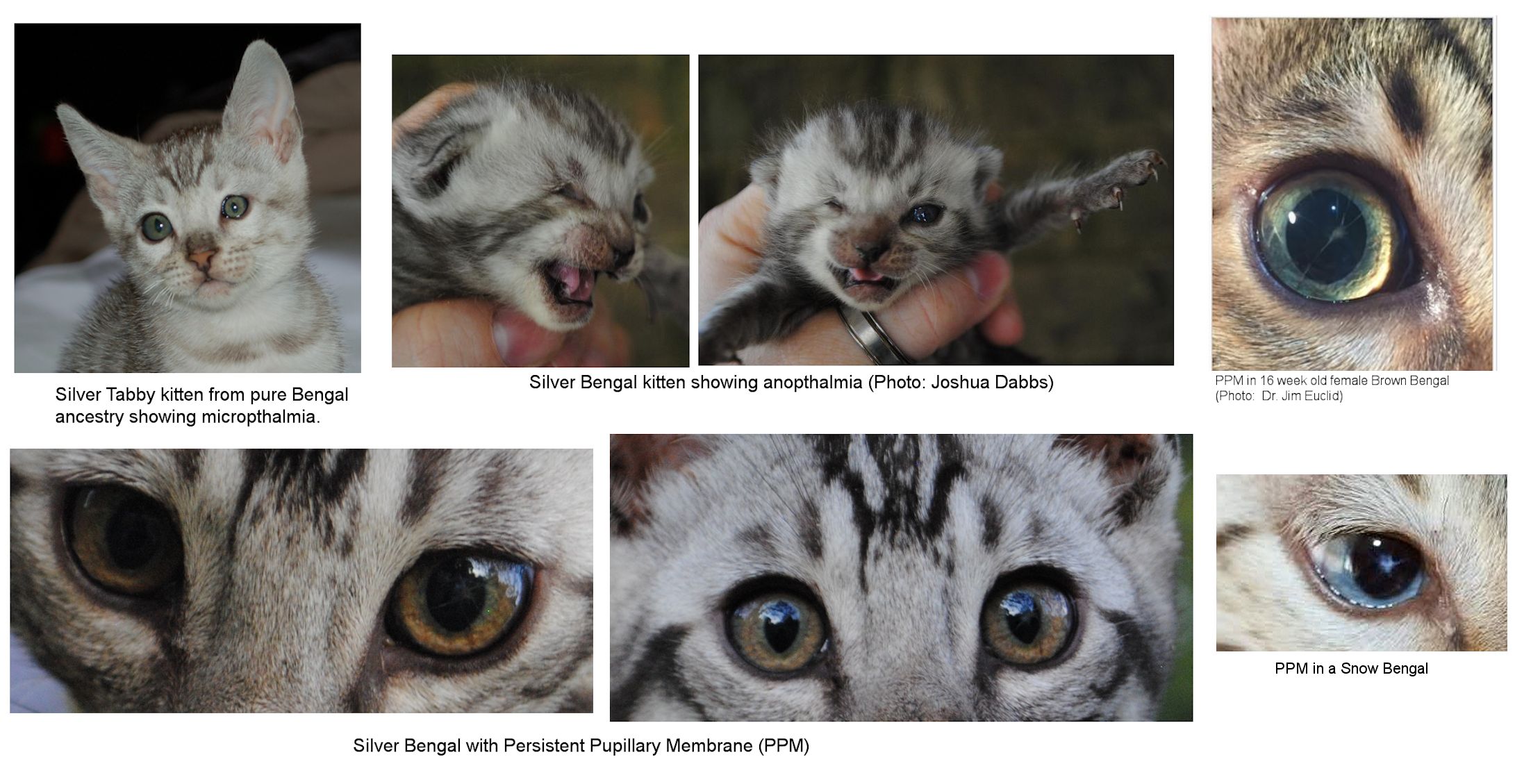
The image top left shows a silver tabby kitten from purebred Bengal parents. The micropthalmia may have been caused when its mother received medication during pregnancy. He is otherwise perfectly healthy.
In 2012, Joshua Dabbs sent me photos of a silver tabby kitten with unilateral anopthalmia. The kitten’s face is also misshapen: slightly sunken on the side without the eye. When he was given his intranasal vaccinations, it was found that the kitten had one functioning nostril. This suggests one side if the skull and face are underdeveloped. However, the kitten is healthy and thriving and will be able to live a happy, healthy life.
The images to the right show a Brown Bengal with PPM and below it a Snow Bengal with PPM. In the latter case, the membrane remnants appear blue rather than golden.
In 2016, Joshua Dabbs had a kitten with PPM. At first look, it was alarming because the strange shapes across the iris looked like some kind of internal eye parasite. He has not seen a case before or since this kitten, but he is breeding its parents together again and will pay close attention to their eyes in case it isn’t a fluke occurrence. It sounded like a developmental issue rather than a genetic issue, but it’s hereditary in some other species. There isn’t enough data in cats to know whether there is a genetic cause.
The PPM-affected kitten was only mildly affected and, when tested, his vision was not adversely affected. The worst part, according to Dabbs, was peering into the affected pupil and seeing a monstrous starburst shape that looked alarmingly like a parasite.
Dabbs has now seen micropthalmia, anopthalmia and PPD [Posterior polymorphous dystrophy - a bilateral, inherited condition affecting the corneal endothelium and Descemet's membrane that lies between corneal layers] in the same maternal line, each case being one generation apart from one another. The 1st gen produced the micropthalmia, the 2nd generation produced the anopthalmia and the 3rd gen produced the PPD. This suggests some kind of predisposition toward eye abnormalities.
The female that produced the microphthalmia had a total of ten litters from ten different sires. She has been used in line-breeding (breeding relatives, but not parent/offspring or sibling/sibling) without seeing any increase in eye issues and with few other issues. While the frequency of issues may seem too low to worry about, it is better to investigate at an early stage.
The males that sired offspring with these different defects are related to the females. It is hard to find unrelated males because the whole Silver Bengal breed in the USA is based upon a single American Shorthair outcross in the late 1990's. Silver requires a very controlled gene pool in order to maintain high contrast and low tarnish in a breed that normally shows a lot of rufousing. This lack of unrelated cats means inbreeding is a concern.
Dabbs has been monitoring the Coefficient of Inbreeding in the silver lines over the years, it's not uncommon to see some of the more developed silver lines have upwards twenty or more occurrences of that same American Shorthair outcross in their pedigrees, giving a Coefficient of Inbreeding between the mid twenties to low thirties. Some breeders have been concerned enough to develop a new outcross line using the American Shorthair and Egyptian Mau to reduce the level of inbreeding. When it comes to combatting inbreeding, TICA allows these normally non-permissible outcrosses with a no-showing restriction for the first three generations.
Some of the older Bengal breeders have also mentioned "eye problems", most often progressive retinal atrophy, which UC Davis now has a test for in Bengals, but occasionally also microphthalmia, anophthalmia and PPM. Even with degree of relatedness of cats, the frequency is low enough to suggest an additional trigger factor, such an environmental cause, is needed.

THE AMERICAN SHORTHAIR CONNECTION?
There is known to be an autosomal incomplete dominant gene causing the American Shorthair “head defect”. Cats with 1 copy of the gene had defects including eye defects such as coloboma (fissure of the eye socket due to eyelid plate not developing properly) or abnormally wide-spaced eyes. The more severe eye abnormalities that occurred were associated with lethal defects. This came about in the 1970s/80s when breeding for domed heads; early warning cosmetic issues were overlooked because they occurred in cats with superior conformation, leading to the further spread of the defect.
It's possible that something from the ASH made its way into the Bengal gene pool. When the silver was introduced in the late 1990's early 2000's, it had a positive impact on the Bengal breed. The breed shifted away from its early focus toward high amounts of rufism in favour of contrast and coat clarity, traits which were selectively bred into silver lines. Bloodlines from the silvers were spread throughout the breed and there are few modern breeding lines have not been touched by them.
However, in Silver Bengals, a lot of the work began at the 2nd and 3rd generation from the original American Shorthair outcross, meaning it could have been something already in the gene pool of the Bengals used in the outcross programme. Because all ancestry led to that one outcross cat, breeders of Silver Bengals have had to alternate between breeding to other silvers to maintain traits, or breeding to other colours to maintain health. By the mid-2000's, breeders were left with few alternatives but to inbreed, resulting in increased health issues (such as eye issues, predisposition to FIP, scoliosis (abnormally curved spine) and Flat Chested Kitten Syndrome). Although these occur at low frequency, the affected cats, and those cats passing on these traits need to be removed from the gene pool.
Outbreeding to brown Brown Bengals meant increased tarnish and poorer contrast. These cats also get removed from the gene pool because they are poorer specimens. This also narrows the gene pool.
While the frequency of issues may seem too low to worry about, it is better to investigate and contain any potential problem at an early stage and not go down the same route as the American Shorthair where apparently minor cosmetic issues were actually early warning signs.